box 2.1| The Cognitive Neuroscientist’s Toolkit
Navigating the Brain
Until now, we have been talking about only one or two neurons at a time. This approach is useful in understanding how neurons transmit information, but it is only one part of how the nervous system and the brain function. Neural communication depends on patterns of connectivity in the nervous system, the neural “highways” along which information travels from one place to another. Identifying patterns of connectivity in the nervous system is tricky because most neurons are not wired together in simple, serial circuits. Instead, neurons are extensively connected in both serial and parallel circuits.
A single cortical neuron is likely to be innervated by (i.e., receive inputs from) a large number of neurons: A typical cortical neuron has between 1,000 and 5,000 synapses, while a Purkinje neuron in the cerebellum may have up to 200,000 synapses. The axons from these input neurons can originate in widely distributed regions. Thus, there is tremendous convergence in the nervous system, but also divergence, in which a single neuron can project to multiple target neurons in different regions.
Localized interconnected neurons form what is known as a microcircuit. They process specific kinds of information and can accomplish sophisticated tasks such as processing sensory information, generating movements, and mediating learning and memory. For example, microcircuits in the retina of the eye process information from small regions of visual space. The patterns of light entering the eye strike photoreceptors, and the information from adjacent photoreceptors is processed through the microcircuitry of the retina to encode spatial information. This encoding permits a much smaller number of retinal ganglion cells to transmit the coded information down the optic nerve.
Although most axons are short projections from neighboring cortical cells, some are quite long, originating in one region of the brain and projecting to another at some distance. For example, the ganglion cells of the retina project via the optic nerve to the lateral geniculate nucleus of the thalamus, and the lateral geniculate neurons project via the optic radiations to primary visual cortex. Axons from neurons in the cortex may reach distant targets by descending below the cortical sheath into the white matter, traveling through long fiber tracts, and then entering another region of cortex, subcortical nucleus, or spinal layer to synapse on another neuron.
These long-distance connections between various brain regions connect to form more complex neural networks, which are macrocircuits that are made up of multiple embedded microcircuits. Neural networks support more complex analyses, integrating information processing from many microcircuits. Connections between two cortical regions are referred to as corticocortical connections, following the convention that the first part of the term identifies the source and the second part identifies the target. Inputs that originate in subcortical structures such as the thalamus would be referred to as thalamocortical connections; the reverse are corticothalamic or, more generally, corticofugal projections (projections extending from more central structures, like cortex, outward toward the peripheral nervous system).
Ultimately, neural networks are organized into neural systems. So, for example, the microcircuits of the retina, the lateral geniculate nucleus of the thalamus, and the patches of visual cortex are organized to create neural networks such as the thalamocortical network. This progression ultimately leads to the organization of the visual system as a whole, with its numerous cortical areas and subcortical components. (We will discuss the organization of the visual system in detail in Chapter 5.) In the next section we go on a brief anatomical tour of the brain. Early in each of Chapters 4 through 14, a section highlights the anatomy most relevant to the cognitive functions discussed in that chapter. The anatomy presented here and in the coming chapters will help you see which structures of the brain are related to which of its functions.
The two main divisions of the nervous system are the central nervous system (CNS), consisting of the brain and spinal cord, and the peripheral nervous system (PNS), consisting of the nerves (bundles of axons and glial cells) and ganglia (clumps of nerve cell bodies) outside of the CNS (Figure 2.17). The CNS can be thought of as the command-and-control center of the nervous system. The PNS represents a courier network that delivers sensory information to the CNS and carries motor commands from the CNS to the muscles. These activities are accomplished through two subsystems: the somatic motor system that controls the voluntary muscles of the body, and the autonomic motor system that controls the automated visceral functions. Before we concentrate on the CNS, a word about the autonomic nervous system.
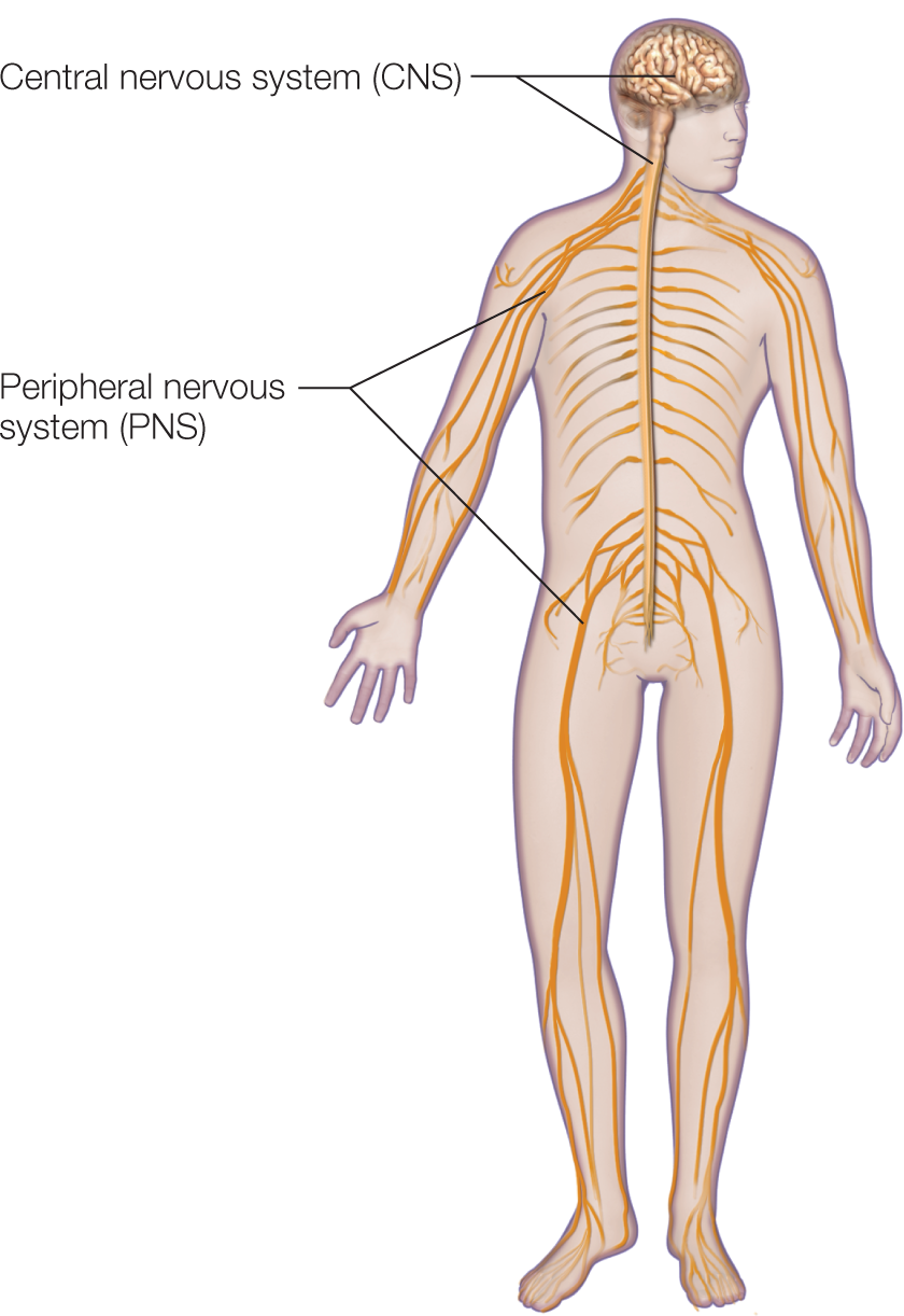
FIGURE 2.17 The peripheral and central nervous systems of the human body.
The nervous system is generally divided into two main parts. The central nervous system (CNS) includes the brain and spinal cord. The peripheral nervous system (PNS), comprising the sensory and motor nerves and associated nerve cell ganglia (groups of neuronal cell bodies), is located outside the central nervous system.
The autonomic nervous system (also called the autonomic motor system or visceral motor system) is involved in controlling the involuntary action of smooth muscles, the heart, and various glands. It also has two subdivisions: the sympathetic and parasympathetic branches (Figure 2.18). In general, the sympathetic system uses the neurotransmitter norepinephrine, and the parasympathetic system uses acetylcholine as its transmitter. The two systems frequently operate antagonistically. For example, activation of the sympathetic system increases heart rate, diverts blood from the digestive tract to the somatic musculature, and prepares the body for action (fight or flight) by stimulating the adrenal glands to release adrenaline. In contrast, activation of the parasympathetic system slows heart rate, stimulates digestion, and in general helps the body with functions germane to maintaining itself (rest and digest).
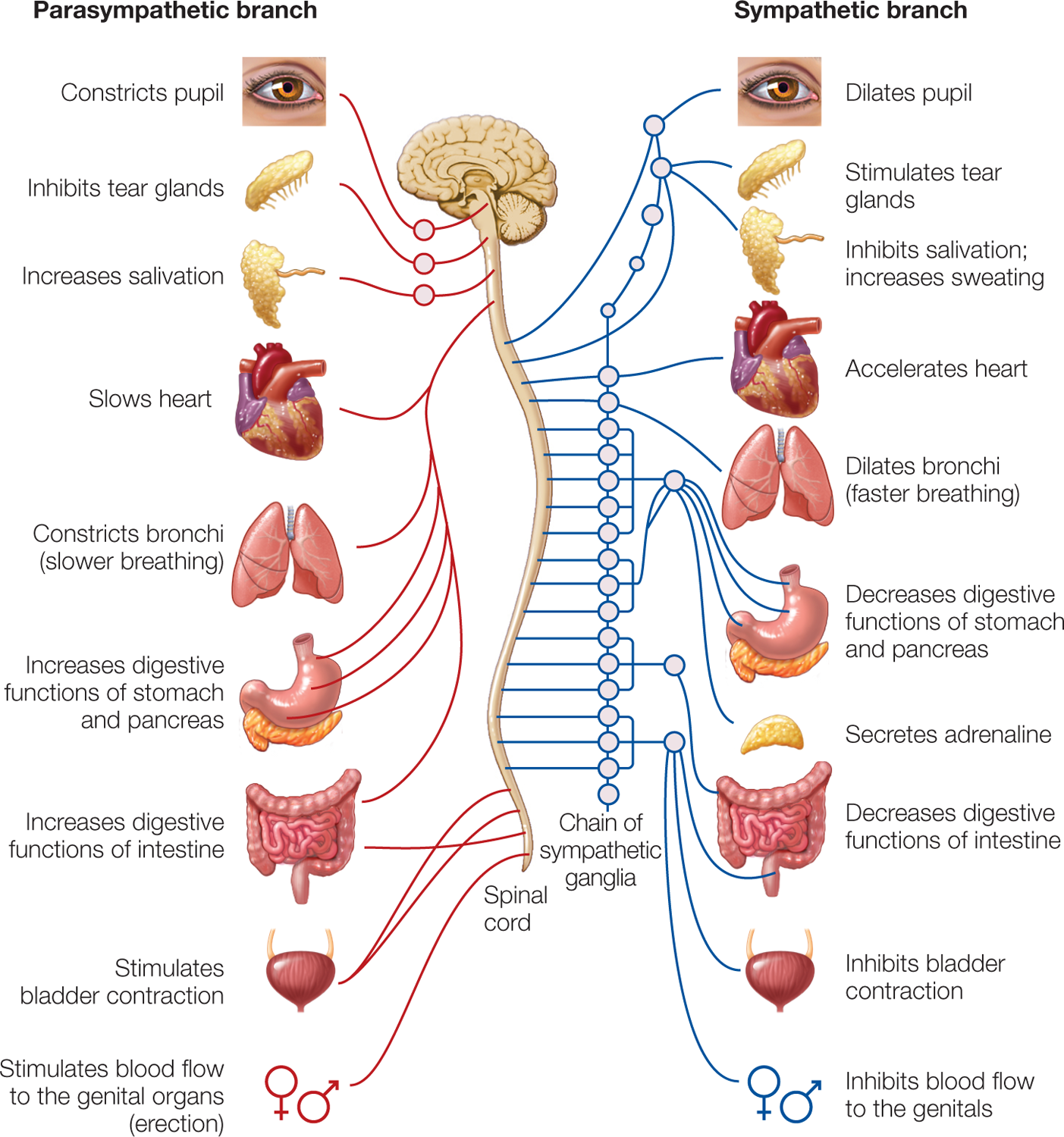
FIGURE 2.18 Organization of the autonomic nervous system.
In the autonomic system, a great deal of specialization takes place that is beyond the scope of this chapter. Still, understanding that the autonomic system is involved in a variety of reflex and involuntary behaviors (mostly below the level of consciousness) is useful for interpreting information presented later in the book. In Chapter 10, on emotion, we will discuss arousal of the autonomic nervous system and how changes in a number of psychophysiological measures tap into emotion-related changes in the autonomic nervous system. For example, changes in skin conductance are related to sweat gland activity, and sweat glands are under the control of the autonomic nervous system.
In the rest of this chapter we focus on the CNS in order to lay the groundwork for the studies of cognition presented throughout the rest of the book. When we talk about brain anatomy, we will use standard terminology to locate parts of the brain in three-dimensional space (see Box 2.1).
box 2.1| The Cognitive Neuroscientist’s Toolkit
Navigating the Brain
For anatomists, the head is merely an appendage of the body, so the terms used to describe the orientation of the head and its brain are in relation to the body. Confusion arises because of differences in how the head and body are arranged in animals that walk on four legs versus humans, who are upright. Let’s first picture the body of the cutest kind of dog, an Australian shepherd, looking off to the left of the page (Figure 2.21a). The front end is the rostral end, meaning “nose.” The opposite end is the caudal end, the “tail.” Along the dog’s back is the dorsal surface, just as the dorsal fin is on the back of a shark. The bottom surface along the dog’s belly is the ventral surface.
We can use the same coordinates to describe the dog’s nervous system (Figure 2.21b). The part of the brain toward the front is the rostral end (toward the frontal lobes); the posterior end is the caudal end (toward the occipital lobe). Along the top of the dog’s head is the dorsal surface, and the bottom surface of the brain is the ventral surface.
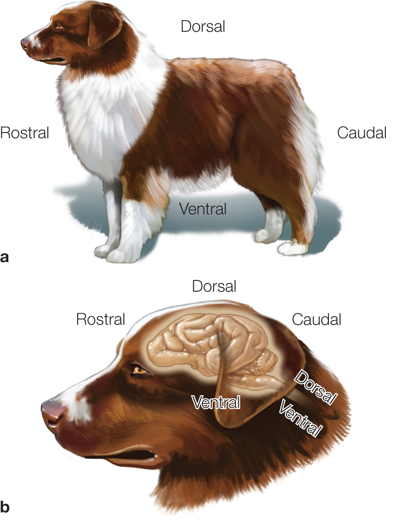
FIGURE 2.21 A dog brain in relation to the body.
We humans are atypical animals because we stand upright and therefore tilt our heads forward in order to be parallel with the ground. Thus, the dorsal surfaces of the human body and brain are at right angles to each other (Figure 2.22). In humans, we also use the terms superior to refer to the dorsal surface of the brain and inferior to refer to the ventral surface of the brain. Similarly, the terms anterior and posterior are used to refer to the front (rostral) and back (caudal) ends of the brain, respectively (Figure 2.22b). When we consider the spinal cord, the coordinate systems align with the body axis. Thus, in the spinal cord rostral means “toward the brain,” just as it does in the dog.
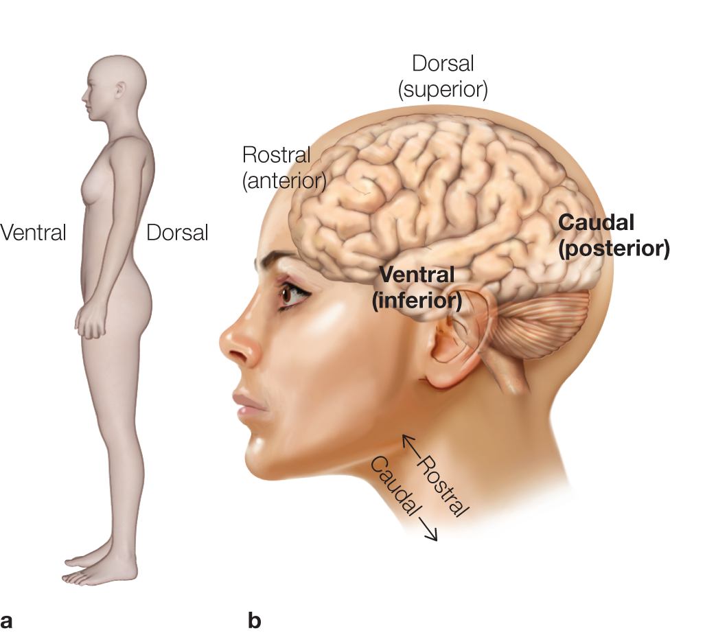
FIGURE 2.22 Navigating the human brain.
Throughout this book, pictures of brain slices are usually in one of three planes (Figure 2.23). If we slice the brain from nose to tail, we get a sagittal section. When we slice directly through the middle, we get a midsagittal or medial section. If we slice off to the side, we get a lateral sagittal section. If we slice perpendicular to a midsagittal section, separating the front of the brain from the back, we get a coronal section. When we slice in a plane that separates dorsal from ventral, we get a section that is described as axial, transverse, or horizontal.
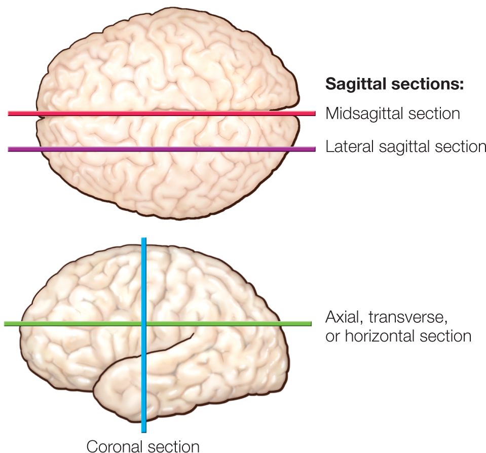
FIGURE 2.23 Three types of orthogonal planes through the brain.
The CNS is made up of the brain and spinal cord, and each is covered with three protective membranes, the meninges. The outer membrane is the thick dura mater; the middle is the arachnoid mater; and the inner and most delicate is the pia mater, which firmly adheres to the surface of the brain. Between the arachnoid membrane and the pia mater is the subarachnoid space, which is filled with cerebrospinal fluid (CSF), as are the brain’s ventricles, cisterns, and sulci, along with the central canal of the spinal cord. The brain actually floats in the CSF, which offsets the pressure and damage that would be present if it were merely sitting and scraping on the base of the skull. CSF also reduces shock to the brain and spinal cord during rapid accelerations or decelerations, such as when we fall, ride roller coasters, or are struck on the head.
Within the brain are four large interconnected cavities called ventricles (Figure 2.19). The largest are the two lateral ventricles in the cerebrum, which are connected to the more caudal third ventricle in the brain’s midline and the fourth ventricle in the brainstem below the cerebellum. The walls of the ventricles contain a system of specialized cells and capillaries, the choroid plexus, which produces CSF from blood plasma. The CSF circulates through the ventricles and on to either the subarachnoid space surrounding the brain or the spinal canal. It is reabsorbed in the brain by the arachnoid villi, protrusions into the venous system in the sagittal sinus.
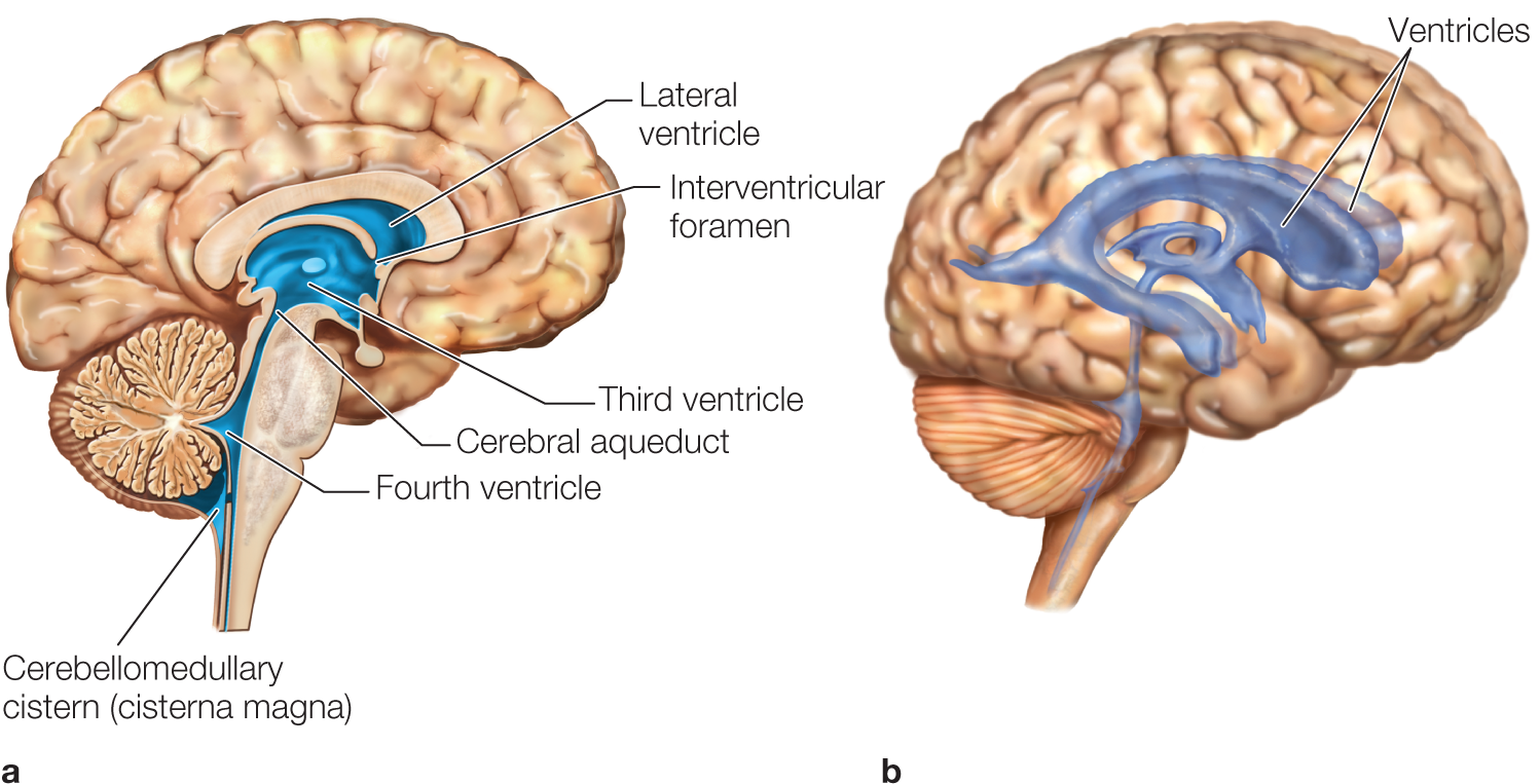
FIGURE 2.19 Ventricles of the human brain.
(a) Midsagittal section, showing the medial surface of the left hemisphere. (b) Transparent brain, showing the ventricular system in 3-D view.
In the CNS, neurons are bunched together in various ways (Figure 2.20). Two of the most common organizational clusters are the nucleus and the layer. A nucleus is a relatively compact arrangement of nerve cell bodies and their connections, ranging in size from hundreds to millions of neurons with functionally similar inputs and outputs. Nuclei are located throughout both the brain and the spinal cord.
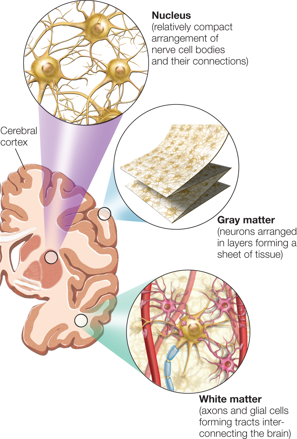
FIGURE 2.20 Organization of neurons in the CNS.
In the CNS, neurons can be organized in clumps called nuclei (top—not to be confused with the nucleus inside each neuron), which are most commonly found in subcortical and spinal structures, or sheets called layers (middle), which are most commonly found in the cortex. The cell bodies of glial cells are located in the white matter (bottom—e.g., oligodendrocytes), and in the cortex.
The cerebral cortex of the brain, on the other hand, has billions of neurons. They are arranged in a sheet containing several layers of neurons, folded across the surfaces of the cerebral hemispheres like a handkerchief. When we look at a slice of the brain, we see the cerebral cortex as a thin grayish layer overlying a whitish interior. The cerebellum is the other structure of the brain that is highly layered, containing billions of neurons, also having gray and white regions. The gray matter in these layers is composed of neuronal cell bodies, whereas the white matter consists of axons and glial cells.
Much like nerves in the PNS, the axons forming the white matter are grouped together in tracts that run from one cortical region to another within a hemisphere (association tracts), or that run to and from the cerebral cortex to the deeper subcortical structures and the spinal cord (projection tracts). Finally, axons may project from one cerebral hemisphere to the other in bundles that are called commissures. The largest of these interhemispheric projections is the main commissure crossing between the hemispheres, the corpus callosum.
The brain needs energy and oxygen, which it extracts from blood. Approximately 20% of the blood flowing from the heart is pumped to the brain. A constant flow of blood is necessary because the brain has no way of storing glucose or extracting energy without oxygen. If the flow of oxygenated blood to the brain is disrupted for even a few minutes, unconsciousness and death can result.
Two sets of arteries bring blood to the brain: the vertebral arteries, which supply blood to the caudal portion of the brain, and the internal carotid arteries, which supply blood to wider brain regions (Figure 2.24). Although the major arteries sometimes join together and then separate again, little mixing of blood occurs between the rostral and caudal arterial supplies or between the right and left sides of the rostral portion of the brain. As a safety measure, in the event of a blockage or ischemic attack, the circulatory system can reroute blood to reduce the probability of a disruption in blood supply; in practice, however, this rerouting of the blood supply is relatively poor.
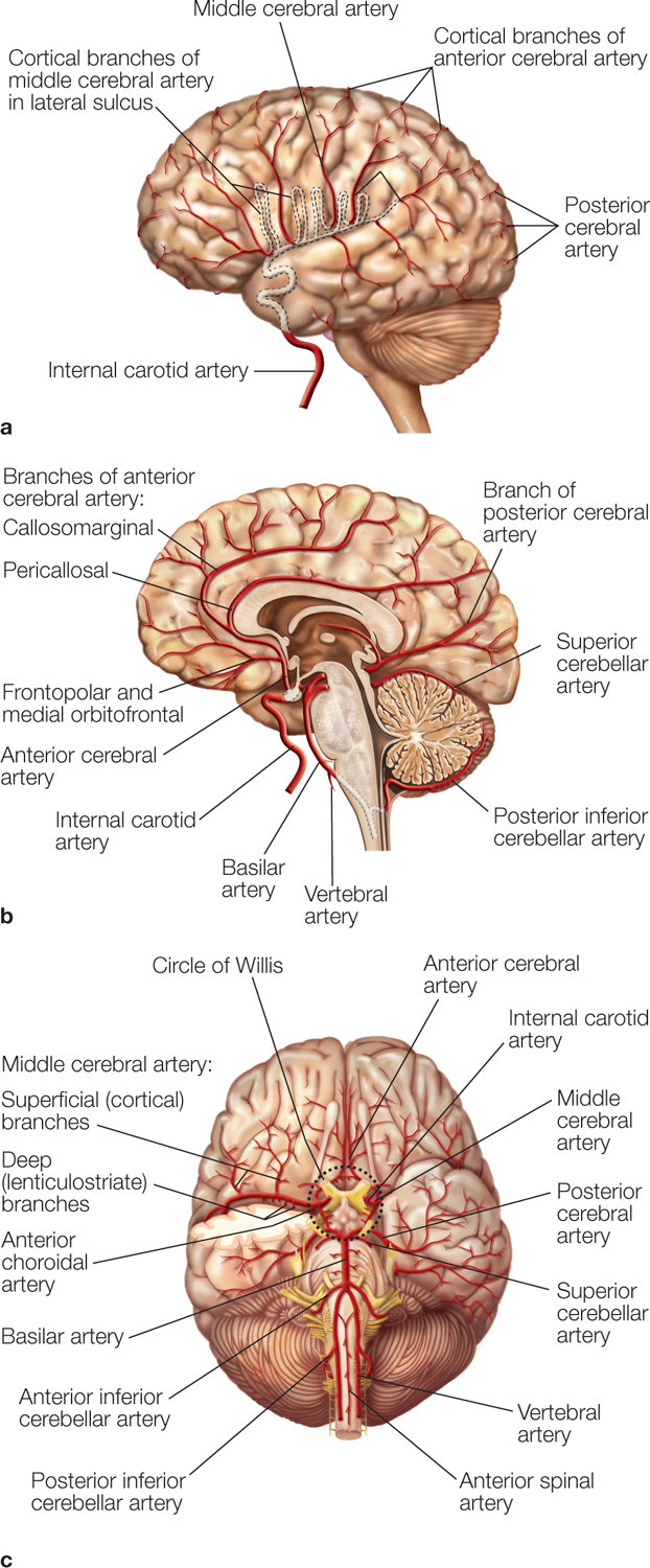
FIGURE 2.24 Blood supply and the brain.
(a) Blood supply to the lateral aspect of the cortex. (b) The midsagittal section reveals branches of the anterior cerebral artery, which extend from the anterior aspect of the circle of Willis and a portion of the posterior cerebral artery, which extends from the posterior aspect of the circle of Willis. (c) Ventral view of brain, showing the circle of Willis, the arteries encircling the base of the brain. The circle of Willis is supplied with arterial blood from the right and left internal carotid arteries, which extend the right and left common carotid artery and the basilar artery formed by the right and left vertebral arteries, which are branches of the subclavian artery.
Blood flow in the brain is tightly coupled with metabolic demand of the local neurons. Hence, increases in neuronal activity lead to a coupled increase in regional cerebral blood flow. The primary purpose of increased blood flow is not to increase the delivery of oxygen and glucose to the active tissue, but rather to hasten removal of the resultant metabolic by-products of the increased neuronal activity. The precise mechanisms for altering blood flow, however, remain hotly debated. These local changes in blood flow permit regional cerebral blood flow to be used as a measure of local changes in neuronal activity, serving as the basis for some types of functional neuroimaging, such as positron emission tomography and functional magnetic resonance imaging.
Take-Home Messages