When Darwin returned from his expedition on the HMS Beagle, he sketched in a notebook a simple tree—above which he jotted the phrase, “I think.” The concept he was attempting to think through was that of evolution: the idea that all living species arise from common ancestors, like twigs that bud from existing tree branches. Some of these twigs are strong and vigorous, and they grow into great branches from which new twigs—or species—will sprout; others die and fall to the ground, like species that go extinct but leave behind remains that are preserved in the fossil record. Although each branch of this tree is unique, all are related and form a family tree that connects the explosion of life forms that have spread across our planet and—as Darwin wrote in The Origin of Species—”covers the surface with its ever branching and beautiful ramifications.”
In this section, we outline—in broad terms—the diversity of organisms on our planet. We discuss the defining characteristics that distinguish the major branches of life’s family tree and offer a brief description of the lifestyles and lineages of the organisms that constitute each of these divisions. Because the genetic blueprints for all of these organisms are written in the universal language of DNA—information that modern sequencing methods allow us to easily read—we now have the ability to accurately characterize and catalog the ways in which living things differ and the ways in which they are alike. By studying these relationships, we can begin to assign every organism—the ones alive today and those that lived in the past—its proper place on the tree of life.
The Tree of Life Has Three Major Divisions
For most of human history, the living world was classified on the basis of outward appearances. A fish has jaws, eyes, a backbone, and brain; humans do, too—but a worm does not. A rosebush looks more like an apple tree than it does a blade of grass. Characterizing such visible features is certainly a good place to begin a study of the natural world. Such observations are what pointed Darwin toward his revolutionary theory of evolution.
But as the differences between organisms become larger, their relationships become harder to decipher. Is the fuzzy mold growing in that jar of jam more like a sunflower or a snow leopard? Is a shark more closely related to a starfish or a sperm whale? And what happens when organisms look remarkably similar? When it comes to bacteria, for example, one tiny rod-shaped cell can be almost impossible to distinguish from another—at least by sight.
The solution lies in their shared chemistry: because all organisms store genetic information in the form of DNA, analyzing the genome of an organism provides a simple yet powerful way to assess its kinship. The complete genome sequences of thousands of different organisms have been collected and are stored in publicly accessible databases. With the help of computers, DNA sequences from one organism can be quickly and easily compared to that of any other. Because DNA is subject to random mutations that alter these sequences, examining the differences between the DNA sequences of any two organisms can provide a direct measure of the evolutionary distance that separates them, a topic we revisit in greater depth in Chapter 9.
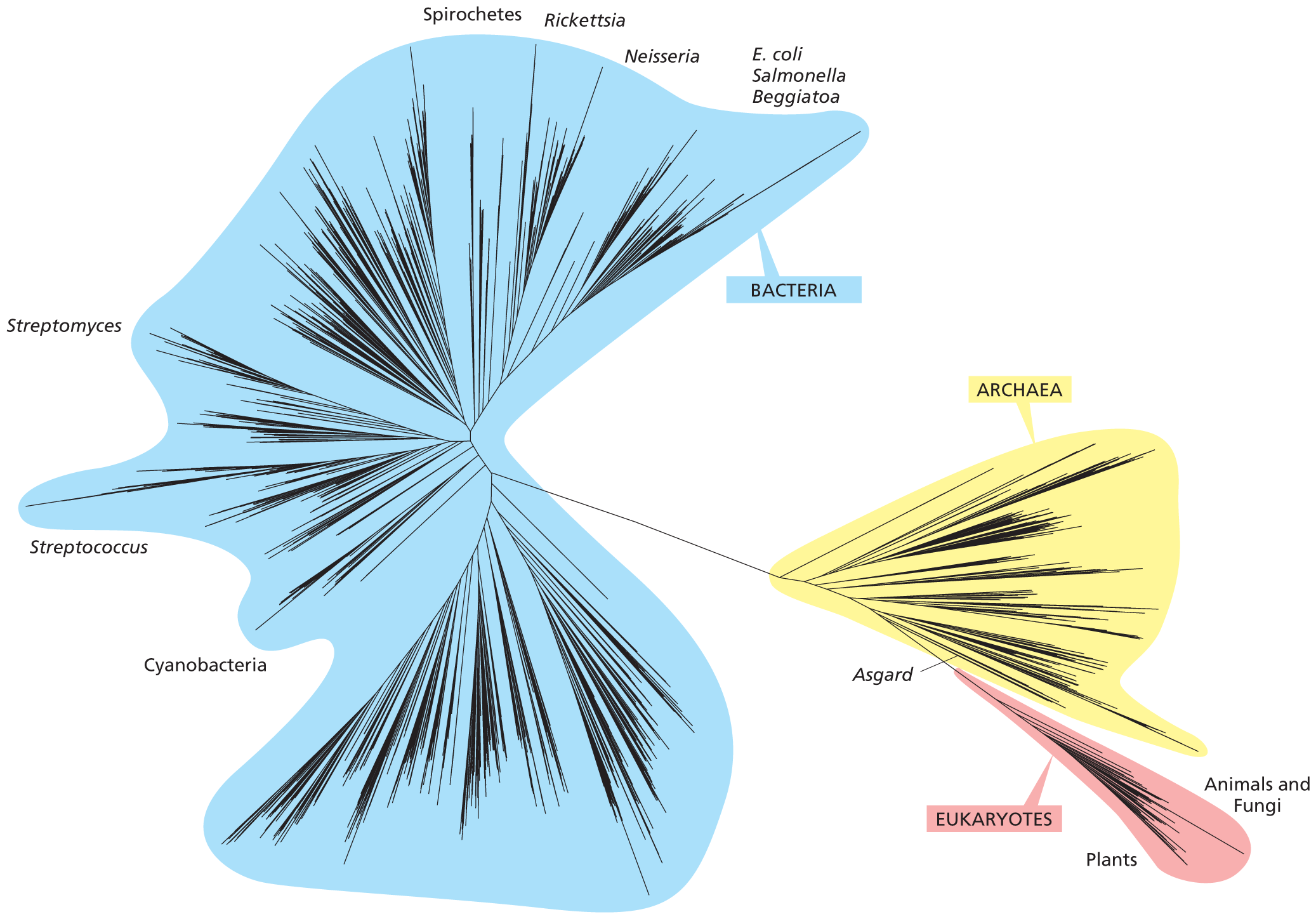
This approach has revealed that the living world consists of three major divisions, or domains: eukaryotes, bacteria, and archaea (Figure 1–10). Although eukaryotes are the main focus of this book, in terms of genetic diversity, they constitute the smallest domain within this elaborate, sequence-based tree.
More information
A diagram with lines representing different branches in the tree of life. Most of the branches are in a group highlighted as bacteria. Some specific bacteria are labeled and include Cyanobacteria, Streptococcus, Streptomyces, Spirochetes, Rickettsia, Neisseria, E coli, Salmonella, and Beggiatoa. A line connects Bacteria to two other smaller groups of branches labeled as Archaea and Eukaryotes. Archaea include Asgard. Eukaryotes include plants, animals, and fungi.
Figure 1–10Genome sequence comparisons produce a family tree of life that contains three major divisions. All organisms on Earth can be assigned to one of these domains: bacteria, archaea, and eukaryotes. Of the three domains, bacteria (blue) encompass by far the greatest diversity, commensurate with their ability to colonize nearly every ecological niche on the planet. So many new species of bacteria and archaea (yellow) are being discovered through DNA sequencing of environmental samples that many do not yet have names. Eukaryotes (pink) form a remarkably small slice of overall global diversity—and given their close kinship with archaea, some scientists have proposed combining eukaryotes and archaea into a single domain. In this diagram, the lengths of the branches are proportional to the differences among genomes as assessed by sequencing common genes that can be recognized and compared across many different species (discussed in Chapter 9). The tips of these branches represent individual species, but for clarity, only a small subset of Earth’s species is displayed. Some of the organisms discussed in this chapter and throughout the book are indicated. (Adapted from C.J. Castelle and J.F. Banfield, Cell 172:1181–1197, 2018.)
Eukaryotes Comprise the Domain of Life Most Familiar to Us
The living things we see around us every day are eukaryotes—from the flowers that bloom in our gardens to the face that gazes back at us from the mirror each day. The name comes from the Greek words eu, meaning “well” or “truly,” and karyon, a “kernel” or “nucleus.” Eukaryotic cells store their DNA in a membrane-enclosed organelle called the nucleus. Because this structure can be seen using a light microscope, its presence (or absence) was an obvious and simple way to begin to classify living organisms: those possessing a nucleus were called eukaryotes and those lacking a nucleus were called prokaryotes (from the Greek pro, meaning “before”). Thanks to DNA sequencing, we now know that the world of prokaryotes actually includes both bacteria and archaea—the other two domains of life.
Eukaryotic cells are typically much larger than those of bacteria and archaea. Their genomes run much larger, too: humans and corals each have more than 20,000 genes, whereas the bacterium Escherichia coli operates on a mere 4300.
Although we often think of animals when we contemplate eukaryotes (perhaps because we are animals, too), an astonishing array of single-celled, microscopic life forms are also eukaryotic—including many of the pond-dwelling creatures that entertained van Leeuwenhoek centuries ago. The eukaryotic family also includes fungi (such as mushrooms and the yeasts we use to make beer and bread) and plants. In fact, most of Earth’s biomass is found in plants. DNA sequencing, combined with other advanced techniques, suggests that our planet supports some 550 gigatons of carbon—carbon being the element on which the chemistry of life is based, as we discuss in Chapter 2. Although we usually measure chemicals in very small amounts—grams or even milligrams or micrograms—the amount of carbon on the planet is unimaginably large: a single gigaton is equal to 1015 grams. (If an elephant has a mass of about 5 × 10⁶ grams, 1 gigaton would be the equivalent of 200,000,000 elephants!) Of the more than 500 gigatons of carbon on Earth, 70 gigatons is found in bacteria, 7 gigatons in archaea, and a whopping 450 gigatons in plants. Animals constitute only 2 gigatons of Earth’s biomass—a number that is humbling.
Bacteria Comprise the Most Diverse Collection of Organisms on Earth
In reconstructing the tree of life based on DNA sequences, one of the biggest surprises was how much more evolutionarily diverse the world of bacteria is compared to that of eukaryotes. We now know that this immense diversity reflects the early appearance of bacteria in the evolutionary history of the planet: evidence suggests that bacteria have been around for at least 3.5 billion years, since Earth was in its infancy.
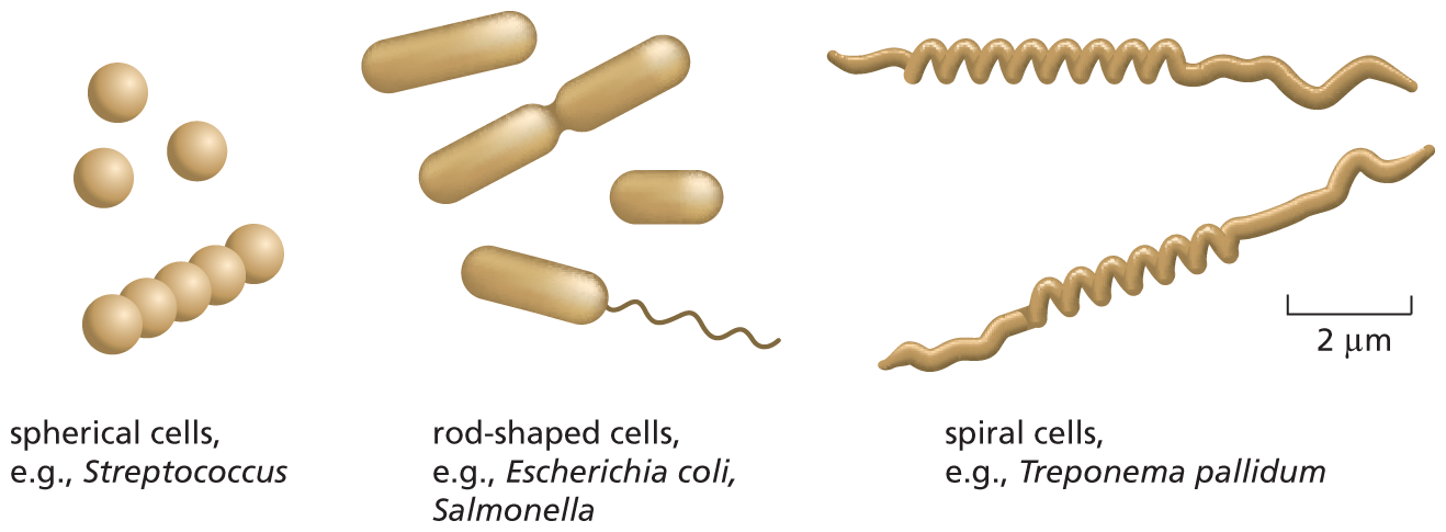
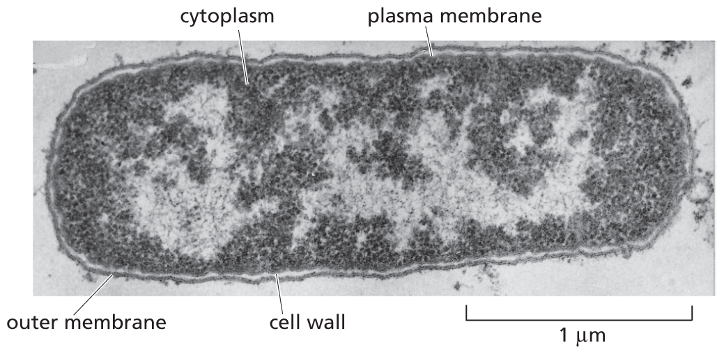
Bacteria (singular, bacterium) are generally very small: only a few micrometers long, they are invisible to the naked eye. Most live as single-celled organisms, although some join together to form chains, clusters, or other organized, multicellular structures. Bacteria are typically spherical, rodlike, or corkscrew-shaped (Figure 1–11). They often have a tough protective coat, or cell wall, surrounding the plasma membrane, which encloses a single compartment containing the cytoplasm and the DNA. Viewed with an electron microscope, the cell interior typically appears as a matrix of varying texture, without any obvious organized internal structure (Figure 1–12).
More information
An illustration shows three types of bacteria with examples. They are on the order of 1 to 10 micrometers in length. The first bacteria are spherical cells 1 micrometer in diameter. Example: Streptococcus. The second bacteria are rod-shaped cells 2 to 3 micrometers in length. Example: Escherichia coli and Salmonella. The third bacteria are spiral cells approximately 10 micrometers in length. Example: Treponema pallidum.
Figure 1–11Bacteria come in different shapes and sizes. Typical spherical, rodlike, and spiral-shaped bacteria are drawn to scale. The spiral cells shown are the organisms that cause syphilis.
More information
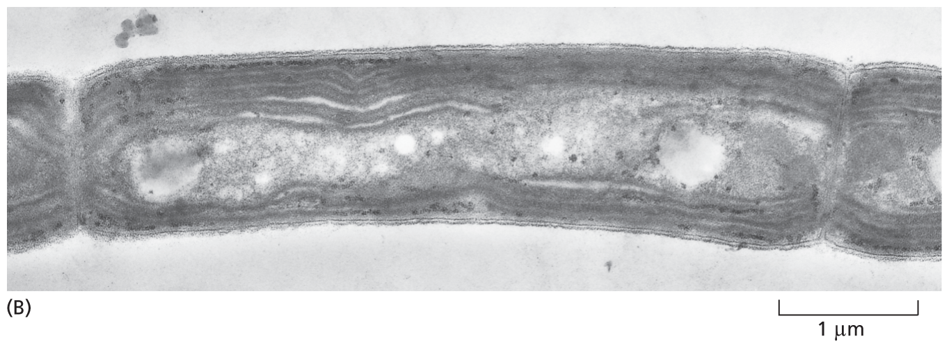
A micrograph shows a longitudinal section of an Escherichia coli bacterium. The bacterium is rod shaped; the labeled parts are as follows: outer membrane, cell wall, plasma membrane, and cytoplasm. The center of the cell is lightly stained. The scale reads 1 micrometer. with the cell being about 2 and a half times that in length.
Figure 1–12The bacterium Escherichia coli (E. coli) has served as an important model organism. An electron micrograph of a longitudinal section is shown here; the cell’s DNA is concentrated in the lightly stained region. Note that E. coli has an outer membrane and an inner (plasma) membrane, with a thin cell wall in between. The many flagella distributed over its surface are not visible in this micrograph. (Courtesy of E. Kellenberger.)
QUESTION 1–4
A bacterium weighs about 10–12 g and can divide every 20 minutes. If a single bacterial cell carried on dividing at this rate, how long would it take before the mass of bacteria would equal that of the Earth (6 × 1024 kg)? Contrast your result with the fact that bacteria originated at least 3.5 billion years ago and have been dividing ever since. Explain the apparent paradox. (The number of cells N in a culture at time t is described by the equation N = N0 × 2t/G, where N0 is the number of cells at zero time, and G is the time it takes for the population to double in size.)
Although simple in shape and structure, in terms of their chemistry bacteria are incredibly sophisticated. Some are aerobic, using oxygen to oxidize food molecules; some are strictly anaerobic and are killed by the slightest exposure to oxygen. Virtually any organic, carbon-containing material—from wood to petroleum—can be used as food by one sort of bacterium or another. Even more remarkably, some can live entirely on inorganic substances: they can get their carbon from CO2 in the atmosphere, their nitrogen from atmospheric N2, and their oxygen, hydrogen, sulfur, and phosphorus from air, water, and inorganic minerals. Some of these bacteria perform photosynthesis, using energy from sunlight to produce organic molecules from CO2, a strategy we commonly associate with plants (Figure 1–13); others derive energy from the chemical reactivity of inorganic substances in the environment (Figure 1–14). In either case, such bacteria play a unique and fundamental part in the nutritional “economics” of life on Earth, as other living organisms depend on the organic compounds that these versatile cells generate from inorganic materials.
More information
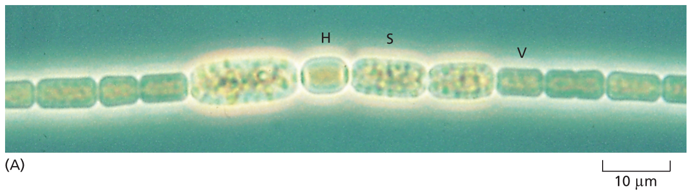
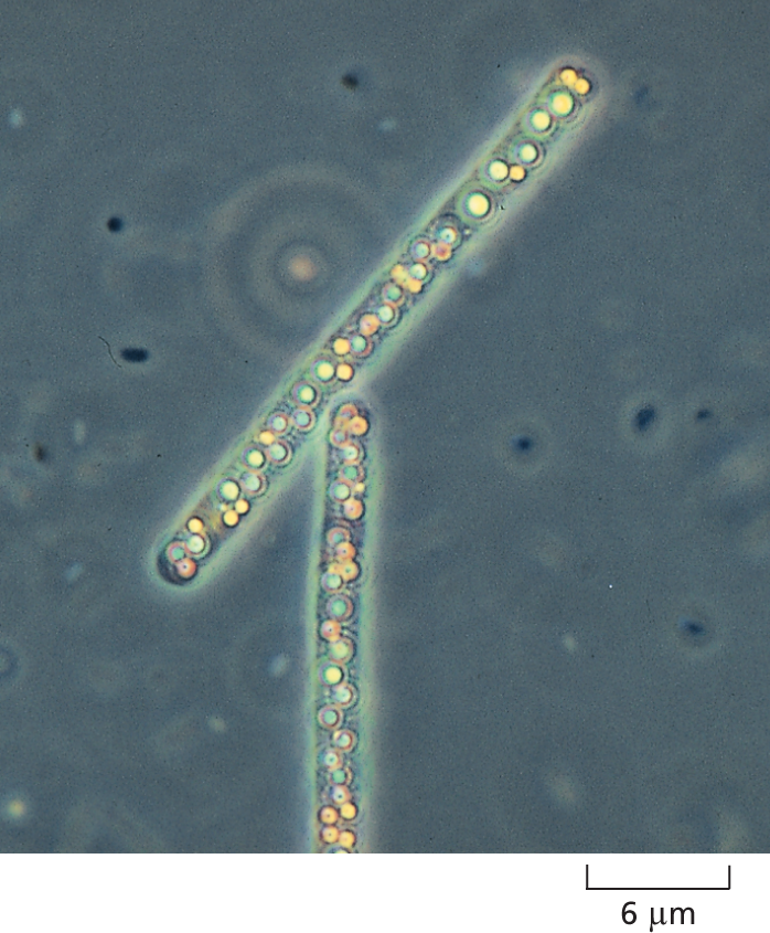
Micrograph A shows Anabaena cylindrical. The structure shows a string of beaded cells on scale of 10 micrometers. Four specialized cells in the string are larger of which two are labeled as H and S. The adjacent smaller cell is labeled V.
More information
Micrograph B shows Phormidium laminosum. The structure shows a rod-shaped cell with several intercellular membranes. The scale reads 1 micrometer.
Figure 1–13Some bacteria are photosynthetic. (A) Anabaena cylindrica forms long, multicellular chains. This light micrograph shows specialized cells that either fix nitrogen (that is, capture N2 from the atmosphere and incorporate it into organic compounds; labeled H), fix CO2 through photosynthesis (labeled V), or become resistant spores (labeled S) that can survive under unfavorable conditions. (B) An electron micrograph of a related species, Phormidium laminosum, shows the intracellular membranes where photosynthesis occurs. As shown in these micrographs, some prokaryotes can have intracellular membranes and form simple multicellular organisms. (A, courtesy of David Adams; B, courtesy of D.P. Hill and C.J. Howe.)More information
A micrograph shows Beggiatoa. A cylindrical shaped cell is shown with several rounded deposits inside the cell body. The scale reads 6 micrometers.
Figure 1–14A sulfur bacterium gets its energy from hydrogen sulfide (H2S).Beggiatoa, a bacterium that lives in sulfurous environments, oxidizes H2S to produce sulfur and can fix carbon even in the dark. In this light micrograph, yellow deposits of sulfur can be seen inside two of these bacterial cells. (Courtesy of Ralph W. Wolfe.)
Bacteria also exploit an enormous range of habitats, from the hot springs in Yellowstone National Park to the insides of other living cells and organisms. Some of these relationships are harmful. In the Middle Ages, bacteria that caused the bubonic plague wiped out half the population of Europe; today, different bacteria cause a variety of human diseases from cholera and whooping cough to tuberculosis. But some bacteria are actually beneficial. Thousands of different bacterial species reside on our skin and in our gut, where they promote a healthy immune response. And as we discuss later in this chapter, mitochondria—the organelles that generate energy in eukaryotic cells—are thought to have evolved from aerobic bacteria that took to living inside an anaerobic ancestral cell. Thus our own metabolism can be regarded as a product of the activity of an organelle whose evolutionary birthright we can trace to a bacterial cell.
Bacteria vastly outnumber all eukaryotic organisms on Earth not only because they are small and have been around for much longer, but because they reproduce so quickly. Under optimum conditions, when food is plentiful, a bacterial cell can divide in two about every 20 minutes. In only 11 hours, a single bacterium can therefore give rise to more than 8 billion progeny (which exceeds the total number of people currently on Earth). Thanks to their large numbers, rapid proliferation, and ability to exchange bits of genetic material by a process akin to sex, bacterial populations can evolve fast, rapidly acquiring the ability to use a new food source or to resist being killed by a new antibiotic (as we discuss in Chapter 9).
The World of Archaea Is the Most Mysterious of Life’s Domains
Of the three domains of life, that of archaea (singular, archaeon) remains the most poorly understood. Most of its members have been identified only by DNA sequencing of environmental samples, including those skimmed from the ocean surface or hauled up from the boiling hot hydrothermal vents that penetrate Earth’s crust along the darkest depths of the ocean floor. Like bacteria, the archaea we know most about are small and lack the internal, membrane-enclosed organelles that distinguish the eukaryotes. But archaea also differ from bacteria in many ways, including the chemistry of their cell walls, the types of lipids that make up their membrane, and the range of chemical reactions they can carry out.
More information
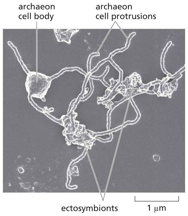
A micrograph of an archaeon and two ectosymbionts. The archaeon has a cell body about half a micrometer wide. Several long protrusions extend out from the body like strands. These strands wrap around two different ectosymbionts.
Figure 1–15An Asgard archaeon is caught clutching a pair of prokaryotic partners. A scanning electron micrograph shows that an elaborate network of membranous branches springs from the body of this cultured archaeal cell. These protrusions are wrapped around two other prokaryotic species—one bacterial, one archaeal—that were isolated as ectosymbionts along with the Asgard cell. This unusual archaeon was cultured for more than 2000 days in a bioreactor that mimicked conditions on the seabed floor; during this period, the Asgard cells divided once about every 20 days—compared to bacteria such as E. coli, which divide once every 20 minutes. (From H. Imachi et al., Nature 577:519–525, 2020.)
At first biologists believed that archaea occupied only the most extreme environments on Earth: the hot acid of volcanic springs, the airless depths of marine sediments, the sludge of sewage treatment plants, the icy pools that lie beneath the frozen surface of Antarctica, or the acidic, oxygen-free environment of a cow’s stomach, where they break down ingested cellulose and generate methane gas. Many of these inhospitable environments resemble the harsh conditions that must have existed on the primitive Earth, when living things first evolved. But we now know that archaea live everywhere, even on our skin. Archaea are believed to be the predominant form of life in soil and seawater, and they play a major role in recycling nitrogen and carbon, two of the most important elements for the biology of all cells.
DNA sequencing revealed an additional surprise regarding archaeal biology: although archaea resemble bacteria in their outward appearance, their genomes are much more closely related to those of eukaryotes. The archaea with the most eukaryotic-like genes belong to a lineage called Asgard (see Figure 1–10). These archaea were initially identified by sequencing DNA fragments collected from Loki’s Castle, a hydrothermal vent in the seabed off the coast of Greenland. While this sequence immediately revealed Asgard’s close kinship with eukaryotes, the most stunning discovery would not come until scientists managed to grow Asgard cells in the lab—a heroic feat that took 12 years of carefully tending to a custom-made bioreactor filled with seafloor muck and bathed in methane gas. Examining these cultured archaea in an electron microscope, the researchers were treated to a view of a cell that looks like no other living prokaryote: a jumble of long, meandering branches extends from its small cell body. But even more astonishing, the Asgard archaeon was observed to harbor a pair of ectosymbionts: one bacterium and an unrelated archaeon that appear to be entangled in the strange creature’s spindly embrace (Figure 1–15). The discovery of this unusual archaeon—with its prokaryotic associates—suggests a mechanism by which an ancestral cell might have ultimately captured and established a relationship with an aerobic bacterium: the step that initiated the evolution of the eukaryotic lineage.
Microscopic organism that is a member of one of the two divisions of prokaryotes; some species cause disease. The term is sometimes used to refer to any prokaryotic microorganism, although the world of prokaryotes also includes archaea, which are only distantly related. (See alsoarchaeon.)
Microscopic organism that is a member of one of the two divisions of prokaryotes; often found in hostile environments such as hot springs or concentrated brine. (See alsobacterium.)