eResearch Activity 2
How Do Bacteria Make a Magnet Chain?
Magnetotactic bacteria (see Figure 2.45) can orient themselves like a compass along Earth’s magnetic field. The vesicles of magnetite (Fe3O4) line up so as to maximize their overall magnetic moment, allowing the cell to behave like a compass needle. But how does the cell build magnetosomes and form the chain of vesicles that hold them in place? This extraordinary program of organelle development (organellogenesis) has been a mystery.
Surprising clues to the mystery emerge from the work of Mauricio Toro-Nahuelpan and colleagues at the University of Bayreuth, Germany (Fig. ERA 2.1). Toro-Nahuelpan observed magnetosome development in the bacterium Magnetospirillum gryphiswaldense. For clues to the development pathway, he constructed mutants lacking genes that encode various proteins needed to form magnetosomes.
More information
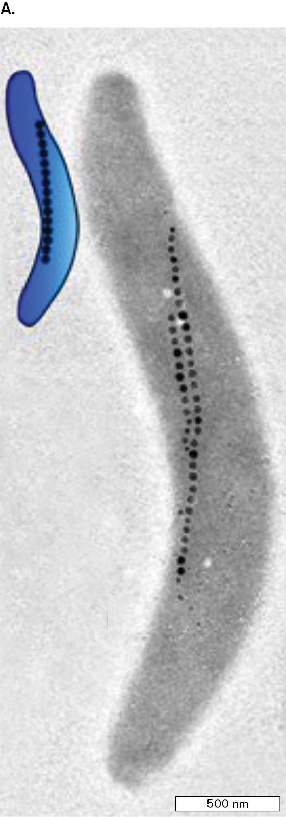
A micrograph of wild type Magnetospirillum gryphiswaldense. The bacterium appears as a grey spirochete with magnetosomes aligned along the axis. The magnetosomes appear as two lines of black dots. The bacterium is approimately 2,000 nanometers long.

More information
A photo of Maruicio Toro Nahuelpan. He has dark hair, a goatee, dark glasses and a white button up shirt with a dark jacket.
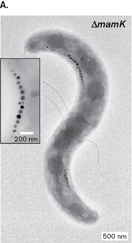
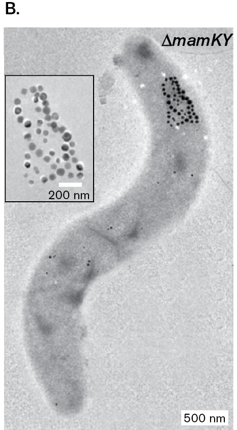
The TEM images of the various M. gryphiswaldense mutants show intriguing differences (Fig. ERA 2.2). The mutant that lacks MamK protein (panel A) makes fragmentary chains of magnetosomes that lie at the cell’s curvature instead of along its central axis. A mutant lacking both MamK and MamY (panel B) makes chains that are tangled, with no connection to the cell membrane. Another mutant possesses MamK and MamY, but fails to express MamJ (panel C). This mamJ deletion mutant forms aggregations of magnetosomes with no linear chains.
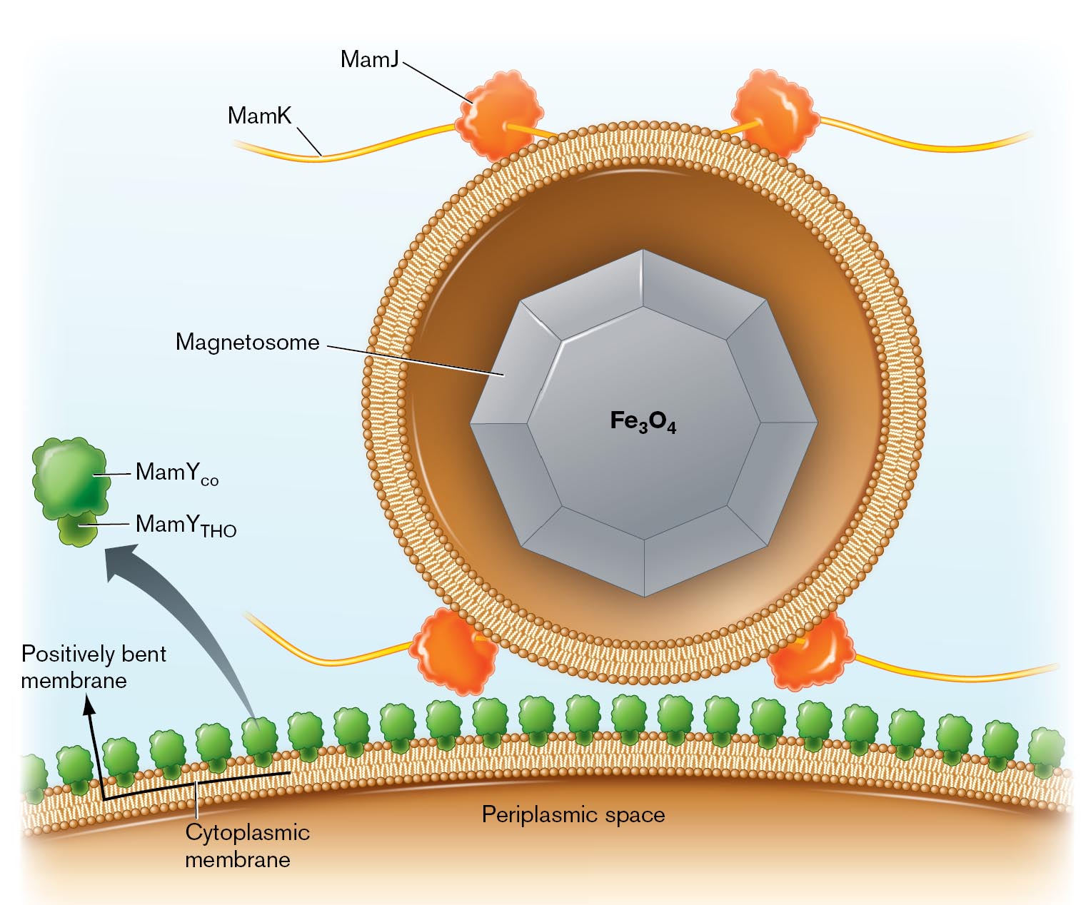
The data from Fig ERA 2.2 were combined with other molecular and genetic experiments that used fluorescence microscopy and cryo-electron tomography. On the basis of these experiments, Toro-Nahuelpan and colleagues propose a model for magnetosome development (Fig. ERA 2.3). In this model, MamY (green) is proposed to be a transmembrane protein that forms a line along the inner curve of cytoplasmic membrane. MamJ proteins (orange) hold the magnetosome vesicles in place along a filament of MamK proteins (yellow). Then the MamJ proteins tether the string of magnetosomes to the MamY line along the inner curve of the cell. By fixing to points on the inner curve, the filament is maintained overall at an axial position in the cell.
More information
One of three micrographs of mutant strains of Magnetospirillum. Each bacterium is approximately 3,000 nanometers long. This figure is Part A. It is strain delta m a m K. The magnetosomes are seen along the curves of the bacterium.
More information
One of three micrographs of mutant strains of Magnetospirillum. This figure is Part B. It is strain delta m a m K Y. The magnetosomes are seen clustered at the top of the bacterium.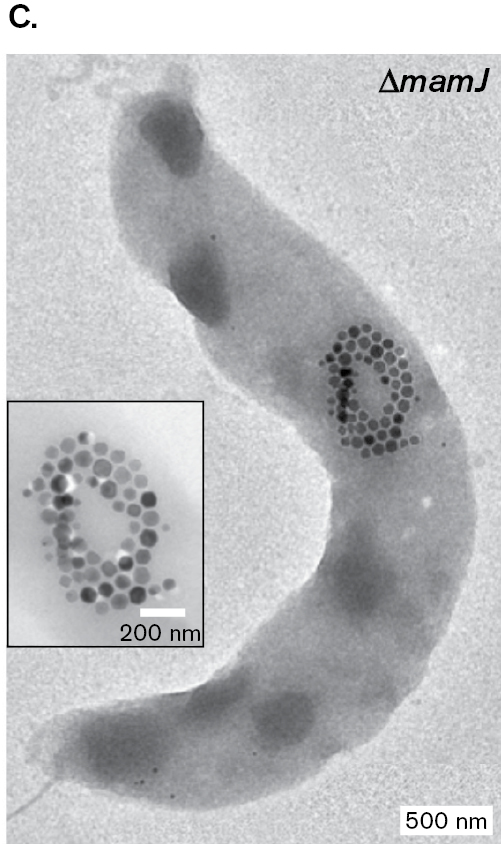
More information
An illustration of the model for magnetosome development. M a m Y is shown as a green, lightbulb like shape on the left of the illustration and attached to the cell’s cytoplasmic membrane. A magnetosome is shown in the middle of the image as a large, grey octagon surrounded by a cytoplasmic membrane. M a m J proteins shown as orange blobs around the outside of the magnetosome’s cytoplasmic membrane. M a m J protein filaments are shown in yellow with each of the M a m J proteins. The cell’s cytoplasmic membrane and periplasmic space is shown at the bottom.
Further Exploration
What do you think of the model proposed in Fig ERA 2.3? Is it consistent with the data? What aspects of magnetosome development are not addressed? How might you test other questions?
Toro-Nahuelpan, Mauricio, G. Giacomelli, O. Raschdorf, S. Borg S, J. M. Plitzko, et al. 2019. MamY is a membrane-bound protein that aligns magnetosomes and the motility axis of helical magnetotactic bacteria. Nature Microbiology 4:1978–1989.
Glossary
- Figure 2.45:
-
More information
Three images show increasing detail of the structure of Magnetospirillum magneticum based on cryo-electron tomography.
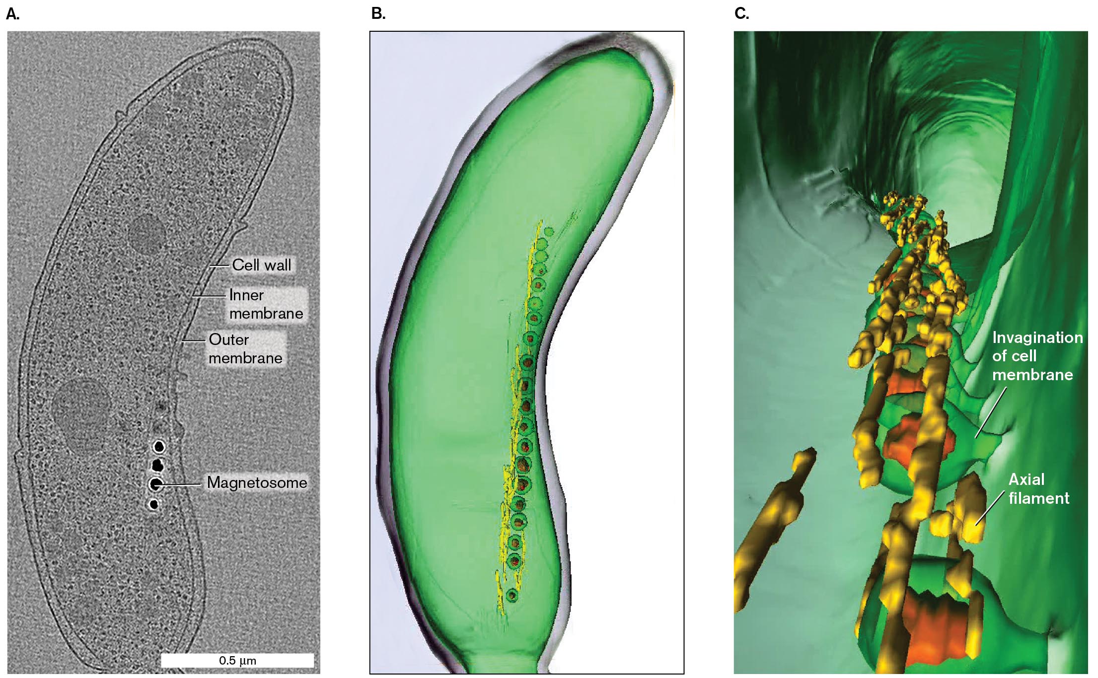
A cryo-electron tomograph shows the labeled structure of Magnetospirillum magneticum. The labeled parts are cell wall, inner membrane, outer membrane, and the four dots near to the inner membrane are labeled as magnetosomes. The cell is a bean shaped structure of about 1.5 micrometers in length and 0.5 micrometer in width.
A 3 D structure of M. magneticum with several magnetosomes. It is green and bean shapes. The magnetosomes form a vertical chain inside on one side.
A 3 D model of the cell interior of M. magneticum. The internal structure appears as a hollow tunnel with several magnetosomes embedded in the cell membrane. The point where the magnetosomes embed in the cell membrane is labeled, invagination of cell membrane. Axial filaments are identified between magnetosomes.
FIGURE 2.45 ■ Magnetotactic cell visualized by cryo-electron tomography. A. A single cryo-EM scan lengthwise through Magnetospirillum magneticum. B. 3D model of M. magneticum based on multiple scans. C. Expanded view of the cell interior.AAAS. ARASH KOMEILI ET AL. SCIENCE 311:242–245, FIG. 1NIH, THE JENSEN LABORATORYNIH, THE JENSEN LABORATORY