LEARNING GOAL
Explain how neurons communicate during the three phases of neural communication.
LEARNING GOAL
Explain how neurons communicate during the three phases of neural communication.
No matter what you are doing, at every moment of your life your neurons are communicating with each other to let your nervous system receive information, process it, and respond to it. Without neural communication, you would not be able to smell a lily, think about your weekend plans, or hold the hand of someone you love.

FIGURE 2.4 Jack Osbourne and MS
Jack Osbourne is one of the estimated 2.5 million people with multiple sclerosis (MS). This disease damages neurons in the nervous system and disrupts neural communication. The result is sensory and motor impairment.
Some people experience a breakdown in this neural communication, including Jack Osbourne, the son of the rocker Ozzy Osbourne. Jack was diagnosed with multiple sclerosis (MS) at age 26 (Figure 2.4). After the birth of his daughter, Osbourne noticed he was having strange problems with his vision, such as black dots limiting his sight. Doctors determined that Jack was in the early stages of MS, a disorder of the nervous system that is typically diagnosed in people between ages 20 and 40 (Dobson & Giovannoni, 2019). MS affects the brain and the spinal cord, and people with MS lose the ability to coordinate their actions. Their movements become jerky, and gradually their abilities to move, see, and think become severely impaired.
To understand how neurons typically communicate, and how this process breaks down in disorders such as MS, you need to understand the three phases of neural communication, summarized in Figure 2.5. During the transmission phase of neural communication (Figure 2.5, Phase 1), electrical signals created in the cell body travel along the axon and neurotransmitters are released from the terminal buttons into the synapse. During the reception phase (Figure 2.5, Phase 2), the dendrites of other neurons receive these signals from sending neurons. During the integration phase (Figure 2.5, Phase 3), neurons assess and integrate the incoming signals. Then this neural communication process repeats, with signals transmitted to other neurons, which receive the signals and integrate them. The process is similar to the tweeting and re-tweeting that occur on Twitter: People tweet to their followers, who receive the tweet in their Twitter feed and possibly re-tweet it to their own followers, who then process the re-tweet and may decide to forward it to even more people.
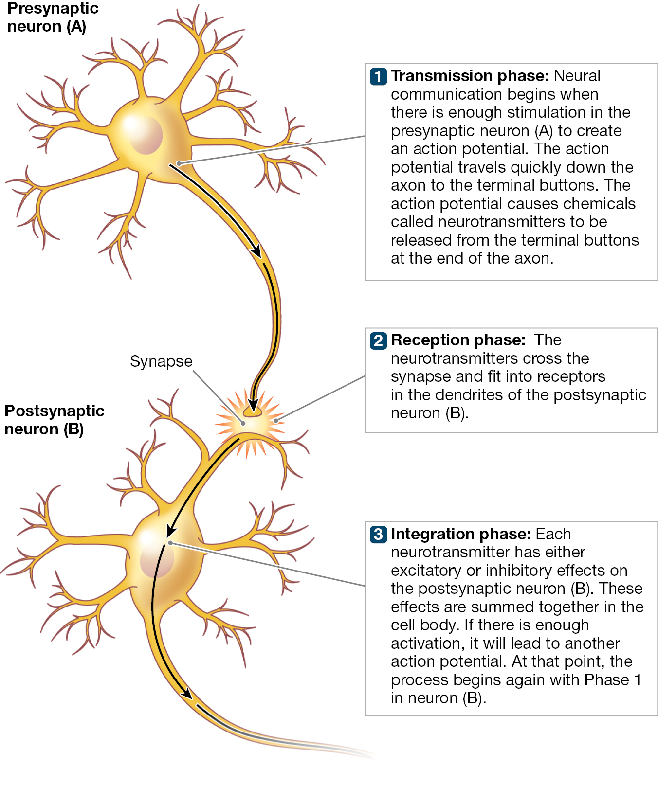
A diagram of a presynaptic neuron and postsynaptic neuron that are connected with a synapse. It explains the process of the action potential traveling through both neurons in three boxes. It states 1. Transmission phase: Neural communication begins when there is enough stimulation in the presynaptic neuron (A) to create an action potential. The action potential travels quickly down the myelinated axon to the terminal buttons. The action potential causes chemicals called neurotransmitters to be released from the terminal buttons at the end of the axon. 2. Reception phase: The neurotransmitters cross the synapse and fit into receptors in the dendrites of the postsynaptic neuron (B). 3. Integration phase: Each neurotransmitter has either excitatory or inhibitory effects on the postsynaptic neuron (B). These effects are summed together in the cell body. If there is enough activation, it will lead to another action potential. At that point, the process begins again with Phase 1 in the neuron (B).
FIGURE 2.5 Three Phases of Neural Communication
The three phases of neural communication are (1) the transmission phase, (2) the reception phase, and (3) the integration phase. Which phase involves neuro transmitters crossing the synapse?
What lets a neuron communicate with other neurons? The answer to this question lies in the electrical properties of neurons and how neurons change when they are stimulated.
Electrical Properties of Neurons Just as your body is covered with skin, the neuron is covered with a thin covering called a membrane. This barrier separates the inside of the neuron from the outside environment. The membrane is semipermeable. In other words, some substances move through the membrane. These substances are electrically charged chemicals called ions. For example, sodium and potassium ions may move from the outside to the inside of the neuron or from the inside of the neuron to the outside. The movement of these ions across the membrane lets neurons communicate. The membrane contributes to neural communication by regulating the neuron’s electrical activity.
It is important to know that the inside of the neuron and the outside of a neuron have different electrical charges. The neuron begins in a resting state. During this state, the electrical charge inside the neuron is more negative than the electrical charge outside. This difference in electrical charge results from the balance of various ions (charged molecules) inside and outside the neuron. Not much is happening during the resting state, just as not much is happening when you are on the couch in your own resting state.
Now imagine that the neuron receives stimulation from nearby neurons. This stimulation causes positively charged sodium ions to move through the membrane and into the neuron. The inside of the neuron becomes more positively charged as sodium enters. If the neuron is stimulated enough, it fires an action potential down the axon to the terminal buttons, where neurotransmitters are released into the synapse. Think of receiving texts from your friends to go out and be social. When you receive enough texts, you finally spring into action and go to meet them.
During an action potential, sodium ions continue to enter the neuron and potassium ions leave the neuron. Within milliseconds, sodium ions stop entering and potassium ions stop leaving. In addition, sodium is pumped out of the neuron while potassium is pumped back in. This sodium potassium pump helps return the neuron to the resting state. During this refractory period, in which the neuron returns to its resting state, the neuron is less responsive to incoming stimulation and is less likely to fire an action potential. This might be how you feel after a long night of socializing—that is, you are less likely to be willing to go out again that night even if your friends once again text you to do so. During your refractory period, you are less responsive to your friends’ texts. Of course, at the neural level this whole process from action potential to refractory period happens in milliseconds rather than over the course of an entire evening.
Through action potentials, one neuron is able to send a message to another neuron. Let’s examine the process of neural communication in more detail.
Action Potentials Let Neurons Communicate As Figure 2.5 shows, a neuron is stimulated by signals from other neurons. The stimulated neuron may transmit this information to other neurons through an action potential (see Phase 1). When a neuron is stimulated, positively charged sodium ions enter the neuron. The inside of the neuron becomes more positive as sodium enters. If the electrical charge changes enough, the action potential begins. Traveling along the axon like a wave, the action potential moves away from the cell body. It moves down the axon toward the terminal buttons.
The action potential travels quickly along the axon. For most neurons, this fast movement down the axon is made possible by the fatty layer that insulates the axon. The fatty casing is called the myelin sheath (see Figure 2.3 on p. 50). Because the myelin sheath makes neural communication so quick, most people are able to perform complicated actions, such as learning the newest TikTok dance. However, recall that in some diseases, such as MS, neural communication is disrupted. In MS, neural communication is affected because the myelin sheath surrounding the diseased axons deteriorates, and the action potential slows as it moves down to the terminal buttons. In short, when neurons lose the myelin sheath surrounding the axon, they lose the ability to communicate quickly. Doctors determined that Jack Osbourne was in the early stages of MS when affected neurons disrupted his ability to process visual information.
To communicate, a neuron fires an action potential. A neuron cannot fire just a little bit: Either it fires or it does not. How often a neuron fires an action potential can change, though, depending on how much stimulation the neuron receives. To understand this idea, suppose you are playing a video game in which you fire missiles at invading aliens by pressing a button. Every time you press the button, a missile is launched at the same speed as the previous missile. The missile speed remains the same no matter how hard you press the button. However, if you press the button faster, missiles will fire more rapidly one after another. Now suppose the missile launcher is a neuron in your visual system. The neuron receives information that a light is bright. The neuron might respond to that stimulation by firing more often than when it receives information that the light is dim. But whether the light is bright or dim, however many times the neuron fires, the strength of the action potential is the same every time.
Neurotransmitters in the Synapse Neurons communicate with each other by firing an action potential down to the terminal buttons. However, recall that neurons do not touch one another, because there is a gap between neurons called the synapse (see Figure 2.3). How can neurons communicate information from an action potential without touching each other? Just as ships transport crops across the seas from one country to another, neurons communicate with each other by sending chemical information across the synapse from one neuron to another. Thus, in the first phase of neural communication—transmission—action potentials cause a neuron to release neurotransmitters (see Figure 2.5, Phase 1). In the second phase—reception—these chemicals travel across the synapse and connect to, or bind with, the receiving neuron’s dendrites (see Figure 2.5, Phase 2). The neuron that sends the signal is called the presynaptic neuron, and the one that receives the signal is called the postsynaptic neuron. The Learning Tip will help you remember these terms.
LEARNING TIP Communication From Presynaptic Neuron to Postsynaptic Neuron
It will be easy to understand how neurons communicate with each other if you remember the following.
|
When you see |
Please think |
Meaning |
|
Presynaptic |
Before the synapse |
Something that occurs in the sending neuron before the synapse (the gap between neurons) |
|
Postsynaptic |
After the synapse |
Something that occurs in the receiving neuron after the synapse (the gap between neurons) |
How do these chemical signals work (Figure 2.6)? When an action potential arrives at the end of the axon, the terminal buttons release neurotransmitters that spread across the synapse. The neurotransmitters then attach (bind) to receptors on the postsynaptic neuron. Receptors are specialized structures that respond to certain types of neurotransmitters. In much the same way as a lock opens only with the correct key, each receptor can be influenced by only one type of neurotransmitter. Here is an analogy: When ships filled with crops travel across the sea, a landing port might allow only ships from certain companies to dock. All other ships are blocked from docking.
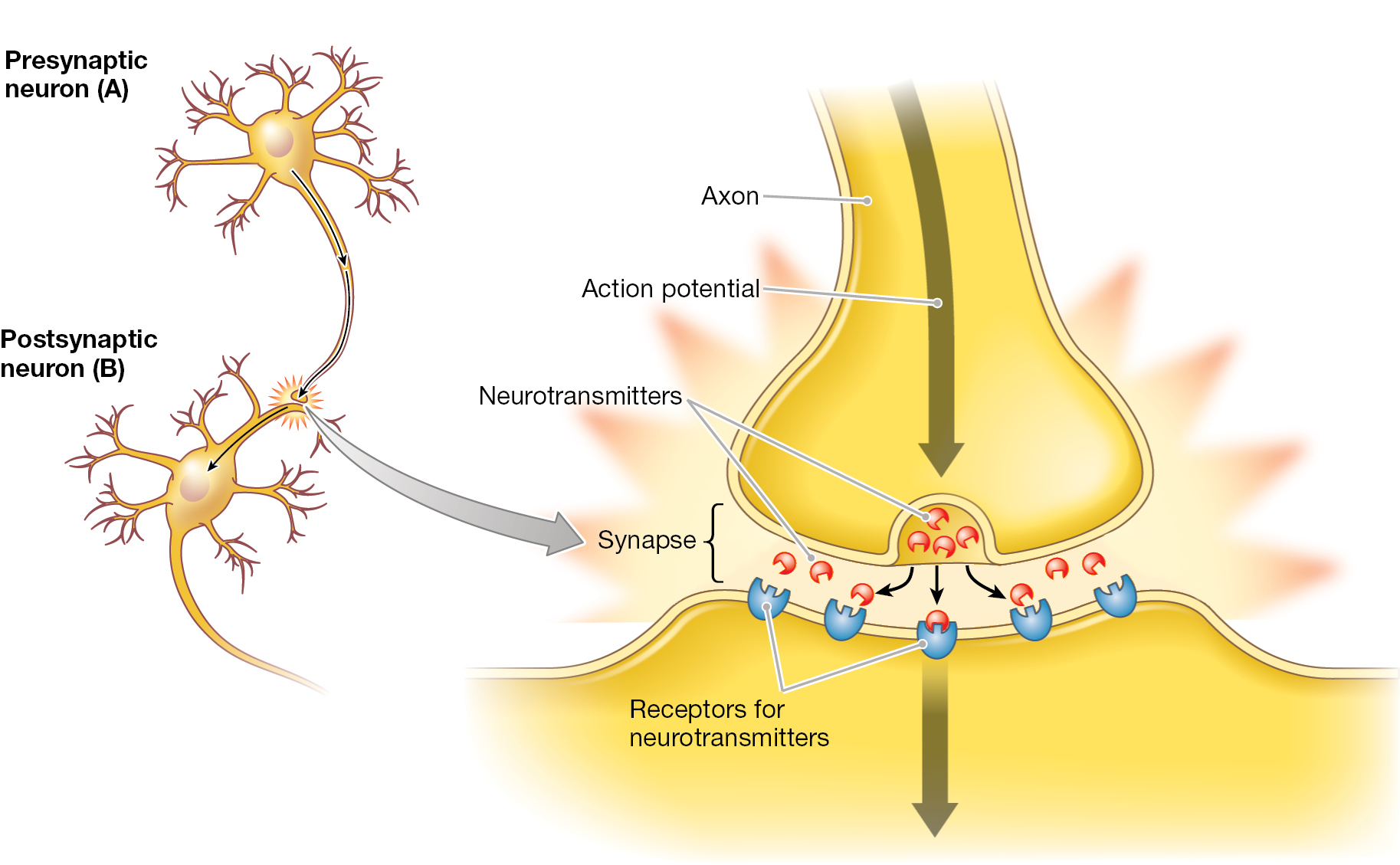
A diagram of a presynaptic neuron (A) and postsynaptic neuron (B) that are connected with a synapse is shown. The action potential shows as a downward arrow reaches the end of the axon which results in the release of neurotransmitters. These neurotransmitters cross an area of space between the presynaptic neuron and the dendrites of the postsynaptic neuron called the synapse. They reach receptors for the neurotransmitters on the dendrite of the postsynaptic neuron.
FIGURE 2.6 Neurotransmitters Move Across the Synapse
When the action potential in the presynaptic neuron reaches the terminal buttons, neurotransmitters are released into the synapse between the neurons. These neurotransmitters cross the synapse and attach (bind) to specific receptors on the dendrites of the postsynaptic neuron.
Once neurotransmitters bind with specific receptors by attaching to them, they stimulate these receptors. After binding and passing along their signal, the neurotransmitters quickly release back into the synapse. Once released, they can bind again with postsynaptic receptors. To prevent constant stimulation, the neurotransmitter needs to be removed from the synapse in one of two major ways. The first occurs when the neurotransmitters are reabsorbed by the presynaptic neuron in a process called reuptake. In the second way, enzymes destroy the neurotransmitters while they are in the synapse. Enzymes are chemicals that break down other substances. Different enzymes break down different neurotransmitters in a process called enzyme degradation. (To degrade something means to break it down.)
Excitatory and Inhibitory Signals In integration—the third phase of neural communication—the postsynaptic neuron processes incoming signals (see Figure 2.5, Phase 3). The attaching, or binding, of neurotransmitters with their receptors on the postsynaptic neuron can produce signals of two types: excitatory or inhibitory. As the name indicates, excitatory signals excite the neuron. That is, they increase the likelihood that it will fire an action potential. Inhibitory signals inhibit the neuron—they decrease the likelihood that it will fire an action potential.
Any individual signal received by the neuron has little impact on whether the neuron fires. Instead, the thousands of excitatory and inhibitory signals that are received are added together within the cell body of the neuron. If the total amount of excitatory input goes past a certain threshold, the postsynaptic neuron fires an action potential. After firing, the neuron returns to its resting state. The process repeats hundreds of times per second.
How do your neurotransmitters affect how you think and behave? You will learn the answer in the next study unit.
LEARNING GOAL CHECK: REVIEW