LEARNING GOAL
Summarize the functions of the hindbrain and midbrain.
LEARNING GOAL
Summarize the functions of the hindbrain and midbrain.
Processes that aid basic survival keep us alive and allow us to maintain the species through reproduction. To survive, we need a beating heart, lungs to take in oxygen, and the ability to control muscles so we can eat food and drink fluids. Many of these basic survival processes rely on brain regions at the lower back of your skull that extend into the spinal cord.
Spinal Cord When your foot is itchy, that message had to travel from your foot to your brain. Deciding to scratch your foot requires a message sent from the brain to your hand. The spinal cord helps you to scratch that itch.
The spinal cord is the gateway for information traveling between the brain and the body. It carries sensory information up to the brain and carries motor signals from the brain to the body parts to initiate action. The spinal cord also coordinates reflexes, such as the way your leg moves when a doctor taps your knee and the way your arm moves when you jerk your hand away from a flame.
Let’s learn about two important parts of the brain: the hindbrain and midbrain.
Hindbrain At the base of the skull, the spinal cord thickens and becomes more complex. At this point, the spinal cord becomes the hindbrain. The hindbrain contains structures that control body functions that are essential for survival.
The hindbrain has three main structures (Figure 2.15). The first is the medulla. The medulla controls the most basic functions of survival, including heart rate, breathing, swallowing, vomiting, and urination. A significant blow directly to the medulla can cause death. Have you ever gagged on something? If so, that gagging was caused by your medulla. Gagging is a reflex that prevents us from choking on something, so it is crucial for survival.
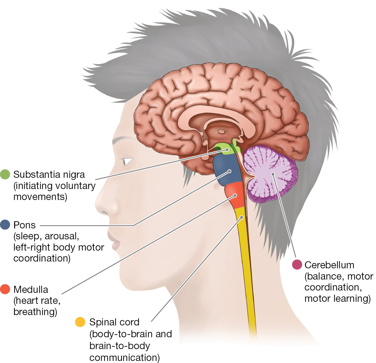
The cerebellum is at the back lower part of the brain. A label reads cerebellum (balance, motor coordination, and motor learning). The substantia nigra is the tip of the medulla that reaches out of the hindbrain and into the midbrain. A label reads substantia nigra (initiating voluntary movements). The Pons is directly in front of the cerebellum and around the medulla. A label reads pons (sleep and arousal). The medulla is long and thin and is between the cerebellum and pons. A label reads medulla (heart rate, breathing).
FIGURE 2.15 The Hindbrain and the Midbrain
This drawing shows where the spinal cord meets the three hindbrain structures (medulla, pons, and cerebellum). It also shows the location of the midbrain (including the substantia nigra). The view shows the brain as though you could see inside to its middle. Which brain region is involved in sleep?
The second structure of the hindbrain, the pons, plays an important role in sleep and arousal and in coordinating movements between the left and right sides of the body (see Figure 2.15).
The third structure of the hindbrain is a large extension called the cerebellum. It is located behind the medulla and pons (see Figure 2.15). Its size and convoluted surface make it look like an extra brain. In fact, the name cerebellum comes from the Latin word for “little brain.”
The cerebellum is essential for proper motor function. Damage to the different parts of the cerebellum produces very different effects. Damage to the very bottom causes problems with head tilt and balance. Damage to the ridge that runs up the back of the cerebellum affects walking. Damage to the lobes on either side causes a loss of coordination in the limbs. For example, a person with this kind of damage could not reach out smoothly to pick up a pen.
The cerebellum is involved in motor learning and motor memory. For example, the cerebellum makes it possible for you to ride a bicycle effortlessly—and to do so while planning your next meal. The cerebellum is also involved in cognitive processes such as making plans, remembering events, using language, and experiencing emotion. It helps organize these cognitive and emotional processes in much the same way it helps organize motor behavior (Schmahmann et al., 2019).
Midbrain The midbrain is located above the pons. It consists of several structures that are involved in the reflexive movement of the eyes and body. One structure in particular, the substantia nigra, is important for initiating voluntary movements (see Figure 2.15). This region is critical for the production of dopamine, the neurotransmitter that motivates behavior and controls normal motor function. Parkinson’s disease is caused by the death of substantia nigra cells and the resulting loss of dopamine produced by those cells. Thus the slurred speech and shuffling walk that Muhammad Ali experienced as symptoms of Parkinson’s disease resulted from a loss of substantia nigra cells in his midbrain.
LEARNING GOAL CHECK: APPLY