LEARNING GOAL
Explain how changes in brain activity produce changes in consciousness.
LEARNING GOAL
Explain how changes in brain activity produce changes in consciousness.
Philosophers have long debated the nature of consciousness. One aspect of this debate is the mind/body problem. Are the mind and the body separate and distinct? Or is the mind simply a person’s personal experience of the physical brain’s activity? In the 1600s, the philosopher René Descartes viewed the mind as separate from the body. The body, Descartes argued, was nothing more than an organic machine governed by “reflex.” In keeping with the prevailing religious beliefs of the seventeenth century, he concluded that the rational mind was divine and separate from the physical body. This belief is known as dualism. Most psychologists now reject dualism in favor of materialism, the modern idea that the brain and the mind are inseparable. In other words, the processing of information in the brain creates the experiences of the mind.
According to materialism, the activity of neurons in the brain produces consciousness. For example, neurons produce consciousness of the sight of a familiar face or consciousness of the sweet smell of a rose. More specifically, for each experience you have—each sight, each smell, each memory—there is an associated pattern in your brain activity. The activation of particular groups of neurons in certain parts of your brain gives rise to particular conscious experiences.
The Global Workspace Model Despite what you might see in some movies, scientists cannot—yet—read your intimate thoughts by looking at your brain activity. However, psychologists now examine, and even measure, consciousness and other mental states that once were considered too subjective to be studied. For example, in an early study, Tong and colleagues (1998) studied the relationship between consciousness and neural responses in the brain. Participants were shown images of houses that were superimposed on faces (Figure 3.2a). Participants were then asked if they saw a face (Figure 3.2b) or a house (Figure 3.2c). When participants reported seeing a face, neural activity increased within the brain regions associated with face recognition—the fusiform face area. When participants reported seeing a house, neural activity increased within brain regions associated with object recognition. This finding suggests that different areas of the brain process different types of sensory information. Importantly, activity in these brain regions is associated with awareness of particular information (Weaverdyck et al., 2020). Additional research in this area has shown that brain activity patterns can reveal whether you are looking at faces or bodies (O’Toole et al., 2014), which emotions you are experiencing (Kragel & LeBar, 2016), or whether you are thinking of yourself or a close friend (Chavez et al., 2017; Wagner et al., 2019). The takeaway message from these studies is that people share common patterns of brain activity that provide a window into their conscious experiences (Deco et al., 2021).
FIGURE 3.2 The Relationship Between Consciousness and Brain Activity
(a) In a 1998 study by Tong and colleagues, participants were shown images with houses superimposed on faces. Participants were asked to report whether they saw (b) a face or (c) a house. Researchers used fMRI to measure neural responses in participants’ brains when the participants reported seeing a house or a face.
The global workspace model is a psychological theory suggesting that consciousness depends on which brain circuits are active (Baars, 1988; Mashour et al., 2020; Shea & Frith, 2019). To put it another way: You experience your brain regions’ activity as conscious awareness of specific information. For instance, when you listen to music, your conscious experience results from activation in particular brain regions happening at the same time. Those regions are processing the sound of the music, the meaning of the lyrics, perhaps your memories of hearing the song in the past, and the emotional states those memories produce. Your total experience results from the simultaneous activity of all the different brain regions supporting these psychological processes.
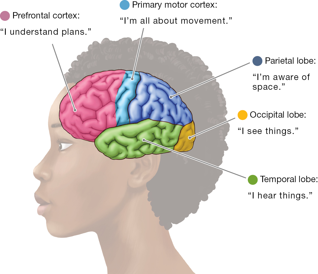
The key idea of the global workspace model is that no one area of the brain is responsible for general “awareness.” Instead, specific areas of the brain process certain types of information. The processing in these brain areas produces conscious experience of the information (Figure 3.3). Unfortunately, the flip side of this perspective is that damage to specific brain regions negatively affects consciousness.

The diagram of the brain model with some lobes and cortices labeled and a small explanation of their functions using first-person statements. The first is the prefrontal cortex which is the first inch of the brain just behind the forehead. The prefrontal cortex’s functional statement is, I understand plans. Next is the primary motor cortex which is located on the last inch of the frontal lobe before it becomes the parietal lobe. The primary motor cortex’s functional statement is, I’m all about movement. Next is the parietal lobe, which is located behind the frontal lobe and above the occipital and temporal lobe. The parietal lobe’s functional statement is, I’m aware of space. Next is the occipital lobe which is the furthest back part of the brain behind and below the parietal lobe. The occipital lobe’s functional statement is, I see things. Last is the temporal lobe which is located under the frontal and parietal lobes. The temporal lobe’s functional statement is, I hear things.
FIGURE 3.3 Global Workspace Model
The global workspace model states that conscious awareness of different aspects of the world is associated with processing in different parts of the brain. This simplified diagram indicates some major cortical regions where processing leads to awareness. Which cortical area is aware of where your body is in space?
Traumatic Brain Injury Athletes who experience extreme hits are sometimes described as having their “bell rung.” In fact, a hard hit to the head can produce the sensation of a ringing sound. Concussions affect consciousness in part because they cause brain damage, and severe concussions cause traumatic brain injury (TBI).
TBI occurs when an external trauma causes changes in consciousness as well as physical damage to the brain. TBIs range from mild to severe, and severe TBIs are responsible for about 30 percent of all injury deaths (Faul et al., 2010). The greater the severity of the injury, the more likely a TBI is to cause negative effects on consciousness with respect to thinking, memory, emotions, and even personality. These effects can last for many years or be permanent.
A concussion used to be considered a mild TBI. After all, most people recover from a concussion within one to two weeks (Baldwin et al., 2016). However, numerous studies have documented the long-term effects of concussions, including an increased risk of multiple sclerosis later in life for adolescents who suffer concussions (Bailes et al., 2013; Montgomery et al., 2017). Each concussion a person experiences can lead to more serious symptoms that are longer lasting (Baugh et al., 2012; Zetterberg et al., 2013).

FIGURE 3.4 Concussions and Young Athletes
There is a risk of concussion among young athletes who participate in competitive sports, such as cheerleading.
In recent years, there has been a growing concern about concussions among young athletes (Figure 3.4). The number of concussions resulting from sports or recreation in American youth under age 18 is estimated at 1 to 2 million each year (Bryan et al., 2016). Millions more are affected during recreational or athletic competition in college (Cheng et al., 2019). For many years, males were the focus of research on concussions, and outcomes in females were less well documented. We now know that females are more vulnerable to concussions and appear to have worse outcomes than males (Koerte et al., 2020). The sports in which females are much more likely than males to have concussions are basketball and especially soccer (Cheng et al., 2019). For this reason, professional soccer player Brandi Chastain has donated her brain to science so that it can be studied after her death for evidence of concussion-related brain injury. These concerns have led to guidelines for reducing the occurrence of such injuries (Harmon et al., 2013). According to the American Academy of Neurology (2013), an athlete should not return to action until all symptoms have cleared up, which often takes several weeks.
Coma Medical advances are helping a greater number of people survive TBIs. For example, doctors now save the lives of many people who previously would have died from injuries sustained in car accidents or on battlefields. However, many of those who sustain TBIs fall into comas or are induced into coma as part of medical treatment to let the brain rest. Those who are in a coma appear unconscious and unresponsive. They seem unaware of their surroundings and do not respond to questions or commands from family or medical staff. What exactly is the relation between coma and consciousness?
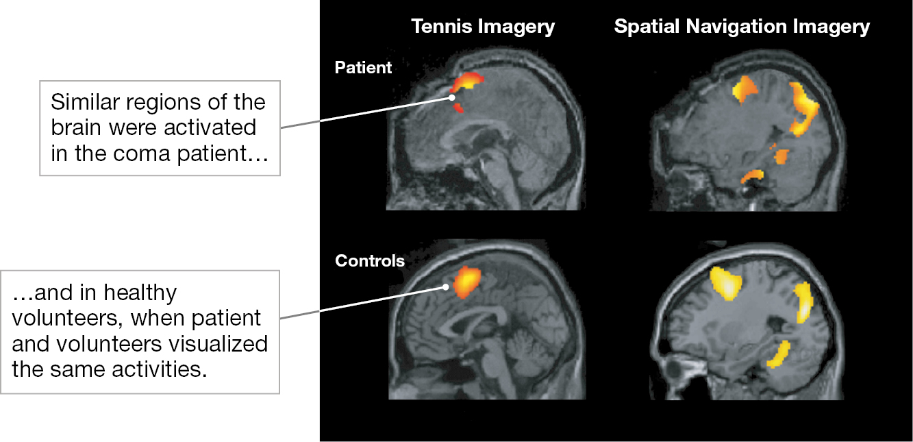
People in comas have sleep/wake cycles—they open their eyes and appear to be awake, close their eyes and appear to be asleep—but they generally do not respond to their surroundings. Because of this activity and inactivity, it is hard to know their level of consciousness. However, brain imaging has shown that some people in comas are aware but unable to respond. For example, a 23-year-old woman in a coma was asked to imagine playing tennis or walking through her house (Owen et al., 2006). This woman’s pattern of brain activity became quite similar to the patterns of control subjects who also imagined playing tennis (Figure 3.5, left) or walking through a house (Figure 3.5, right). The woman could not give outward signs of awareness, but researchers believe she was able to understand language and respond to the experimenters’ requests.

Four states of the brain in a 2 by 2 format. The images on the top are from a coma patient, and those on the bottom are from a control group. The left column is titled, Tennis Imagery, and the right column is titled, Spatial Navigation Imagery. The patient in the coma and the control group were each told to imagine playing tennis and then to imagine walking around. The coma patients’ scans are lit up in the same areas as the control groups, suggesting that the patient was minimally conscious. Near the images of the brain scans are two dialogue boxes, the first pointing at the patient’s scans and reading Similar regions of the brain were activated in the coma patient... and the second dialogue box pointing at the control groups scans and reading ... and in healthy volunteers, when patient and volunteers visualized the same activities.
FIGURE 3.5 In a Coma but Conscious: Minimally Conscious State
The brain images on the top are from the patient, a young woman in a coma who showed no visible signs of consciousness. The images on the bottom are a composite from the control group, which consisted of healthy volunteers. Both the patient and the control group were told to visualize playing tennis (left) and walking around (right). Immediately after the directions were given, the patient’s neural activity appeared similar to the control group’s neural activity. This similarity suggests that the coma patient was in a minimally conscious state. That is, she actually was conscious.
The implications of this finding are extraordinary. Can these methods be used to reach comatose people who are conscious but unable to communicate? Indeed, this research team evaluated 54 coma patients and found 5 who could willfully control brain activity to communicate (Monti et al., 2010). Being able to communicate from comas might allow patients to express thoughts, ask for more medications, and increase the quality of their lives (Fernández-Espejo & Owen, 2013). The use of brain imaging is now recommended to assess whether those in a coma have conscious awareness (Kondziella et al., 2020). These advances add up to one astonishing fact: Some people in comas are conscious! But because these coma patients cannot make their bodies respond, observers think they are unconscious. Instead, they are in a minimally conscious state.
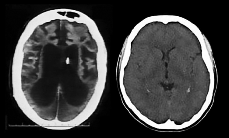
Most people come out of a coma within a few days. Others remain in a coma for weeks, and some people never emerge from the coma. When a coma lasts for more than a month, the person most likely has unresponsive wakefulness syndrome (Laureys et al., 2010). This unresponsive state is not associated with consciousness. Normal brain activity does not occur in this state, in part because much of the person’s brain may be damaged beyond recovery. The longer the unresponsive wakefulness state lasts, the less likely it is that the person will ever recover consciousness or show normal brain activity. Terri Schiavo, a woman living in Florida, spent more than 15 years with unresponsive wakefulness syndrome. Eventually, her husband wanted to terminate her life support, but her parents wanted to continue it. Both sides waged a legal battle. A court ruled in the husband’s favor, and life support was terminated. After Schiavo’s death, an autopsy revealed substantial and irreversible damage throughout her brain (Figure 3.6, left). Schiavo had been unconscious because the cortical areas of her brain that create conscious awareness had been completely destroyed.

Two brain scans. The left brain scan has large black lobes in the middle, within a white circle delineating the skull. The right brain scan is all gray inside of the white circle, delineating the skull.
FIGURE 3.6 In a Coma and Unconscious: Unresponsive Wakefulness Syndrome
Before Terri Schiavo was taken off life support, her parents and their supporters believed she showed some awareness. But the dark areas of her brain scan (left) indicate that her cortex had deteriorated beyond recovery (an undamaged brain is shown on the right). There could not have been activity in these areas.
LEARNING GOAL CHECK: REVIEW