Distinguish between the two basic divisions of the nervous system.
Distinguish between the functions of distinct types of neurons.
Describe the structure of the neuron.
Describe the electrical and chemical changes that occur when neurons communicate.
Describe how agonists and antagonists can influence the action of neurotransmitters.
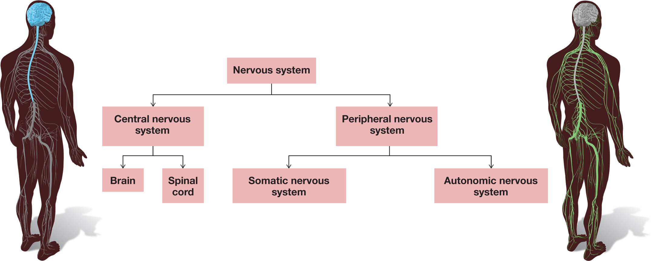
The nervous system’s response to the world around us is responsible for everything we think, feel, or do. Essentially, each of us is a nervous system. The entire nervous system is divided into two basic units: the central nervous system and the peripheral nervous system. The central nervous system (CNS) consists of the brain and the spinal cord (FIGURE 3.1). The peripheral nervous system (PNS) consists of all the other nerve cells in the rest of the body and includes the somatic and autonomic nervous systems. The somatic component is involved in voluntary behavior, such as when you reach for an object. The autonomic component is responsible for the less voluntary actions of your body, such as controlling your heart rate and other bodily functions. The CNS and PNS are anatomically separate, but their functions are highly interdependent. The PNS sends a variety of information to the CNS. The CNS organizes and evaluates that information and then directs the PNS to perform specific behaviors or make bodily adjustments.
FIGURE 3.1
The Basic Divisions of the Nervous System
3.1Neurons Are the Basic Units of the Nervous System
The basic units of the nervous system are the nerve cells, called neurons (FIGURE 3.2), which receive, integrate, and transmit information. Complex networks of neurons sending and receiving signals are the functional basis of all psychological activity. Although the actions of single neurons are simple to describe, human complexity results from billions of neurons. Each neuron communicates with tens of thousands of other neurons. Neurons do not communicate randomly or arbitrarily, however. They communicate selectively with other neurons to form circuits, or neural networks. These networks develop through genetic influence, maturation and experience, and repeated firing.
FIGURE 3.2
Human Neuron
Neurons like this one are the basic units of the human nervous system.
FUNCTIONS OF NEURONS Neurons are specialized for communication with each other. Unlike other cells in the body, nerve cells are excitable: They are powered by electrical impulses and communicate with other nerve cells through chemical signals. During the reception phase, neurons take in chemical signals from neighboring neurons. During integration, incoming signals are assessed. During transmission, neurons pass their own signals to yet other receiving neurons.
There are many types of neurons. Sensory neurons detect information from the physical world and pass that information along to the brain. To get a sense of how fast that process can work, think of the last time you touched something hot or accidentally pricked yourself with a sharp object, such as a tack. Those signals triggered your body’s nearly instantaneous response and sensory experience of the impact. The sensory nerves that provide information from the skin and muscles are called somatosensory nerves. (This term comes from the Greek for “body sense.” It means sensations experienced from within the body.) Motor neurons direct muscles to contract or relax, thereby producing movement. Interneurons act as relay stations facilitating communication between sensory and motor neurons.
Sensory and motor neurons work together to control movement. For instance, if you are using a pen to take notes as you read these words, you are contracting and relaxing your hand muscles and finger muscles to adjust your fingers’ pressure on the pen. When you want to use the pen, your brain sends a message via motor neurons to your finger muscles so they move in specific ways. Receptors in both your skin and your muscles send back messages through sensory neurons to help determine how much pressure is needed to hold the pen. This symphony of neural communication for a task as simple as using a pen is remarkable, yet most of us employ motor control so easily that we rarely think about it. In fact, our reflexes, automatic motor responses, occur before we even think about those responses. For each reflex action, a handful of neurons simply convert sensation into action.
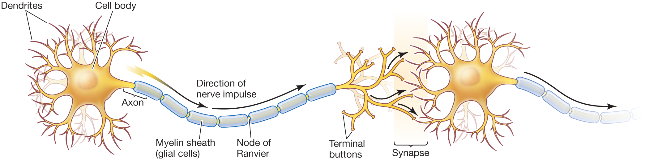
NEURON STRUCTURE In addition to performing different functions, neurons have a wide assortment of shapes and sizes. A typical neuron has four structural regions that participate in communication functions: the dendrites, the cell body, the axon, and the terminal buttons (FIGURE 3.3). The dendrites are short, branchlike appendages that detect chemical signals from neighboring neurons. In the cell body, also known as the soma (Greek for “body”), the information received via the dendrites from thousands of other neurons is collected and integrated.
FIGURE 3.3
Neuron Structure
Messages are received by the dendrites, processed in the cell body, transmitted along the axon, and sent to other neurons via chemical substances released from the terminal buttons across the synapse. (The myelin sheath and the nodes of Ranvier are discussed on p. 73.)
Once the incoming information from many other neurons has been integrated in the cell body, electrical impulses are transmitted along a long, narrow outgrowth known as the axon. Axons vary tremendously in length, from a few millimeters to more than a meter. The longest axons stretch from the spinal cord to the big toe. You may have heard the term nerve in reference to a “pinched nerve.” In this context, a nerve is a bundle of axons that carry information between the brain and other specific locations in the body. At the end of each axon are knoblike structures called terminal buttons.
The site where chemical communication occurs between neurons is called the synapse. Since neurons do not touch one another, they communicate by sending chemicals into the synapse, a tiny gap between the terminal buttons of the “sending” neuron and the dendrites of the “receiving” neurons. Chemicals leave the terminal buttons of one neuron, cross the synapse, and pass signals along to the dendrites of other neurons.
The outer surface of a neuron is a membrane, a fatty barrier that does not dissolve in the watery environment inside and outside the neuron. The membrane is selectively permeable. In other words, some substances move in or out of the membrane, and some do not. Located on the membrane are ion channels. These specialized pores allow ions to pass in and out of the cell when the neuron transmits signals down the axon. Ions are electrically charged molecules, some charged negatively and some charged positively. By controlling the movement of ions, the membrane plays an important role in communication between neurons: It regulates the concentration of electrically charged molecules that are the basis of the neuron’s electrical activity.
All nerve cells in the body that are not part of the central nervous system. The peripheral nervous system includes the somatic and autonomic nervous systems.
The basic units of the nervous system; cells that receive, integrate, and transmit information. They operate through electrical impulses, communicate with other neurons through chemical signals, and form neural networks.
The gap between the terminal buttons of a “sending” neuron and the dendrites of a “receiving” neuron, where chemical communication occurs between the neurons.