 1.1 Innate Recognition of Pathogens
Principles of adaptive immunity.
We come now to the components of adaptive immunity, the antigen-specific lymphocytes. Unless indicated otherwise, we shall use the term ‘lymphocyte’ in the remainder of this chapter to refer only to the antigen-specific lymphocytes. Lymphocytes allow responses against a vast array of antigens from various pathogens encountered during a person’s lifetime and confer the important feature of immunological memory. Lymphocytes make this possible through the highly variable antigen receptors on their surface, by which they recognize and bind antigens. Each lymphocyte matures bearing a unique variant of a prototype antigen receptor, so that the population of lymphocytes expresses a huge repertoire of receptors that are highly diverse in their antigen-binding sites. Among the billion or so lymphocytes circulating in the body at any one time there will always be some that can recognize a given foreign antigen.
A unique feature of the adaptive immune system is that it is capable of generating immunological memory, so that having been exposed once to an infectious agent, a person will make an immediate and stronger response against any subsequent exposure to it; that is, the individual will have protective immunity against it. Finding ways of generating long-lasting immunity to pathogens that do not naturally provoke it is one of the greatest challenges facing immunologists today.
1-8 The interaction of antigens with antigen receptors induces lymphocytes to acquire effector and memory activity.
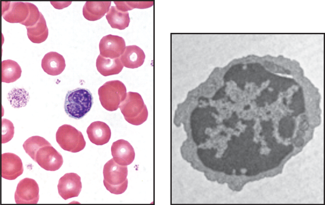
There are two major types of lymphocytes in the vertebrate immune system, the B lymphocytes (B cells) and T lymphocytes (T cells). These express distinct types of antigen receptors and have quite different roles in the immune system, as was discovered in the 1960s. Most lymphocytes circulating in the body appear as rather unimpressive small cells with few cytoplasmic organelles and a condensed, inactive-appearing nuclear chromatin (Fig. 1.12). Lymphocytes manifest little functional activity until they encounter a specific antigen that interacts with an antigen receptor on their cell surface. Lymphocytes that have not yet been activated by antigen are known as naive lymphocytes; those that have met their antigen, become activated, and have differentiated further into fully functional lymphocytes are known as effector lymphocytes.
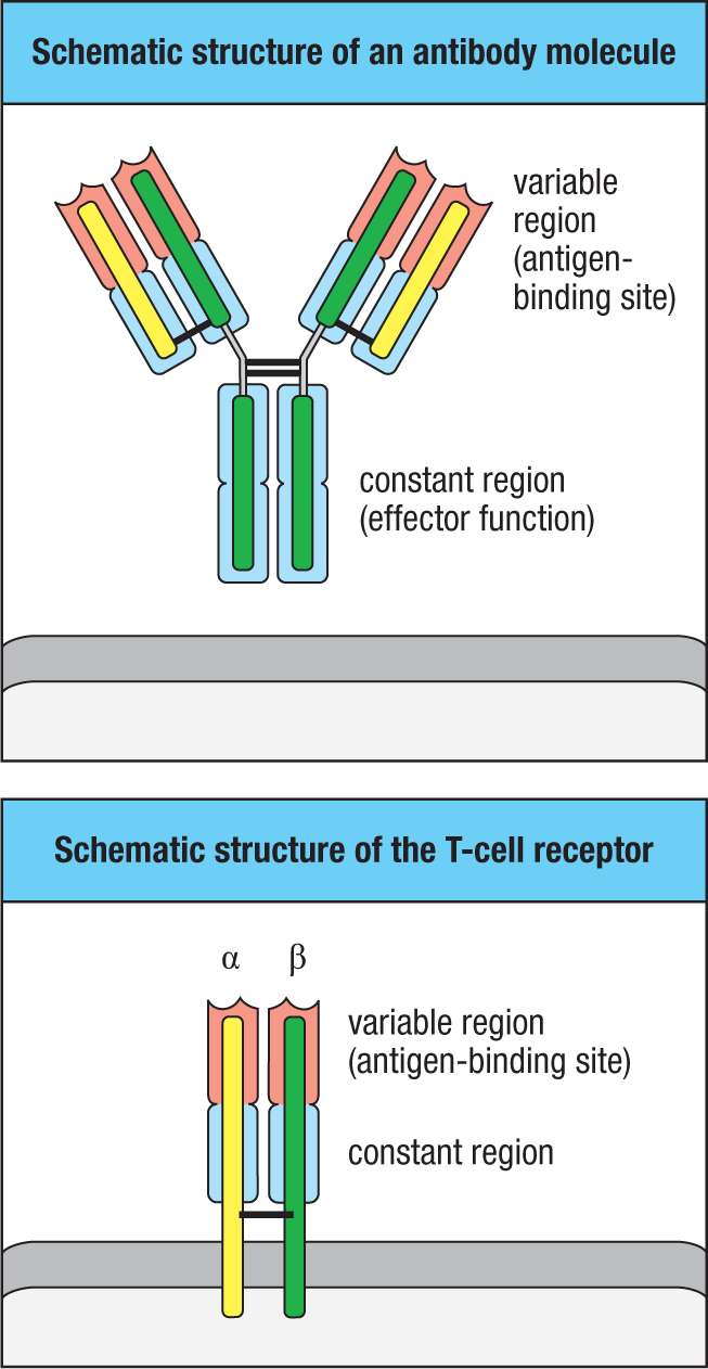
B cells and T cells are distinguished by the structure of the antigen receptor that they express. The B-cell antigen receptor, or B-cell receptor (BCR), is formed by the same genes that encode antibodies, therefore B-cell receptors and antibodies compose the class of proteins known as immunoglobulin (Ig) (Fig. 1.13). Thus, the antigen receptor of B lymphocytes is also known as membrane immunoglobulin (mIg) or surface immunoglobulin (sIg). The T-cell antigen receptor, or T-cell receptor (TCR), is related to the immunoglobulins but is quite distinct in its structure and recognition properties.
After antigen binds to a B-cell receptor, the B cell will proliferate and differentiate into plasma cells. These are the effector form of B lymphocytes, and they secrete antibodies that have the same antigen specificity as the plasma cell’s B-cell receptor. Thus the antigen that activates a given B cell becomes the target of the antibodies produced by that B cell’s progeny.
When a T cell first encounters an antigen that its receptor can bind, it proliferates and differentiates into one of several different functional types of effector T lymphocytes. When effector T cells subsequently detect antigen, they can manifest three broad classes of activity. Cytotoxic T cells kill other cells that are infected with viruses or other intracellular pathogens bearing the antigen. Helper T cells provide signals, often in the form of specific cytokines that activate the functions of other cells, such as B-cell production of antibody and macrophage killing of engulfed pathogens. Regulatory T cells suppress the activity of other lymphocytes and help to limit the possible damage of immune responses. We discuss the detailed functions of cytotoxic, helper, and regulatory T cells in Chapters 9, 11, 12, and 15.
Some of the B cells and T cells activated by antigen will differentiate into memory cells, the lymphocytes that are responsible for the long-lasting immunity that can follow exposure to disease or vaccination. Memory cells will readily differentiate into effector cells on a second exposure to their specific antigen. Immunological memory is described in Chapter 11.
1-9 Antibodies and T-cell receptors are composed of constant and variable regions that provide distinct functions.
Antibodies were studied by traditional biochemical techniques long before recombinant DNA technology allowed the study of the membrane-bound forms of the antigen receptors on B and T cells. These early studies found that antibody molecules are composed of two distinct regions. One is a constant region, also called the fragment crystallizable region, or Fc region, which takes one of only five biochemically distinguishable forms (see Fig. 1.13). The variable region of different antibody molecules, by contrast, can be composed of a vast number of different amino acid sequences that allow antibodies to recognize an equally vast variety of antigens. The basic structure of antibody molecules was determined by Gerald Edelman and Rodney Porter, who shared the 1972 Nobel Prize for their work. These biochemical studies led to subsequent X-ray crystallographic determination of the structure of the antibody Fc region.
The antibody molecule is composed of two identical heavy chain and two identical light chain polypeptides. Heavy and light chains each have variable and constant regions. The variable regions of a heavy chain and a light chain combine to form an antigen-binding site that determines the antigen-binding specificity of the antibody. Thus, both heavy and light chains contribute to the antigen-binding specificity of the antibody molecule. Also, each antibody has two identical variable regions, and so has two identical antigen-binding sites. The constant region determines the effector function of the antibody; that is, how the antibody will interact with various immune cells to dispose of antigen once it is bound.
The T-cell receptor shows many similarities to the B-cell receptor and antibody (see Fig. 1.13). It is composed of two chains, the TCR α and β chains, that are roughly equal in size and which span the T-cell membrane. Like antibody, each TCR chain has a variable region and a constant region, and the combination of the α- and β-chain variable regions creates a single site for binding antigen. The structures of both antibodies and T-cell receptors are described in detail in Chapter 4, and functional properties of antibody constant regions are discussed in Chapters 4 and 10.
1-10 Antibodies and T-cell receptors recognize antigens by fundamentally different mechanisms.
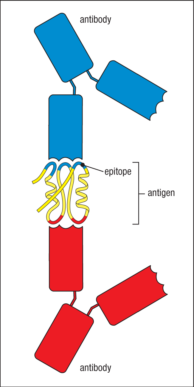
In principle, almost any chemical structure can be recognized as an antigen by the adaptive immune system, but the usual antigens encountered in an infection are the proteins, glycoproteins, and polysaccharides of pathogens. An individual antigen receptor or antibody recognizes a small portion of the antigen’s molecular structure, and the part recognized is known as an antigenic determinant, or epitope (Fig. 1.14). Typically, proteins and glycoproteins have many different epitopes that can be recognized by different antigen receptors.
Antibodies and B-cell receptors directly recognize the epitopes of native antigen in the serum or the extracellular spaces. It is possible for different antibodies to simultaneously recognize an antigen by its different epitopes; such simultaneous recognition increases the efficiency of clearing or neutralizing the antigen.
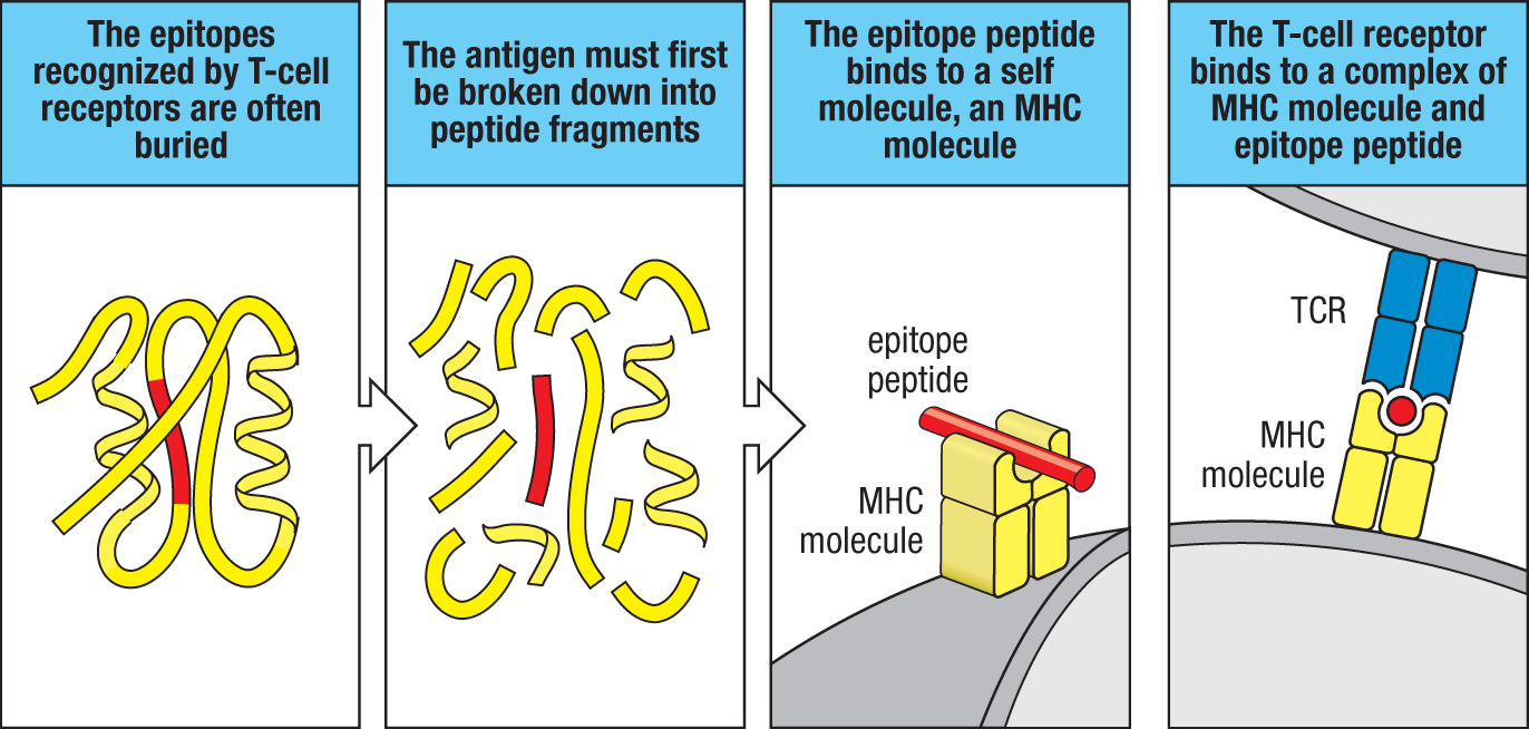
Whereas antibodies can recognize nearly any type of chemical structure, T-cell receptors usually recognize protein antigens and do so very differently from antibodies. The T-cell receptor recognizes a peptide epitope derived from a partially degraded protein, but only if the peptide is bound to specialized cell-surface glycoproteins called MHC molecules (Fig. 1.15). The members of this large family of cell-surface glycoproteins are encoded in a cluster of genes called the major histocompatibility complex (MHC). The antigens recognized by T cells can be derived from proteins arising from intracellular pathogens, such as a virus, or from extracellular pathogens. A further difference from the antibody molecule is that there is no secreted form of the T-cell receptor; the T-cell receptor functions solely to signal to the T cell that it has bound its antigen, and the subsequent immunological effects depend on the actions of the T cells themselves. We will further describe how epitopes from antigens are placed on MHC proteins in Chapter 6 and how T cells carry out their subsequent functions in Chapter 9.
1-11 Antigen-receptor genes are assembled by somatic gene rearrangements of incomplete receptor gene segments.
The innate immune system detects inflammatory stimuli by means of a relatively limited number of sensors, such as the TLR and NOD proteins, numbering fewer than 100 different types of proteins. Antigen-specific receptors of adaptive immunity provide a seemingly infinite range of specificities and yet are encoded by a finite number of genes. The basis for this extraordinary range of specificity was discovered in 1976 by Susumu Tonegawa, for which he was awarded the 1987 Nobel Prize. Immunoglobulin variable regions are inherited as sets of gene segments, each encoding a part of the variable region of one of the immunoglobulin polypeptide chains. During B-cell development in the bone marrow, these gene segments are irreversibly joined by a process of DNA recombination to form a stretch of DNA encoding a complete variable region. A similar process of antigen-receptor gene rearrangement takes place for the T-cell receptor genes during development of T cells in the thymus.
Just a few hundred different gene segments can combine in different ways to generate thousands of different receptor chains. This combinatorial diversity allows a small amount of genetic material to encode a truly staggering diversity of receptors. During this recombination process, the random addition or subtraction of nucleotides at the junctions of the gene segments creates additional diversity known as junctional diversity. Diversity is amplified further by the fact that each antigen receptor has two different variable chains, each encoded by distinct sets of gene segments. In Chapter 5 we will describe the gene-rearrangement process that assembles complete antigen receptors from gene segments.
1-12 Lymphocytes activated by antigen give rise to clones of antigen-specific effector cells that mediate adaptive immunity.
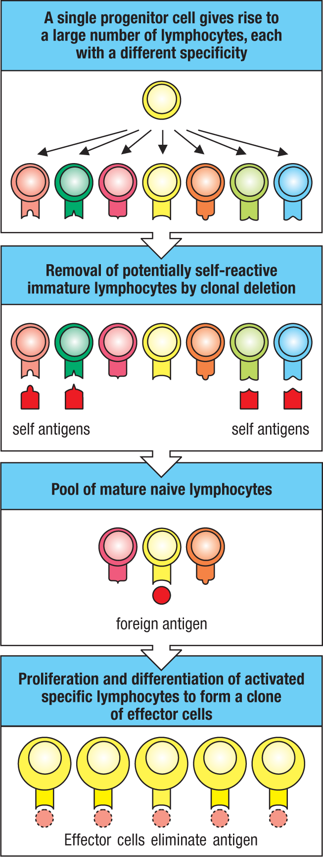
There are two critical features of lymphocyte development that distinguish adaptive immunity from innate immunity. First, the process that assembles antigen receptors from incomplete gene segments, described earlier, is carried out in a manner ensuring that each developing lymphocyte expresses only one receptor specificity. Whereas the cells of the innate immune system express many different pattern-recognition receptors and recognize features shared by many pathogens, the antigen-receptor expression of lymphocytes is ‘clonal,’ so that each mature lymphocyte differs from others in the specificity of its antigen receptor. Second, because the gene-rearrangement process irreversibly changes the lymphocyte’s DNA, all its progeny inherit the same receptor specificity. Because this specificity is inherited by a cell’s progeny, the proliferation of an individual lymphocyte forms a clone of cells with identical antigen receptors.
There are lymphocytes of at least 108 different specificities in an individual human at any one time, composing the lymphocyte receptor repertoire of the individual. These lymphocytes are continually undergoing a process similar to natural selection: only those lymphocytes that encounter an antigen to which their receptor binds will be activated to proliferate and differentiate into effector cells. This selective mechanism was first proposed in the 1950s by Frank Macfarlane Burnet, who postulated the preexistence in the body of many different potential antibody-producing cells, each displaying on its surface a membrane-bound version of the antibody that served as a receptor for the antigen. On binding antigen, the cell is activated to divide and to produce many identical progeny, a process known as clonal expansion; this clone of identical cells can now secrete clonotypic antibodies with a specificity identical to that of the surface receptor that first triggered activation and clonal expansion (Fig. 1.16). Burnet called this the clonal selection theory of antibody production; its four basic postulates are listed in Fig. 1.17. Clonal selection of lymphocytes is the single most important principle in adaptive immunity.
|
Postulates of the clonal selection hypothesis |
|
Each lymphocyte bears a single type of receptor with a unique specificity |
|
Interaction between a foreign molecule and a lymphocyte receptor capable of binding that molecule with high affinity leads to lymphocyte activation |
|
The differentiated effector cells derived from an activated lymphocyte will bear receptors of identical specificity to those of the parental cell from which that lymphocyte was derived |
|
Lymphocytes bearing receptors specific for ubiquitous self molecules are deleted at an early stage in lymphoid cell development and are therefore absent from the repertoire of mature lymphocytes |
Fig. 1.17 The four basic principles of clonal selection.
1-13 Lymphocytes with self-reactive receptors are normally eliminated during development or are functionally inactivated.
When Burnet formulated his theory, nothing was known of the antigen receptors or indeed the function of lymphocytes themselves. In the early 1960s, James Gowans discovered that removal of the small lymphocytes from rats resulted in the loss of all known adaptive immune responses, which were restored when the small lymphocytes were replaced. This led to the realization that lymphocytes must be the units of clonal selection, and their biology became the focus of the new field of cellular immunology.
Clonal selection of lymphocytes with diverse receptors elegantly explained adaptive immunity, but it raised one significant conceptual problem. With so many different antigen receptors being generated randomly during the lifetime of an individual, there is a possibility that some receptors might react against an individual’s own self antigens. How are lymphocytes prevented from recognizing native antigens on the tissues of the body and attacking them? Ray Owen had shown in the late 1940s that genetically different twin calves with a common placenta, and thus a shared placental blood circulation, were immunologically unresponsive, or tolerant, to one another’s tissues. Peter Medawar then showed in 1953 that exposure to foreign tissues during embryonic development caused mice to become immunologically tolerant to these tissues. Burnet proposed that developing lymphocytes that are potentially self-reactive are removed before they can mature, a process known as clonal deletion. Medawar and Burnet shared the 1960 Nobel Prize for their work on tolerance. This process was demonstrated experimentally in the late 1980s. Some lymphocytes that receive either too much or too little signal through their antigen receptor during development are eliminated by a form of cell suicide called apoptosis—derived from a Greek word meaning the falling of leaves from trees—or programmed cell death. Other types of mechanisms of immunological tolerance have been identified since then that rely on the induction of an inactive state, called anergy, as well as mechanisms of active suppression of self-reactive lymphocytes. Chapter 8 will describe lymphocyte development and tolerance mechanisms that shape the lymphocyte receptor repertoire. Chapters 14 and 15 will discuss how immune-tolerance mechanisms can sometimes fail.
1-14 Lymphocytes mature in the bone marrow or the thymus and then congregate in lymphoid tissues throughout the body.
Lymphocytes circulate in the blood and the lymph and are also found in large numbers in lymphoid tissues or lymphoid organs, the latter being organized aggregates of lymphocytes in a framework of nonlymphoid cells. Lymphoid organs can be divided broadly into the central (or primary) lymphoid organs, where lymphocytes are generated, and the peripheral (or secondary) lymphoid organs, where mature naive lymphocytes are maintained and adaptive immune responses are initiated. The central lymphoid organs are the bone marrow and the thymus, an organ in the upper chest. The peripheral lymphoid organs comprise the lymph nodes, the spleen, and the mucosal lymphoid tissues of the gut, the nasal and respiratory tract, the urogenital tract, and other mucosa. The locations of the main lymphoid tissues are shown schematically in Fig. 1.18; we describe the individual peripheral lymphoid organs in more detail later in the chapter. Lymph nodes are interconnected by a system of lymphatic vessels, which drain extracellular fluid from tissues, carry it through the lymph nodes, and deposit it back into the blood.
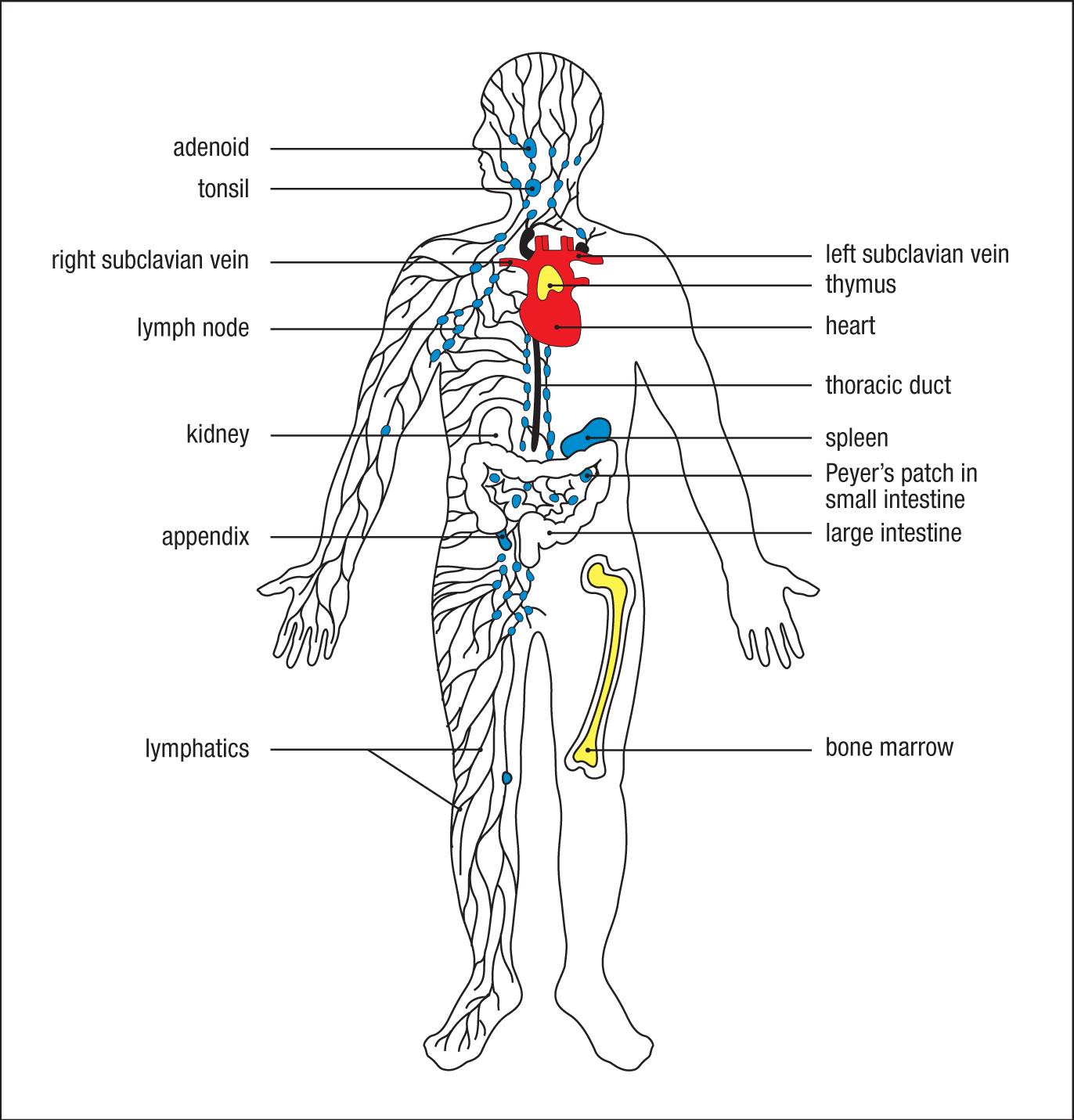
The progenitors that give rise to B and T lymphocytes originate in the bone marrow. B cells complete their development within the bone marrow. Although the ‘B’ in B lymphocytes originally stood for the bursa of Fabricius, a lymphoid organ in young chicks in which lymphocytes mature, it is a useful mnemonic for bone marrow. The immature precursors of T lymphocytes migrate to the thymus, from which they get their name, and complete their development there. Once they have completed maturation, both types of lymphocytes enter the bloodstream as mature naive lymphocytes and continually circulate through the peripheral lymphoid tissues.
1-15 Adaptive immune responses are initiated by antigen and antigen-presenting cells in peripheral lymphoid tissues.
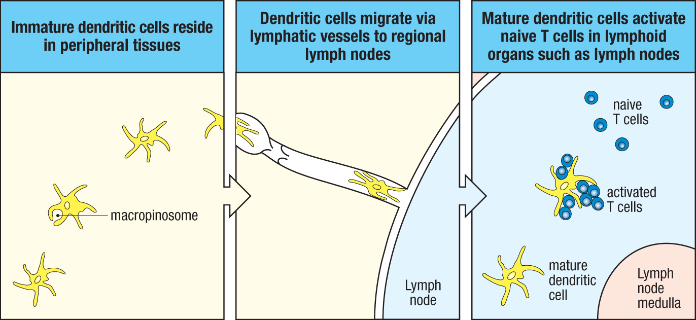
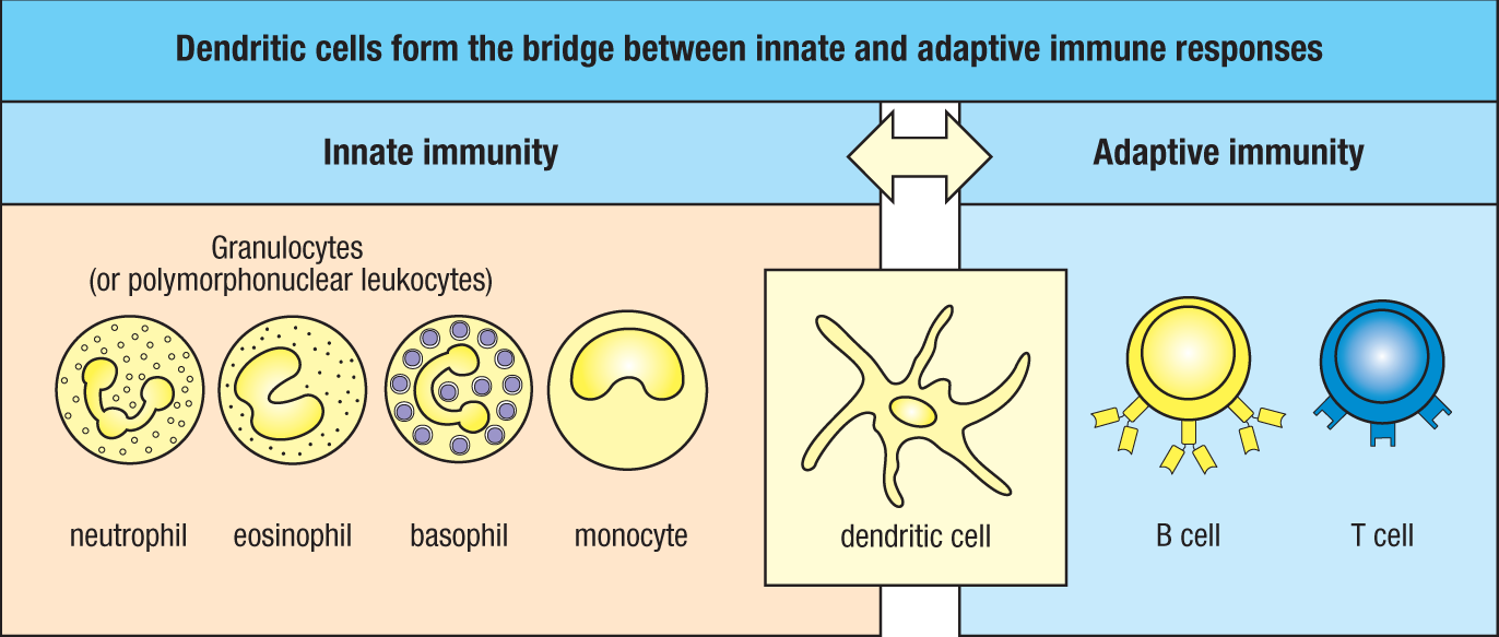
Adaptive immune responses are initiated when B or T lymphocytes encounter antigens for which their receptors have specific reactivity, provided that there are appropriate inflammatory signals to support activation. For T cells, this activation occurs via encounters with dendritic cells that have picked up antigens at sites of infection and migrated to peripheral (also called secondary) lymphoid organs. Activation of the dendritic cells’ PRRs by PAMPs at the site of infection stimulates the dendritic cells in the tissues to engulf the pathogen and degrade it intracellularly. They also take up extracellular material, including virus particles and bacteria, by receptor-independent macropinocytosis. These processes lead to the display of peptide antigens on the MHC molecules of the dendritic cells, a display that activates the antigen receptors of lymphocytes. Activation of PRRs also triggers the dendritic cells to express cell-surface proteins called co-stimulatory molecules, which support the ability of the T lymphocyte to proliferate and differentiate into its final, fully functional form (Fig. 1.19). For these reasons dendritic cells are also called antigen-presenting cells (APCs), and as such, they form a crucial link between the innate immune response and the adaptive immune response (Fig. 1.20). In certain situations, macrophages and B cells can also act as antigen-presenting cells, but dendritic cells are the cells that are specialized in initiating the adaptive immune response. Free antigens can also stimulate the antigen receptors of B cells, but most B cells respond better to antigens on the surface of cells, such as pathogens, and in addition require ‘help’ from activated helper T cells for optimal antibody responses. The activation of naive T lymphocytes is therefore an essential first stage in virtually all adaptive immune responses. Chapter 6 returns to dendritic cells to discuss how antigens are processed for presentation to T cells. Chapters 7 and 9 discuss co-stimulation and lymphocyte activation. Chapter 10 describes how T cells help in activating B cells.
1-16 Lymphocytes encounter and respond to antigen in the peripheral lymphoid organs.
Antigen and lymphocytes eventually encounter each other in the peripheral lymphoid organs—the lymph nodes, spleen, and mucosal lymphoid tissues (see Fig. 1.18). Mature naive lymphocytes are continually recirculating through these tissues, to which pathogen antigens are carried from sites of infection, primarily by dendritic cells. The peripheral lymphoid organs are specialized to trap antigen-bearing dendritic cells and to facilitate the initiation of adaptive immune responses. Peripheral lymphoid organs are composed of aggregations of lymphocytes in a framework of nonleukocyte stromal cells. The stromal cells provide both the basic structural organization of the tissue and survival signals to help sustain the life of the lymphocytes. Besides lymphocytes, peripheral lymphoid organs also contain resident macrophages and dendritic cells.
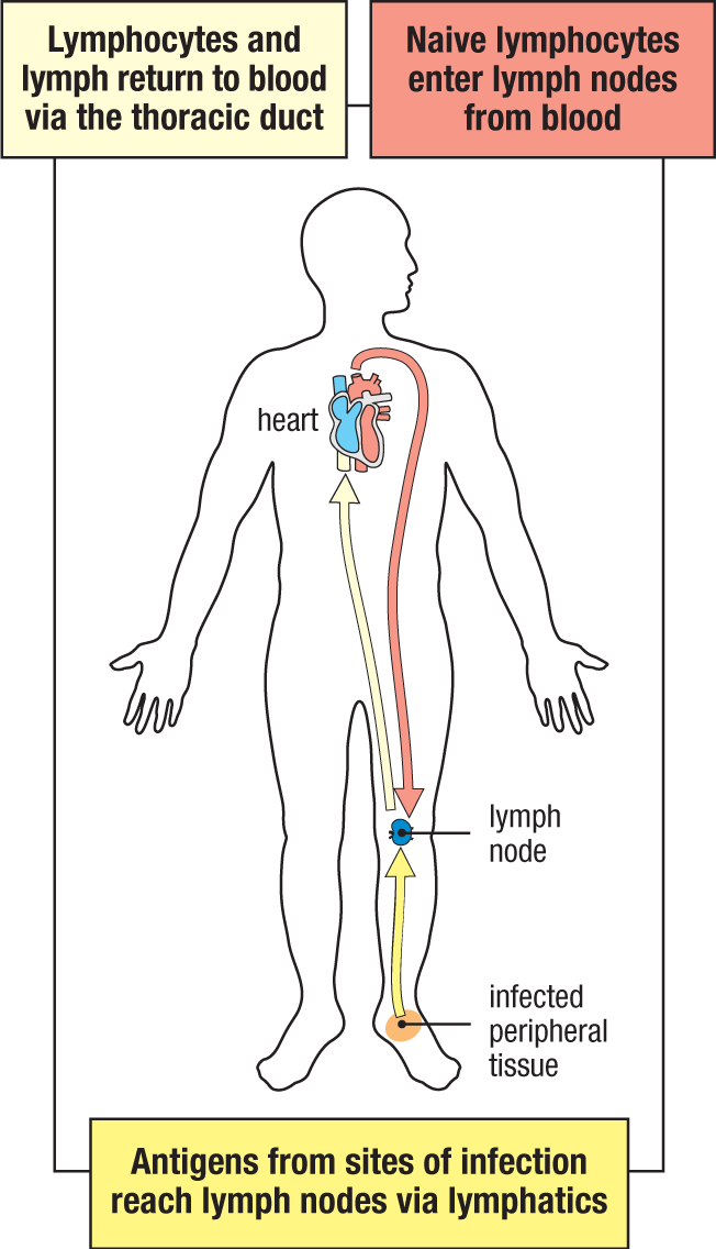
When an infection occurs in a tissue such as the skin, free antigen and antigen-bearing dendritic cells travel from the site of infection through the afferent lymphatic vessels into the draining lymph nodes (Fig. 1.21)—peripheral lymphoid organs in which the free antigen and antigen-bearing dendritic cells activate antigen-specific lymphocytes. The activated lymphocytes then undergo a period of proliferation and differentiation, after which most leave the lymph nodes as effector cells via the efferent lymphatic vessel. This eventually returns them to the bloodstream (see Fig. 1.18), which then carries them to the tissues where they will act. This whole process takes about 4–6 days from the time that the antigen is recognized, which means that an adaptive immune response to an antigen that has not been encountered before does not become effective until about a week after infection (see Fig. 1.7). Naive lymphocytes that do not recognize their antigen also leave through the efferent lymphatic vessel and are returned to the blood, from which they continue to recirculate through lymphoid tissues until they recognize antigen or die.
The lymph nodes are highly organized lymphoid organs located at the points of convergence of vessels of the lymphatic system, which is the extensive system that collects extracellular fluid from the tissues and returns it to the blood (see Fig. 1.18). This extracellular fluid is produced continually by filtration from the blood and is called lymph. Lymph flows away from the peripheral tissues under the pressure exerted by its continual production and is carried by lymphatic vessels, or lymphatics. One-way valves in the lymphatic vessels prevent a reverse flow, and the movements of one part of the body in relation to another are important in driving the lymph along.
As noted above, afferent lymphatic vessels drain fluid from the tissues and carry pathogens and antigen-bearing cells from infected tissues to the lymph nodes (Fig. 1.22). Free antigens simply diffuse through the extracellular fluid to the lymph node, while the dendritic cells actively migrate into the lymph node, attracted by chemokines. The same chemokines also attract lymphocytes from the blood, and these enter lymph nodes by squeezing through the walls of specialized blood vessels called high endothelial venules (HEVs), named for their thicker, more rounded appearance relative to flatter endothelial cells in other locations. In the lymph nodes, B lymphocytes are localized in follicles, which make up the outer cortex of the lymph node, with T cells more diffusely distributed in the surrounding paracortical areas, also referred to as the deep cortex or T-cell zones (see Fig. 1.22). Lymphocytes migrating from the blood into lymph nodes enter the paracortical areas first, and because they are attracted by the same chemokines, antigen-presenting dendritic cells and macrophages also become localized there. Free antigen diffusing through the lymph node can become trapped on these dendritic cells and macrophages. This juxtaposition of antigen, antigen-presenting cells, and naive T cells in the T-cell zone creates an ideal environment in which naive T cells can bind their specific antigen and thus become activated.
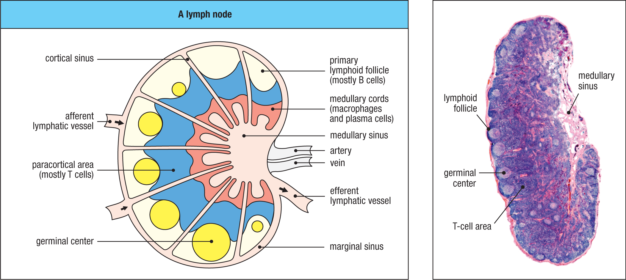
As noted earlier, activation of B cells usually requires not only antigen, which binds to the B-cell receptor, but also the cooperation of activated helper T cells, a type of effector T cell. The location of B cells and T cells within the lymph node is dynamically regulated by their state of activation. When they become activated, T cells and B cells both move to the border of the follicle and T-cell zone, where T cells can first provide their helper function to B cells. Some of the B-cell follicles include germinal centers, where activated B cells are undergoing intense proliferation and differentiation into plasma cells. These mechanisms are described in detail in Chapter 10.
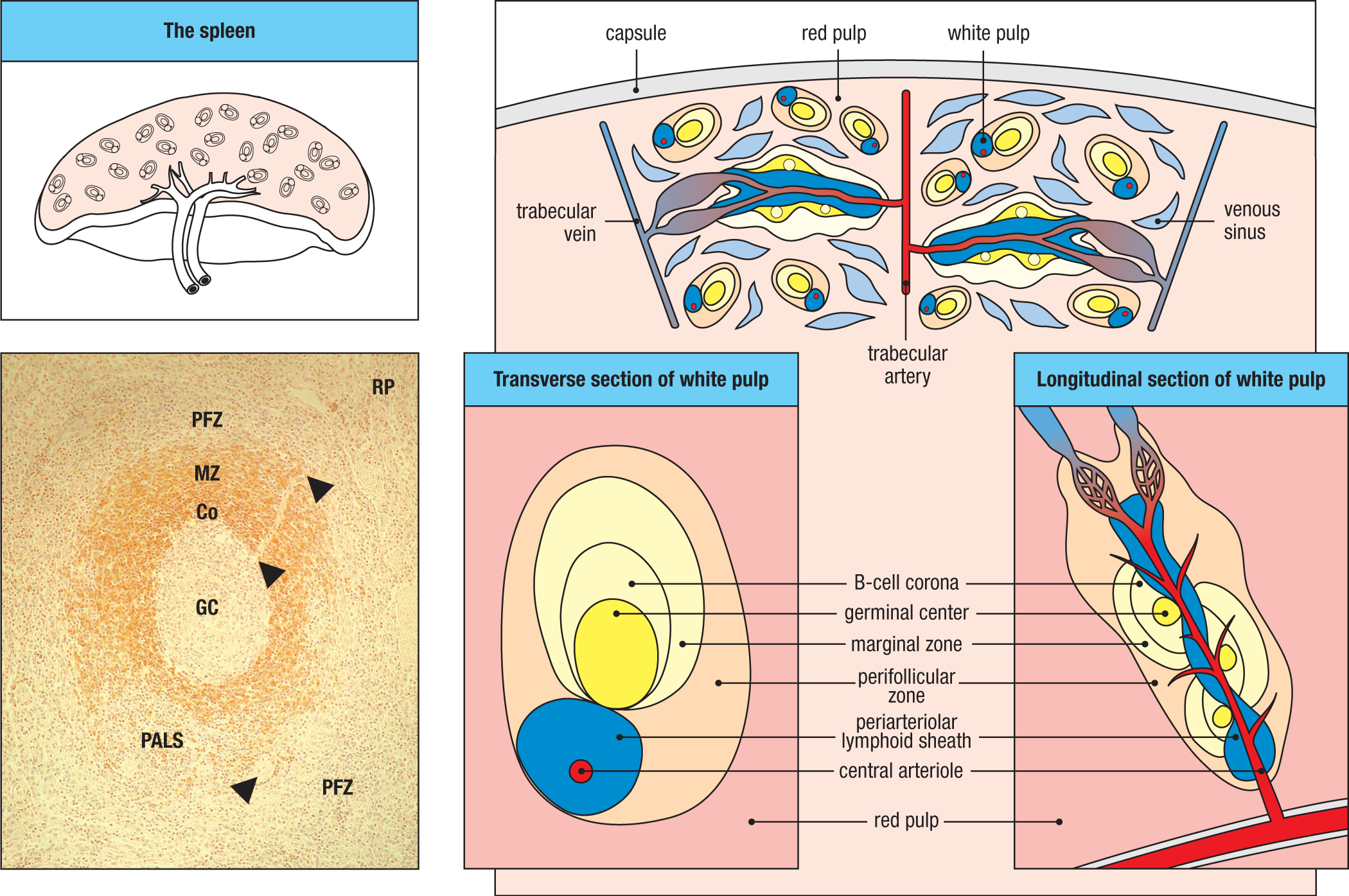
In humans, the spleen is a fist-sized organ situated just behind the stomach (see Fig. 1.18). It has no direct connection with the lymphatic system; instead, it collects antigen from the blood and is involved in immune responses to blood-borne pathogens. Lymphocytes enter and leave the spleen via blood vessels. The spleen also collects and disposes of senescent red blood cells. Its organization is shown schematically in Fig. 1.23. The bulk of the spleen is composed of red pulp, which is the site of red blood cell disposal. The lymphocytes surround the arterioles running through the spleen, forming isolated areas of white pulp. The sheath of lymphocytes around an arteriole is called the periarteriolar lymphoid sheath (PALS) and contains mainly T cells. Lymphoid follicles occur at intervals along it, and these contain mainly B cells. An area called the marginal zone surrounds the follicle; it has few T cells, is rich in macrophages, and has a resident, noncirculating population of B cells known as marginal zone B cells. These B cells are poised to rapidly produce antibodies that have low affinity to bacterial capsular polysaccharides. These antibodies, which are discussed in Chapter 8, provide some degree of protection before the adaptive immune response is fully activated. Blood-borne microbes, soluble antigens, and antigen:antibody complexes are filtered from the blood by macrophages and immature dendritic cells within the marginal zone. Like the migration of immature dendritic cells from peripheral tissues to the T-cell areas of lymph nodes, dendritic cells in the marginal zones in the spleen migrate to the T-cell areas after taking up antigen and becoming activated; here they are able to present the antigens they carry to T cells.
1-17 Mucosal surfaces have specialized immune structures that orchestrate responses to environmental microbial encounters.
Most pathogens enter the body through mucosal surfaces, which are also exposed to a vast load of other potential antigens from the air, food, and the natural microbial flora of the body. Mucosal surfaces are protected by an extensive system of lymphoid tissues known generally as the mucosal immune system or mucosa-associated lymphoid tissue (MALT). Collectively, the mucosal immune system is estimated to contain as many lymphocytes as all the rest of the body, and these lymphocytes form a specialized set of cells obeying somewhat different rules of recirculation from those of lymphocytes in the other peripheral lymphoid organs. The gut-associated lymphoid tissue (GALT) includes the tonsils, adenoids, appendix, and specialized structures in the small intestine called Peyer’s patches, and they collect antigen from the epithelial surfaces of the gastrointestinal tract. In Peyer’s patches, which are the most important and highly organized of these tissues, the antigen is collected by specialized epithelial cells called microfold cells, or M cells (Fig. 1.24). The lymphocytes form a follicle consisting of a large central dome of B lymphocytes surrounded by smaller numbers of T lymphocytes. Dendritic cells resident within the Peyer’s patch present the antigen to T lymphocytes. Lymphocytes enter Peyer’s patches from the blood and leave through efferent lymphatics. Effector lymphocytes generated in Peyer’s patches travel through the lymphatic system and into the bloodstream, from where they are disseminated back into mucosal tissues to carry out their effector actions.
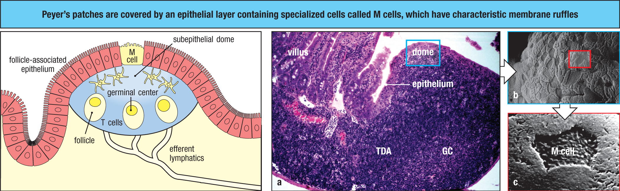
Similar but more diffuse aggregates of lymphocytes are present in the respiratory tract and other mucosa: nasal-associated lymphoid tissue (NALT) and bronchus-associated lymphoid tissue (BALT) are present in the respiratory tract. Like the Peyer’s patches, these mucosal lymphoid tissues are also overlaid by M cells, through which inhaled microbes and antigens that become trapped in the mucous covering of the respiratory tract can pass. The mucosal immune system is discussed in Chapter 12.
Although very different in appearance, the lymph nodes, spleen, and mucosa-associated lymphoid tissue all share the same basic architecture. They all operate on the same principle, trapping antigens and antigen-presenting cells from sites of infection in order to present antigen to migratory small lymphocytes, thus inducing adaptive immune responses. The peripheral lymphoid organs also provide sustaining signals to lymphocytes that do not encounter their specific antigen immediately, so that they survive and continue to recirculate.
Because they are involved in initiating adaptive immune responses, the peripheral lymphoid organs are not static structures but vary quite markedly, depending on whether or not infection is present. The diffuse mucosal lymphoid tissues may appear in response to infection and then disappear, whereas the architecture of the organized tissues changes in a more defined way during an infection. For example, the B-cell follicles of the lymph nodes expand as B lymphocytes proliferate to form germinal centers (see Fig. 1.22), and the entire lymph node enlarges, a phenomenon familiarly known as swollen glands.
Finally, specialized populations of lymphocytes and innate lymphoid cells can be found distributed throughout particular sites in the body rather than being found in organized lymphoid tissues. Such sites include the liver and the lamina propria of the gut, as well as the base of the epithelial lining of the gut, reproductive epithelia, and, in mice but not in humans, the epidermis. These lymphocyte populations seem to have an important role in protecting these tissues from infection and are described further in Chapters 8 and 12.
1-18 Lymphocytes activated by antigen proliferate in the peripheral lymphoid organs, generating effector cells and immunological memory.
The great diversity of the lymphocyte receptor repertoire means that there will usually be some lymphocytes bearing a receptor for any given foreign antigen. Recent experiments suggest this number to be perhaps a few hundred per mouse, certainly not enough to mount a response against a pathogen. To generate sufficient antigen-specific effector lymphocytes to fight an infection, a lymphocyte with an appropriate receptor specificity is activated first to proliferate. Only when a large clone of identical cells has been produced do these finally differentiate into effector cells, a process that requires 4–6 days. This means the adaptive immune response to a pathogen occurs nearly a week after the initial infection has occurred and been detected by the innate immune system.
On recognizing its specific antigen on an activated antigen-presenting cell, a naive lymphocyte stops migrating, the volume of its nucleus and cytoplasm increases, and new mRNA molecules and new proteins are synthesized. Within a few hours, the cell looks completely different and is known as a lymphoblast. Dividing lymphoblasts are able to duplicate themselves two to four times every 24 hours over the course of 3–5 days, so that a single naive lymphocyte can produce a clone of around 1000 daughter cells of identical specificity. These then differentiate into effector cells. In the case of B cells, the differentiated effector cells are the plasma cells, which secrete antibody. In the case of T cells, the effector cells are either cytotoxic T cells, which are able to destroy infected cells, or helper T cells, which activate other cells of the immune system (see Section 1-8).
Effector lymphocytes do not recirculate like naive lymphocytes. Some effector T cells detect sites of infection and migrate into them from the blood; others stay in the lymphoid tissues to activate B cells. Some antibody-secreting plasma cells remain in the peripheral lymphoid organs, but most plasma cells generated in the lymph nodes and spleen will migrate to the bone marrow and take up residence there, secreting large amounts of antibodies into the blood system. Effector cells generated in the mucosal immune system generally stay within the mucosal tissues. Most lymphocytes generated by clonal expansion in an immune response will eventually die. However, a significant number of activated antigen-specific B cells and T cells persist after antigen has been eliminated. These cells are known as memory cells and form the basis of immunological memory. They can be reactivated much more quickly than naive lymphocytes, which ensures a more rapid and effective response on a second encounter with a pathogen and thereby usually provides lasting protective immunity.
The characteristics of immunological memory are readily observed by comparing an individual’s antibody response to a first or primary immunization with the response to a secondary (or booster) immunization with the same antigen. As shown in Fig. 1.25, the secondary antibody response occurs after a shorter lag phase and achieves a markedly higher level than in the primary response. During the secondary response, antibodies can also acquire higher affinity, or strength of binding, for the antigen because of a process called affinity maturation, which takes place in the specialized germinal centers within B-cell follicles (see Section 1-16). Importantly, helper T cells are required for the process of affinity maturation, but T-cell receptors do not undergo affinity maturation. Compared with naive T cells, memory T cells show a lower threshold for activation, but as a result of changes in the responsiveness of the cell and not because of changes in the receptor. We describe the mechanisms of antibody affinity maturation in Chapter 10.
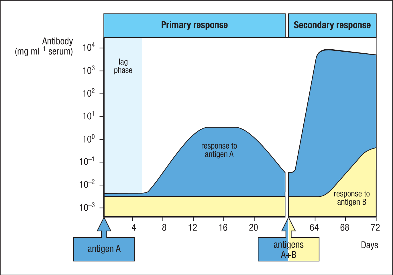
The cellular basis of immunological memory is the clonal expansion and clonal differentiation of cells that have a specific attraction for the eliciting antigen, and the memory is therefore entirely antigen-specific. It is immunological memory that enables successful vaccination and prevents reinfection with pathogens that have been repelled successfully by an adaptive immune response. In Chapter 11, we will return to immunological memory, which is perhaps the most important biological consequence of adaptive immunity.
Summary.
While the innate immune system relies on invariant pattern-recognition receptors to detect common microbial structures or the damage they cause, the adaptive immune system relies on a repertoire of antigen receptors to recognize structures that are specific to individual pathogens. This feature provides adaptive immunity with greater sensitivity and specificity. The clonal expansion of antigen-reactive lymphocytes also confers the property of immunological memory, which enhances protection against reinfection by the same pathogen.
Adaptive immunity relies on two major types of lymphocytes. B cells mature in the bone marrow and are the source of circulating antibodies. T cells mature in the thymus and recognize peptides from pathogens presented by MHC molecules on infected cells or antigen-presenting cells. An adaptive response involves the selection and amplification of clones of lymphocytes bearing receptors that recognize the foreign antigen. This clonal selection provides the theoretical framework for understanding all the key features of an adaptive immune response.
Each lymphocyte carries cell-surface receptors of a single antigen specificity. These receptors are generated by the random recombination of variable receptor gene segments and the pairing of distinct variable protein chains: heavy and light chains in immunoglobulins or the two chains of T-cell receptors. The large antigen-receptor repertoire of lymphocytes can recognize virtually any antigen. Adaptive immunity is initiated when an innate immune response fails to eliminate a new infection, and activated antigen-presenting cells—typically dendritic cells that bear antigens from pathogens and co-stimulatory receptors—migrate to the draining lymphoid tissues.
Immune responses are initiated in several peripheral lymphoid organs. The spleen serves as a filter for blood-borne infections. Lymph nodes draining various tissues and the mucosa-associated and gut-associated lymphoid tissues (MALT and GALT) are organized into specific zones where T and B cells can be activated efficiently by antigen-presenting cells or helper T cells. When a recirculating lymphocyte encounters its corresponding antigen in these peripheral lymphoid organs, it proliferates, and its clonal progeny differentiate into effector T and B lymphocytes that can eliminate the infectious agent. A subset of these proliferating lymphocytes differentiates into memory cells, ready to respond rapidly to the same pathogen if it is encountered again. The details of these processes of recognition, development, and differentiation form the main material of the central three parts of this book.
Glossary
- immunological memory
- The ability of the immune system to respond more rapidly and more effectively on a second encounter with an antigen. Immunological memory is specific for a particular antigen and is long-lived.
- B lymphocytes (B cells)
- One of the two types of antigen-specific lymphocytes responsible for adaptive immune responses, the other being the T cells. The function of B cells is to produce antibodies. B cells are divided into two classes. Conventional B cells have highly diverse antigen receptors and are generated in the bone marrow throughout life, emerging to populate the blood and lymphoid tissues. B-1 cells have much less diverse antigen receptors and form a population of self-renewing B cells in the peritoneal and pleural cavities.
- T lymphocytes (T cells)
-
One of the two types of antigen-specific lymphocytes responsible for adaptive immune responses, the other being the B cells. T cells are responsible for the cell-mediated adaptive immune reactions. They originate in the bone marrow but undergo most of their development in the thymus. The highly variable antigen receptor on T cells is called the T-cell receptor and recognizes a complex of peptide antigen bound to MHC molecules on cell surfaces. There are two main lineages of T cells: those carrying αβ receptors and those carrying γδ receptors. Effector T cells perform a variety of functions in an immune response, acting always by interacting with another cell in an antigen-specific manner. Some T cells activate macrophages, some help B cells produce antibody, and some kill cells infected with viruses and other intracellular pathogens.
- naive lymphocytes
- T cells or B cells that have undergone normal development in the thymus or the bone marrow but have not yet been activated by foreign (or self) antigens.
- effector lymphocytes
- The cells that differentiate from naive lymphocytes after initial activation by antigen and can then mediate the removal of pathogens from the body without further differentiation. They are distinct from memory lymphocytes, which must undergo further differentiation to become effector lymphocytes.
- immunoglobulin (Ig)
- The protein family to which antibodies and B-cell receptors belong.
- membrane immunoglobulin (mIg)
- Transmembrane immunoglobulin present on B cells; it is the B-cell receptor for antigen.
- surface immunoglobulin (sIg)
- The membrane-bound immunoglobulin that acts as the antigen receptor on B cells.
- T-cell antigen receptor, T-cell receptor (TCR)
- The cell-surface receptor for antigen on T lymphocytes. It consists of a disulfide-linked heterodimer of the highly variable α and β chains in a complex with the invariant CD3 and ζ proteins, which have a signaling function. T cells carrying this type of receptor are often called αβ T cells. An alternative receptor made up of variable γ and δ chains is expressed with CD3 and ζ on a subset of T cells.
- T-cell antigen receptor, T-cell receptor (TCR)
- The cell-surface receptor for antigen on T lymphocytes. It consists of a disulfide-linked heterodimer of the highly variable α and β chains in a complex with the invariant CD3 and ζ proteins, which have a signaling function. T cells carrying this type of receptor are often called αβ T cells. An alternative receptor made up of variable γ and δ chains is expressed with CD3 and ζ on a subset of T cells.
- plasma cells
- Terminally differentiated activated B lymphocytes. Plasma cells are the main antibody-secreting cells of the body. They are found in the medulla of the lymph nodes, in splenic red pulp, in bone marrow, and in mucosal tissues.
- regulatory T cells (Treg cells)
- Effector CD4 T cells that inhibit T-cell responses and are involved in controlling immune reactions and preventing autoimmunity. Several different subsets have been distinguished, notably the natural regulatory T-cell lineage that is produced in the thymus, and the induced regulatory T cells that differentiate from naive CD4 T cells in the periphery in certain cytokine environments.
- memory cells
- B and T lymphocytes that mediate immunological memory. They are more sensitive to antigen than are naive lymphocytes and respond rapidly on reexposure to the antigen that originally induced them.
- memory cells
- B and T lymphocytes that mediate immunological memory. They are more sensitive to antigen than are naive lymphocytes and respond rapidly on reexposure to the antigen that originally induced them.
- constant region (C region)
- That part of an immunoglobulin or a T-cell receptor that is relatively constant in amino acid sequence between different molecules. Also known as the Fc region in antibodies. The constant region of an antibody determines its particular effector function. Cf. variable region.
- fragment crystallizable region
- The carboxy-terminal halves of the two heavy chains of an IgG molecule disulfide-bonded to each other by the residual hinge region. It is produced by cleavage of IgG by papain. In the complete antibody this portion is often called the Fc region.
- variable region (V region)
- The region of an immunoglobulin or T-cell receptor that is formed of the amino-terminal domains of its component polypeptide chains. These are the most variable parts of the molecule and contain the antigen-binding sites. Cf. constant region.
- antigen-binding site
- The site at the tip of each arm of an antibody that makes physical contact with the antigen and binds it noncovalently. The antigen specificity of the site is determined by its shape and the amino acids present.
- antigenic determinant
- That portion of an antigenic molecule that is bound by the antigen-binding site of a given antibody or antigen receptor; it is also known as an epitope.
- epitope
- A site on an antigen recognized by an antibody or an antigen receptor. T-cell epitopes are short peptides bound to MHC molecules. B-cell epitopes are typically structural motifs on the surface of the antigen. Also called an antigenic determinant.
- MHC molecules
- Highly polymorphic cell-surface proteins encoded by MHC class I and MHC class II genes involved in presentation of peptide antigens to T cells. They are also known as histocompatibility antigens.
- major histocompatibility complex (MHC)
- A cluster of genes on human chromosome 6 that encodes a set of membrane glycoproteins called the MHC molecules. The MHC also encodes proteins involved in antigen processing and other aspects of host defense. The genes for the MHC molecules are the most polymorphic in the human genome, having large numbers of alleles at the various loci.
- gene segments
-
Sets of short DNA sequences at the immunoglobulin and T-cell receptor loci that encode different regions of the variable domains of antigen receptors. Gene segments of each type are joined together by somatic recombination to form a complete variable-domain exon. There are three types of gene segments: V gene segments encode the first 95 amino acids, D gene segments (in heavy-chain and TCRα chain loci only) encode about 5 amino acids, and J gene segments encode the last 10–15 amino acids of the variable domain. There are multiple copies of each type of gene segment in the germline DNA, but only one of each type is joined together to form the variable domain.
- combinatorial diversity
-
The diversity among antigen receptors generated by combining separate units of genetic information, comprising two types. First, receptor gene segments are joined in many different combinations to generate diverse receptor chains; second, two different receptor chains (heavy and light in immunoglobulins; α and β, or γ and δ, in T-cell receptors) are combined to make the antigen-recognition site.
- junctional diversity
- The variability in sequence present in antigen-specific receptors that is created during the process of joining V, D, and J gene segments and which is due to imprecise joining and insertion of non-templated nucleotides at the joins between gene segments.
- clone
- A population of cells all derived from the same progenitor cell.
- lymphocyte receptor repertoire
- All the highly variable antigen receptors carried by B and T lymphocytes.
- clonal expansion
- The proliferation of antigen-specific lymphocytes in response to antigenic stimulation that precedes their differentiation into effector cells. It is an essential step in adaptive immunity, allowing rare antigen-specific cells to increase in number so that they can effectively combat the pathogen that elicited the response.
- clonotypic
- Describes a feature unique to members of a clone. For example, the distribution of antigen receptors in the lymphocyte population is said to be clonotypic, as the cells of a given clone all have identical antigen receptors.
- clonal selection theory
-
The central paradigm of adaptive immunity. It states that adaptive immune responses derive from individual antigen-specific lymphocytes that are self-tolerant. These specific lymphocytes proliferate in response to antigen and differentiate into antigen-specific effector cells that eliminate the eliciting pathogen and into memory cells to sustain immunity. The theory was formulated by Frank Macfarlane Burnet and in earlier forms by Niels K. Jerne and David Talmage.
- cellular immunology
- The study of the cellular basis of immunity.
- self antigens
- The potential antigens on the tissues of an individual, against which an immune response is not usually made except in the case of autoimmunity.
- tolerant
- Describes the state of immunological tolerance, in which the individual does not respond to a particular antigen.
- clonal deletion
- The elimination of immature lymphocytes when they bind to self antigens, which produces tolerance to self as required by the clonal selection theory of adaptive immunity. Clonal deletion is the main mechanism of central tolerance and can also occur in peripheral tolerance.
- apoptosis
- A form of cell death common in the immune system, in which the cell activates an internal death program. It is characterized by nuclear DNA degradation, nuclear degeneration and condensation, and the rapid phagocytosis of cell remains. Proliferating lymphocytes experience high rates of apoptosis during their development and during immune responses.
- programmed cell death
- A form of cell death common in the immune system, in which the cell activates an internal death program. It is characterized by nuclear DNA degradation, nuclear degeneration and condensation, and the rapid phagocytosis of cell remains. Proliferating lymphocytes experience high rates of apoptosis during their development and during immune responses.
- anergy
- A state of nonresponsiveness to antigen. People are said to be anergic when they cannot mount delayed-type hypersensitivity reactions to a test antigen, whereas T cells and B cells are said to be anergic when they cannot respond to their specific antigen under optimal conditions of stimulation.
- lymphoid tissue
- Tissue composed of large numbers of lymphocytes.
- lymphoid organs
- Organized tissues characterized by very large numbers of lymphocytes interacting with a nonlymphoid stroma. The central, or primary, lymphoid organs, where lymphocytes are generated, are the thymus and bone marrow. The main peripheral, or secondary, lymphoid organs, in which adaptive immune responses are initiated, are the lymph nodes, spleen, and mucosa-associated lymphoid organs such as tonsils and Peyer’s patches.
- thymus
- A central lymphoid organ, in which T cells develop, situated in the upper part of the middle of the chest, just behind the breastbone.
- lymph nodes
- A type of peripheral lymphoid organ present in many locations throughout the body where lymphatic vessels converge.
- spleen
- An organ in the upper left side of the peritoneal cavity containing a red pulp, involved in removing senescent blood cells, and a white pulp of lymphoid cells that respond to antigens delivered to the spleen by the blood.
- bursa of Fabricius
- Lymphoid organ associated with the gut that is the site of B-cell development in chickens.
- co-stimulatory molecules
- Cell-surface proteins on antigen-presenting cells that deliver co-stimulatory signals to naive T cells. Examples are the B7 molecules on dendritic cells, which are ligands for CD28 on naive T cells.
- antigen-presenting cells (APCs)
- Highly specialized cells that can process antigens and display their peptide fragments on the cell surface together with other, co-stimulatory, proteins required for activating naive T cells. The main antigen-presenting cells for naive T cells are dendritic cells, macrophages, and B cells.
- draining lymph node
- A lymph node downstream of a site of infection that receives antigens and microbes from the site via the lymphatic system. Draining lymph nodes often enlarge enormously during an immune response and can be palpated; they were originally called swollen glands.
- afferent lymphatic vessels
- Vessels of the lymphatic system that drain extracellular fluid from the tissues and carry antigen, macrophages, and dendritic cells from sites of infection to lymph nodes or other peripheral lymphoid organs.
- follicles
- An area of predominantly B cells in a peripheral lymphoid organ, such as a lymph node, which also contains follicular dendritic cells.
- cortex
- The outer part of a tissue or organ; in lymph nodes it refers to the follicles, which are mainly populated by B cells.
- paracortical areas
- The T-cell areas of lymph nodes.
- T-cell zones
- Regions of peripheral lymphoid organs that are enriched in naive T cells and are distinct from the follicles. They are the sites at which adaptive immune responses are initiated.
- germinal centers
- Sites of intense B-cell proliferation and differentiation that develop in lymphoid follicles during an adaptive immune response. Somatic hypermutation and class switching occur in germinal centers.
- red pulp
- The nonlymphoid area of the spleen in which red blood cells are broken down.
- white pulp
- The discrete areas of lymphoid tissue in the spleen.
- periarteriolar lymphoid sheath (PALS)
- Part of the inner region of the white pulp of the spleen; it contains mainly T cells.
- marginal zone
- Area of lymphoid tissue lying at the border of the white pulp in the spleen.
- marginal zone B cells
- A unique population of B cells found in the spleen marginal zones; they do not circulate and are distinguished from conventional B cells by a distinct set of surface proteins.
- mucosal immune system
- The immune system that protects internal mucosal surfaces (such as the linings of the gut, respiratory tract, and urogenital tracts), which are the site of entry for virtually all pathogens and other antigens. See also mucosa-associated lymphoid tissue.
- mucosa-associated lymphoid tissue (MALT)
- Generic term for all organized lymphoid tissue found at mucosal surfaces, in which an adaptive immune response can be initiated. It comprises gut-associated lymphoid tissue (GALT), nasal-associated lymphoid tissue (NALT), and bronchus-associated lymphoid tissue (BALT) (when present).
- gut-associated lymphoid tissue (GALT)
- Lymphoid tissues associated with the gastrointestinal tract, comprising Peyer’s patches, the appendix, and isolated lymphoid follicles found in the intestinal wall, where adaptive immune responses are initiated.
- tonsils
- Paired masses of organized peripheral lymphoid tissue situated at the base of the tongue, in which adaptive immune responses can be initiated. They are part of the mucosal immune system. See also palatine tonsils.
- adenoids
- Paired mucosa-associated lymphoid tissue located in the nasal cavity.
- appendix
- A gut-associated lymphoid tissue located at the beginning of the colon.
- Peyer’s patches
- Organized peripheral lymphoid organs under the epithelium in the small intestine, especially the ileum, and in which an adaptive immune response can be initiated. They contain lymphoid follicles and T-cell areas. They are part of the gut-associated lymphoid tissue (GALT).
- microfold cells
- Specialized epithelial cell type in the intestinal epithelium over Peyer’s patches, through which antigens and pathogens enter from the gut.
- M cells
- Specialized epithelial cell type in the intestinal epithelium over Peyer’s patches, through which antigens and pathogens enter from the gut.
- bronchus-associated lymphoid tissue (BALT)
- Organized lymphoid tissue found in the bronchi in some animals. Adult humans do not normally have such organized lymphoid tissue in the respiratory tract, but it may be present in some infants and children.
- lamina propria
- A layer of connective tissue underlying a mucosal epithelium. It contains lymphocytes and other immune-system cells.
- lymphoblast
- A lymphocyte that has enlarged after activation and has increased its rate of RNA and protein synthesis but is not yet fully differentiated.
- primary immunization
- The first encounter with a given antigen, which generates the primary adaptive immune response.
- affinity maturation
- The increase in affinity of antibodies for their specific antigen as an adaptive immune response progresses. This phenomenon is particularly prominent in secondary and subsequent immunizations.
- B-cell antigen receptor, B-cell receptor (BCR)
- The cell-surface receptor on B cells for specific antigen. It is composed of a transmembrane immunoglobulin molecule (which recognizes antigen) associated with the invariant Igα and Igβ chains (which have a signaling function). On activation by antigen, B cells differentiate into plasma cells producing antibody molecules of the same antigen specificity as this receptor.
- B-cell antigen receptor, B-cell receptor (BCR)
- The cell-surface receptor on B cells for specific antigen. It is composed of a transmembrane immunoglobulin molecule (which recognizes antigen) associated with the invariant Igα and Igβ chains (which have a signaling function). On activation by antigen, B cells differentiate into plasma cells producing antibody molecules of the same antigen specificity as this receptor.
- effector T lymphocytes, effector T cells
- The T cells that perform the functions of an immune response (such as cell killing and cell activation) that clear the infectious agent from the body. There are several different subsets, each with a specific role in an immune response.
- effector T lymphocytes, effector T cells
- The T cells that perform the functions of an immune response (such as cell killing and cell activation) that clear the infectious agent from the body. There are several different subsets, each with a specific role in an immune response.
- helper CD4 T cells, helper T cells
- Effector CD4 T cells that stimulate or ‘help’ B cells to make antibody in response to antigenic challenge. TH2, TH1, and the TFH subsets of effector CD4 T cells can perform this function.
- Fc fragment, Fc region
- The carboxy-terminal halves of the two heavy chains of an IgG molecule disulfide-bonded to each other by the residual hinge region. It is produced by cleavage of IgG by papain. In the complete antibody this portion is often called the Fc region.
- heavy chain, H chain
- One of the two types of protein chains in an immunoglobulin molecule, the other being called the light chain. There are several different classes, or isotypes, of heavy chain (α,δ, ε, γ, and μ), each of which confers a distinctive functional activity on the antibody molecule. Each immunoglobulin molecule contains two identical heavy chains.
- light chain, L chain
- The smaller of the two types of polypeptide chains that make up an immunoglobulin molecule. It consists of one V and one C domain and is disulfide-bonded to the heavy chain. There are two classes, or isotypes, of light chain, known as κ and λ, which are produced from separate genetic loci.
- primary lymphoid organs
- The sites of lymphocyte development; in humans, these are the bone marrow and thymus. B lymphocytes develop in bone marrow, whereas T lymphocytes develop within the thymus from bone marrow–derived progenitors. Also called the primary lymphoid organs and tissues.
- secondary lymphoid organs
- The lymph nodes, spleen, and mucosa-associated lymphoid tissues, in which adaptive immune responses are induced, as opposed to the central lymphoid organs, in which lymphocytes develop. They are also called secondary lymphoid organs and tissues.
- lymphatic vessels, lymphatics
- Thin-walled vessels that carry lymph.
- lymphatic vessels, lymphatics
- Thin-walled vessels that carry lymph.
- high endothelial cells, high endothelial venules (HEVs)
- Specialized small venous blood vessels in lymphoid tissues. Lymphocytes migrate from the blood into lymphoid tissues by attaching to the high endothelial cells in the walls of the venules and squeezing between them.
- secondary immunization
- A second or booster injection of an antigen, given some time after the initial immunization. It stimulates a secondary immune response.
- nasal- (or nasopharynx-) associated lymphoid tissue (NALT)
- Organized lymphoid tissues found in the upper respiratory tract. In humans, NALT consists of Waldeyer’s ring, which includes the adenoids and palatine and lingual tonsils, plus other similarly organized lymphoid tissue located around the pharynx. It is part of the mucosal immune system.