Organization of the mucosal immune system.
The main line of defense against invasion by microbes at mucosal tissues is the epithelium that covers their surfaces. In some sites the mucosal epithelium is composed of multiple layers (for example, oral cavity, nasal cavity, nasopharynx), but in sites such as the intestines and the lower airways of the respiratory tract, the epithelium is a single cell layer thick to facilitate nutrient uptake and gas exchange, respectively. Especially in these sites, where the physiological role of the tissue depends on free exchange of molecules between the ‘outside’ and ‘inside,’ the epithelium can be breached relatively easily. Epithelial barrier function is therefore supplemented by molecules provided by the epithelium itself, such as mucus and antimicrobial peptides, as well as the cells and molecules of the mucosal immune system. Each of these components is integrated into a layered defense that restrains the entry of microbes while allowing physiological activities to proceed.
The epithelium is not just a passive barrier between the external and internal environments; it is central to sensing the environment and communicating with immune cells to induce mucosal immune responses. Throughout this chapter, we focus on the unique relationship of the mucosal epithelia with the innate and adaptive mucosal immune systems and on how these two arms of immunity are integrated with epithelial function at homeostasis and in host defense. Many of the anatomic and immunological principles underlying the mucosal immune system apply to all mucosal tissues. Here we will focus primarily on the intestines as our main example, as it is the largest and best-studied mucosal immune system. Unique aspects of respiratory mucosa and skin barrier immunity will be considered later.
12-1 The mucosal immune system protects the internal surfaces of the body.
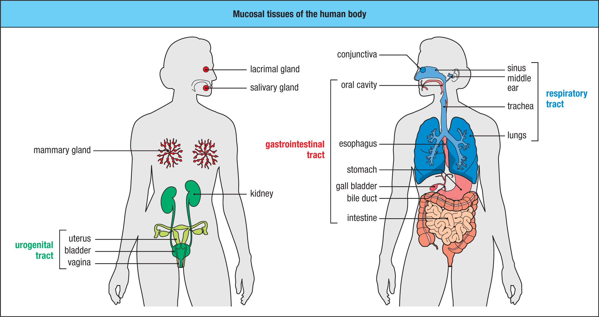
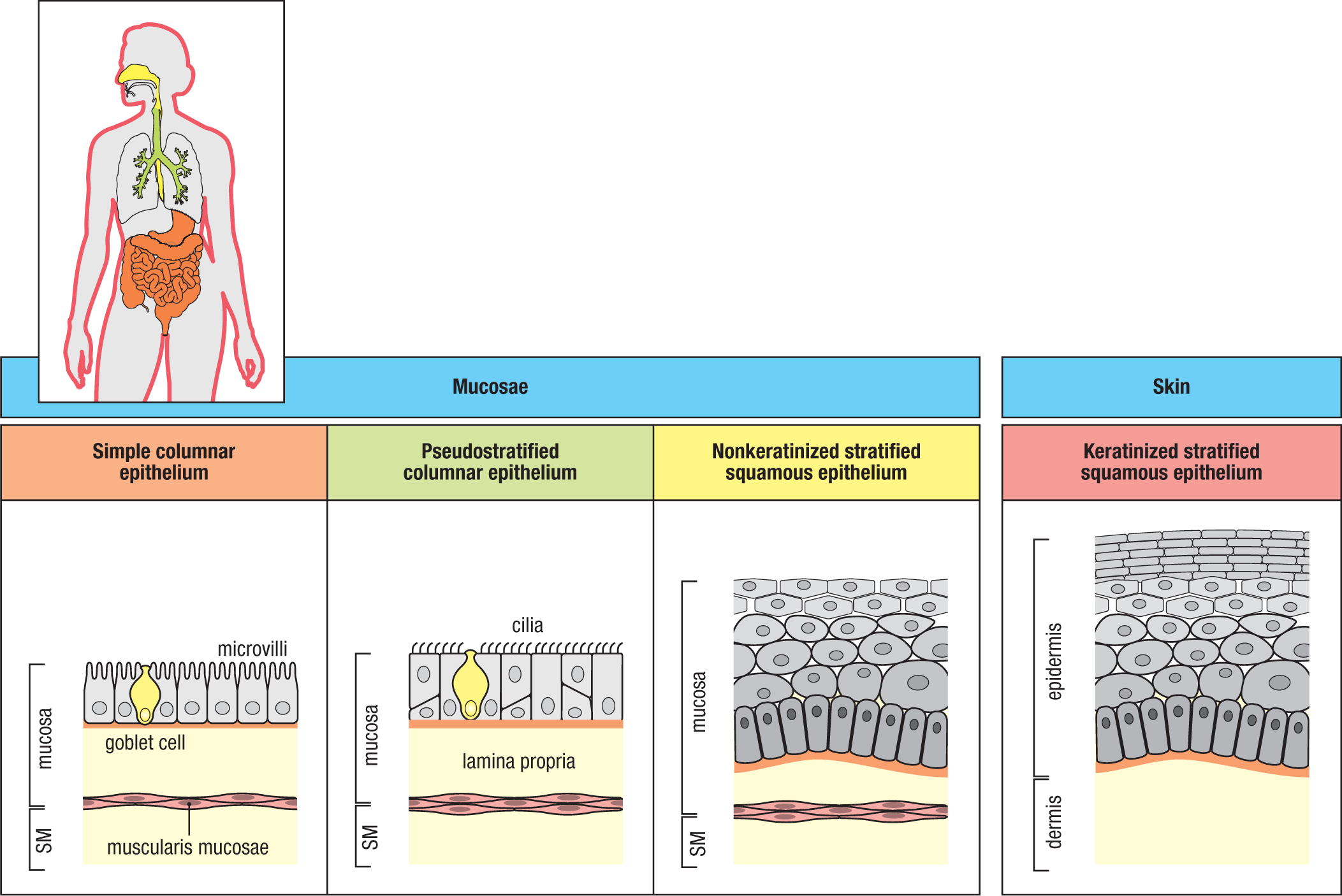
The mucosal immune system comprises the internal body surfaces that are lined by a mucus-covered epithelium—the gastrointestinal (GI) tract, the upper and lower respiratory tract, and the urogenital tract. It also includes the biliary tract, middle ear, and exocrine glands, such as the conjunctivae and lacrimal glands of the eye, the salivary glands, and the lactating breast (Fig. 12.1). The surfaces of mucosal tissues are covered by epithelia that range from a single cell layer thick—such as the simple columnar epithelium composed of enterocytes, or intestinal epithelial cells, that line the intestines—to multiple layers, such as the nonkeratinized stratified squamous epithelium of the esophagus and vagina (Fig. 12.2). The mucosal surfaces represent an enormous area to be protected. Collectively, the mucosal tissues of humans have a surface area of almost 400 m2, roughly 200 times that of the skin. Although the skin is a barrier tissue, it is not moist and mucus-covered, and hence not a mucosal tissue. It is covered instead by layers of dying and dead interdigitating ‘squames’ (literally, scales), or keratinocytes—flattened cells that compose its multilayered keratinized squamous epithelium (see Fig. 12.2), as will be discussed later.
In accord with the large surface area of mucosal tissues, the mucosal immune system forms the largest part of the body’s immune tissues. Approximately three-quarters of all lymphocytes in the body are found in the mucosae and they produce the majority of immunoglobulin in healthy individuals. The mucosae are continually exposed to antigens and other materials that enter from the environment or are produced by resident microbes. When compared with lymph nodes and spleen (which we will refer to as the systemic immune system in this chapter), the mucosal immune system has many unique and unusual features (Fig. 12.3).
|
Distinctive features of the mucosal immune system |
||
|
Anatomic features |
Intimate interactions between mucosal polarized epithelia and lymphoid tissues Discrete compartments of diffuse lymphoid tissue and more organized structures such as Peyer’s patches, isolated lymphoid follicles, and tonsils Specialized antigen-uptake mechanisms, e.g., M cells in Peyer’s patches, adenoids, and tonsils Broad surface area in contact with environmental agents/microbes |
|
|
Effector mechanisms |
Activated/memory T cells predominate even in the absence of infection Multiple activated ‘natural’ effector/regulatory T cells present Production of mucins and mucus Secretory IgA antibodies Production of antimicrobial peptides (AMPs) Presence of distinctive microbiota |
|
|
Immunoregulatory environment |
Active down-regulation of immune responses (e.g., to food and other innocuous antigens) predominates at homeostasis Inhibitory macrophages and tolerance-inducing dendritic cells High number of FoxP3+ Treg cells and FoxP3− TR1 cells |
|
Fig. 12.3 Distinctive features of the mucosal immune system. The mucosal immune system is larger, encounters a wider range of antigens, and encounters them much more frequently than the rest of the immune system—what we call in this chapter the systemic immune system. This is reflected in distinctive anatomic features, specialized mechanisms for the uptake of antigen, and unusual effector and regulatory responses that are designed to prevent unwanted immune responses to food and other innocuous antigens.
Because of their physiological functions in food absorption (the intestines), gas exchange (the lungs), sensory activities (eyes, nose, mouth, and throat), and reproduction (prostate, uterus, and vagina), many mucosal surfaces are thin and permeable barriers to the interior of the body. The importance of these tissues to life means that effective defense mechanisms are essential to protect them from invasion. Equally significant is that their fragility and permeability create obvious vulnerability to infection, and it is not surprising that the vast majority of infectious agents invade via these routes. Diarrheal diseases, acute respiratory infections, pulmonary tuberculosis, measles, whooping cough, and worm infestations continue to be the major causes of death throughout the world, especially in infants in developing countries. To these must be added the human immunodeficiency virus (HIV), a pathogen whose natural route of entry via a mucosal surface is often overlooked, as well as other sexually transmitted infections, such as syphilis (Fig. 12.4).
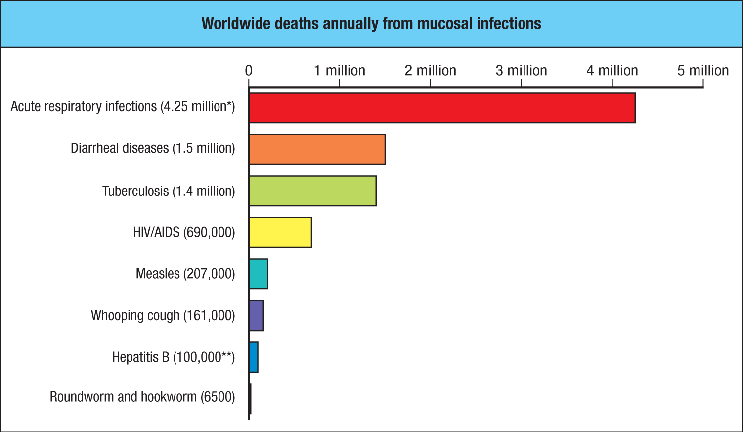
*Note that this estimate predates the SARS-CoV-2 pandemic.
**Does not include deaths from liver cancer or cirrhosis resulting from chronic hepatitis infections.
The mucosal surfaces are also portals of entry for a vast array of foreign antigens that must be tolerated to prevent untoward immune activation and immune-mediated disease. This is perhaps best illustrated by the gut, which is exposed to enormous quantities of food proteins—an estimated 30–35 kg per year per person. Moreover, the healthy large and small intestines are colonized by hundreds of species of microbes that live in symbiosis with their host, known as commensal microorganisms, or commensal microbiota. The microbiota consists of complex communities that have coevolved with their mammalian hosts over hundreds of millions of years. Bacteria dominate the commensal microbiota, and at least 1013 bacteria are present in the colon contents, making them the most numerous cells in the body—outnumbering the total number of cells of the host. Substantial populations of archaea, fungi, and bacterial viruses (bacteriophages) are also found in the healthy intestine, as are parasites such as worms (helminths) in countries with less advanced sanitation. The host provides the microbiota with a warm, nutrient-rich environment, and in return, the microbiota provides key functions that benefit the host. In normal circumstances these organisms do no harm, and many are beneficial to their hosts, having important metabolic functions, as well as being essential for normal immune function and providing a buffer against potential pathogens. Other mucosal tissues are also colonized by substantial populations of resident commensal organisms, as is the skin (Fig. 12.5).
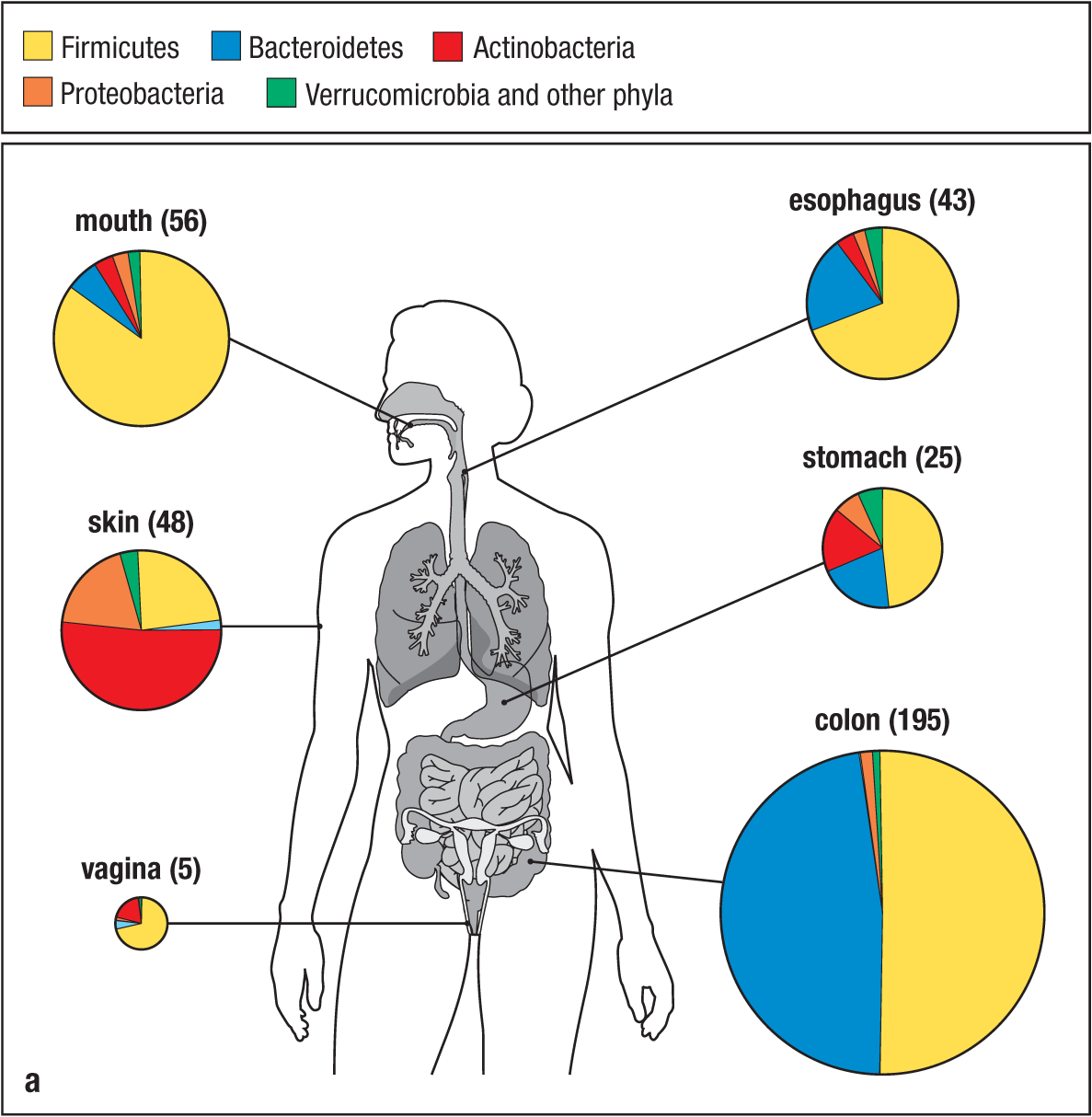
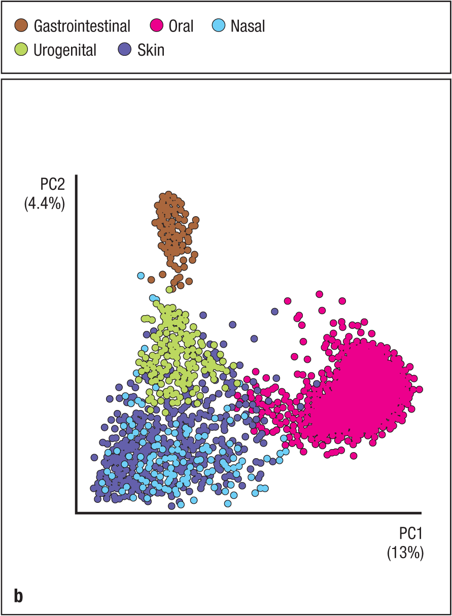
Fig. 12.5 Composition of the commensal microbiota at different barrier surfaces in healthy humans. Panel a: The different sizes of the pie charts for different sites reflect the number of distinct bacterial phyla typically present at those sites. The color key indicates the five bacterial phyla that make up the majority of commensal species. Ubiquitous commensal bacteria include Lactobacillus and Clostridium spp. (Firmicutes), Bacteroides fragilis (Bacteroidetes), Bifidobacterium spp. (Actinobacteria), Escherichia coli (Proteobacteria), and Akkermansia spp. (Verrucomicrobia). The numbers in parentheses denote the number of independent strains. Panel b: Principal component analysis of the microbiota isolated from the indicated human tissues in different individuals, plotting the first and second principal components (PC1 and PC2, respectively). The primary component of microbiota variation between individuals is body area (that is, site location) and accounts for 13% of the variation in microbial identity among site-specific samples taken from different individuals. The colon contains the greatest number of different species (more than 1000 as estimated from individual surveys). Although not shown because it is more difficult to sample, the commensal microbiota of the human small intestine are distinct from and less diverse than those of the large intestine, dominated by members of Firmicutes and Proteobacteria phyla, particularly Lactobacillaceae and Enterobacteriaceae family members, respectively. Note that the values shown are derived from surveys of the Western countries/Western diets; the microbiota composition shifts contingent on diet and other factors.
Because food and the commensal microbiota contain many foreign antigens, they can be recognized by the adaptive immune system and normally induce antigen-specific immune tolerance. Inappropriate responses to these antigens are potentially damaging to the host and are the cause of a number of common diseases, including celiac disease (caused by an aberrant immune response to the wheat protein gluten; discussed in Chapter 14) and inflammatory bowel diseases such as Crohn’s disease (due to a dysregulated response to commensal bacteria; discussed further later and in Chapter 15). As we shall see, the innate and adaptive mucosal immune systems of the intestinal immune system have developed multiple strategies to accommodate these ‘friendly’ antigens while retaining robust host defense against pathogens, including opportunistic pathogens that are normal constituents of the commensal microbiota. Similar issues are faced at other mucosal surfaces, such as the respiratory tract and genital tract. Here, protective immunity against pathogens is essential, but many of the antigens entering these tissues are harmless, being derived from commensal organisms, pollen, other innocuous environmental material, and, in the lower urogenital tract, seminal fluid.
12-2 Immune cells of the mucosal immune system are located both within and outside of specialized lymphoid tissues in proximity to the epithelium.
The deployment of immune cells in mucosal tissues can be divided into inductive and effector sites—that is, sites where immune cells are initially activated and sites where they act directly in support of the barrier function of the epithelium. Like all other tissues in the body, lymphatics that drain the mucosal tissues deliver antigens to regional lymph nodes, such as the mesenteric lymph nodes, which serve as inductive sites for the small intestine and proximal colon, or the cervical lymph nodes, which serve as inductive sites for the oral and nasal cavities, for example. In addition, the mucosae contain inductive sites with organizational features similar to those of other peripheral lymphoid tissues, but they lack afferent lymphatics and receive antigens directly across the overlying epithelium with which they are in direct contact, as will be discussed below; these are collectively referred to as mucosa-associated lymphoid tissue (MALT) (Fig. 12.6). Although organized lymphoid tissue is not typically present in the nose or lower respiratory tract in adult humans (unless infection is present), it is present in children and in some species such as mice; in the nose it is called the nasal- (or nasopharynx-) associated lymphoid tissue (NALT), while in the lower respiratory tract it is known as the bronchus-associated lymphoid tissue (BALT). The best-studied mucosal lymphoid tissue—and the largest in mass—is that associated with the gastrointestinal tract, or gut, known as the gut-associated lymphoid tissue (GALT), the bulk of which resides in the intestines. Because the intestines comprise several anatomically defined segments with distinct functions—extending in the small intestine from the duodenum to jejunum and ileum, and in the large intestine from cecum to colon (Fig. 12.7)—there are regional differences in the environmental exposure and response of the associated immune tissues. There are also microanatomic differences that reflect regional specialization and affect the local immune response.
|
Components of the mucosa-associated lymphoid tissue (MALT) |
||
|
Region |
Components |
Regional lymph nodes |
|
GALT (gut-associated lymphoid tissue) |
Peyer’s patches, cecal patch (mice), appendix (human), and isolated lymphoid follicles (ILFs) or solitary intestinal lymphoid tissue (SILT) |
Mesenteric lymph nodes (small intestine, cecum, proximal and middle colon), caudal and iliac lymph nodes (distal colon) |
|
NALT (nasal-associated lymphoid tissue) |
Tonsils of Waldeyer’s ring (encircling the nasopharynx and oropharynx in humans and composed of nasopharyngeal, tubal, palatine, and lingual tonsils) and ILFs in nasal mucosa. Mice lack tonsils but do have paired NALT structures in floor of nasal cavity |
Cervical lymph nodes |
|
BALT (bronchus-associated lymphoid tissue) |
Not typically present in uninfected adults, as the lower airways are sterile, but develops in respiratory infections |
Peribronchial and mediastinal lymph nodes |
Fig. 12.6 Components of the mucosa-associated lymphoid tissue (MALT).
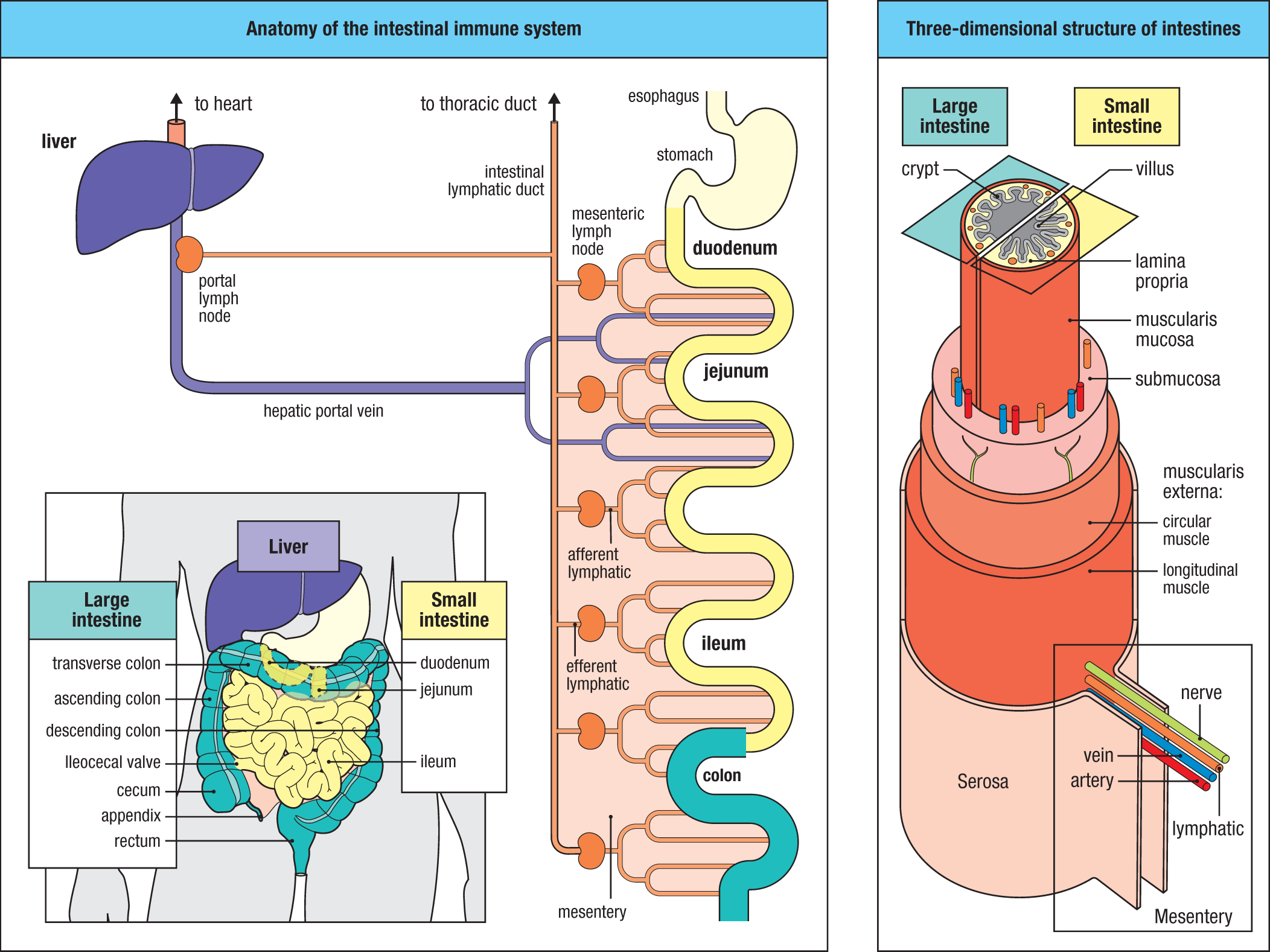
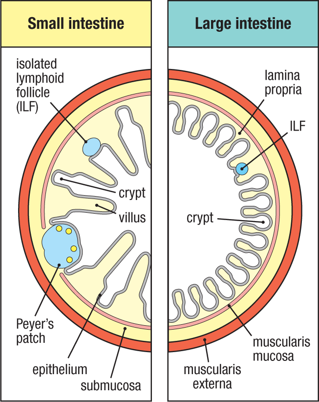
The GALT includes the Peyer’s patches, which are most prevalent in the distal small intestine; isolated lymphoid follicles (ILFs), also known as solitary isolated lymphoid tissue (SILT), which are smaller structures found throughout the small and large intestine; and, in humans, the appendix (Fig. 12.8; and see Fig. 12.7). (Similar to the human appendix is the cecal patch in mice, found near the tip of the blind pouch, or cecum, that is the beginning part of the large intestine; cecum is derived from the Latin for ‘blind.’) Each of these components of the GALT is located within the wall of the intestine; the patches and larger ILFs extend from the intestinal epithelium through the full thickness of the mucosa to the submucosa, whereas smaller ILFs are fully contained within the mucosa; that is, bound below by the muscularis mucosa. In mice, but not typically in humans, Peyer’s patches (and the cecal patch) are visible to the naked eye and have a distinctive appearance, forming dome-like aggregates of lymphoid cells that project into the intestinal lumen (see Fig. 1.24). There are approximately 100–300 Peyer’s patches in the human small intestine, concentrated in the distal small intestine, or ileum. Peyer’s patches are much richer in B cells than the systemic peripheral lymphoid organs, with each consisting of multiple B-cell follicles that contain active germinal centers (that is, secondary B-cell follicles). T-cell zones are situated between and immediately below the follicles. This is in contrast to ILFs, which typically contain a single follicle or a less organized aggregate of B cells, without a distinct T-cell zone. In humans, Peyer’s patches may consist of more than a hundred B-cell follicles, although they vary in size with age, peaking in adolescence and declining thereafter.
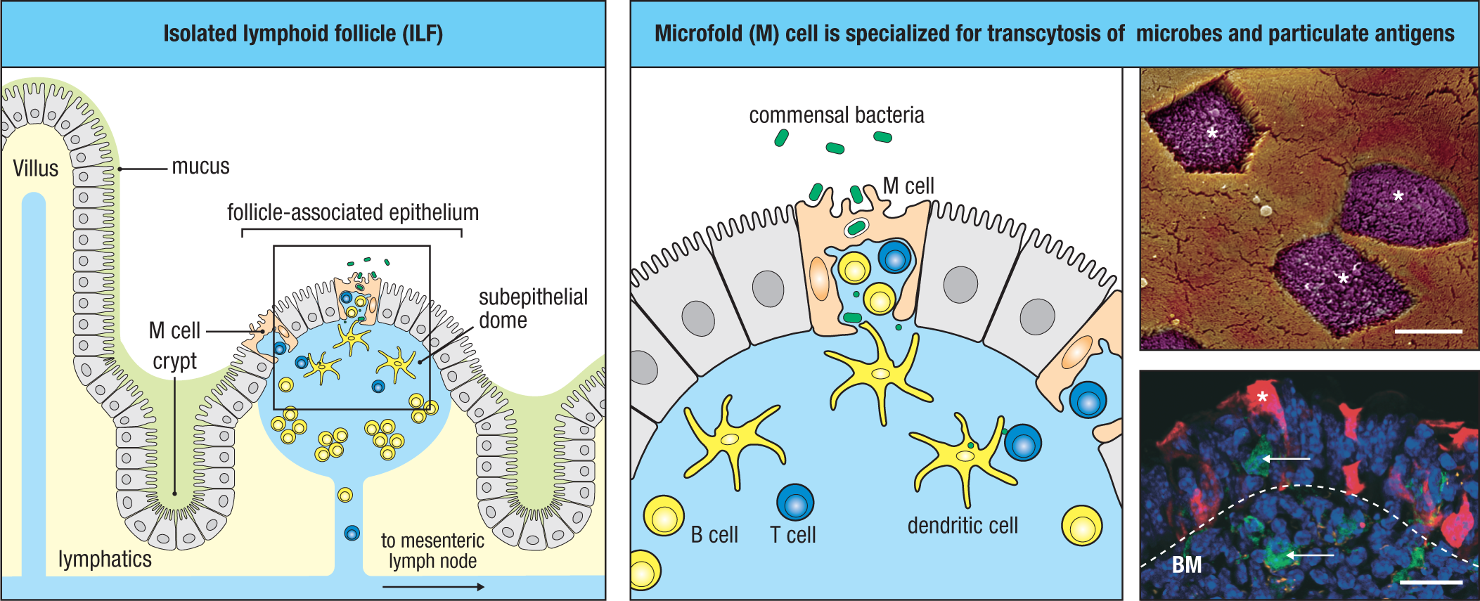
A feature common to all MALT, including GALT, that distinguishes it from conventional lymph nodes is the absence of afferent lymphatics or a capsule. This organization is essential to the function of the GALT as an important site for the initiation of immune responses in the gut, enabling direct sampling of luminal antigens that are transported across the specialized epithelium overlying the Peyer’s patches and ILFs—the follicle-associated epithelium (FAE). Contained within the FAE are specialized epithelial cells called microfold cells (M cells) (Fig. 12.9; and see Fig. 1.24 and Section 12-4), which transport intact bacteria, abiotic particulates, and soluble antigens within vacuoles across their cell bodies. The area immediately beneath the FAE, the subepithelial dome, is rich in dendritic cells and lymphocytes, which are in intimate contact with the FAE. Unlike the enterocytes that make up the rest of the intestinal epithelium, cells that compose the follicle-associated epithelium do not secrete digestive enzymes or mucins, and so lack the thick layer of surface mucus (the glycocalyx) found covering conventional epithelial cells, and the M cells have a folded luminal surface instead of microvilli (see Fig. 1.24 and Section 12.4). These cells are therefore directly exposed to microorganisms and particles within the gut lumen and, because of their ability to transport large antigenic particles and intact microbes across their cell bodies within vesicles, are a preferred route by which microbes and their antigens enter the Peyer’s patches from the lumen.
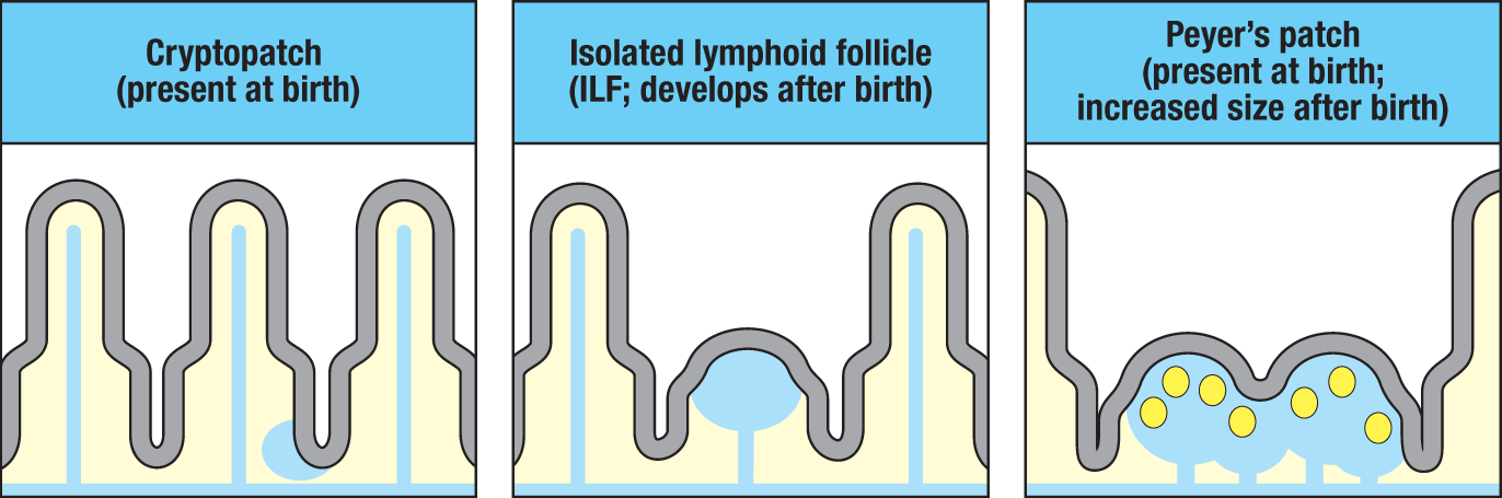
Unlike Peyer’s patches, which are found mainly in the distal ileum, several thousand ILFs can be identified microscopically throughout the small and large intestines, but they are more abundant in the large intestine, correlating with the greater load of commensal microorganisms there. Like Peyer’s patches, these follicles are also covered by a follicle-associated epithelium containing M cells (see Fig. 12.9). However, ILFs contain mainly B cells and develop only after birth in response to antigen stimulation due to colonization of the gut by commensal microorganisms (Fig. 12.10). Peyer’s patches, in contrast, are already present in the fetal gut, although their full development is completed only after colonization by the intestinal microbiota after birth. In the mouse gut, and perhaps in humans, isolated lymphoid follicles arise from small aggregates in the intestinal wall called cryptopatches, which are composed of dendritic cells and lymphoid tissue inducer (LTi) cells, a type of ILC3 (see Section 9-2 and Section 11-3). Cryptopatches undergo maturation into ILFs only after signals from the developing microbiota stimulate the recruitment of B cells. Peyer’s patches and ILFs are connected by efferent lymphatics to regional lymph nodes, such as the mesenteric lymph nodes. This allows antigens taken up across the FAE to stimulate local responses in the Peyer’s patch or ILF, as well as distally in the draining lymph node.
The small intestine and the proximal large intestine drain to the mesenteric lymph nodes, which, as the name implies, are located in the mesentery, a connective tissue that tethers the intestine to the rear wall of the abdomen and contains the vasculature, nerves, and lymphatics that supply the intestines (see Fig. 12.7). Although not technically part of the GALT, these lymph nodes are in communication with the GALT via efferent lymphatics, as indicated earlier. They also drain the effector sites within the intestine, described later. The mesenteric lymph nodes represent the largest collection of lymph nodes in the body and play a crucial role in initiating and shaping immune responses to intestinal antigens. The ILFs and lamina propria of the distal large intestine drain to separate nodes known as the caudal and inguinal lymph nodes found close to the bifurcation of the descending aorta on the posterior wall of the lower abdominal cavity. The mesenteric lymph nodes and Peyer’s patches differentiate independently of the systemic immune system during fetal development, and their development involves distinct chemokines and receptors of the tumor necrosis factor (TNF) family (see Section 9-2). The differences between the GALT and the systemic lymphoid organs are thus imprinted early in life.
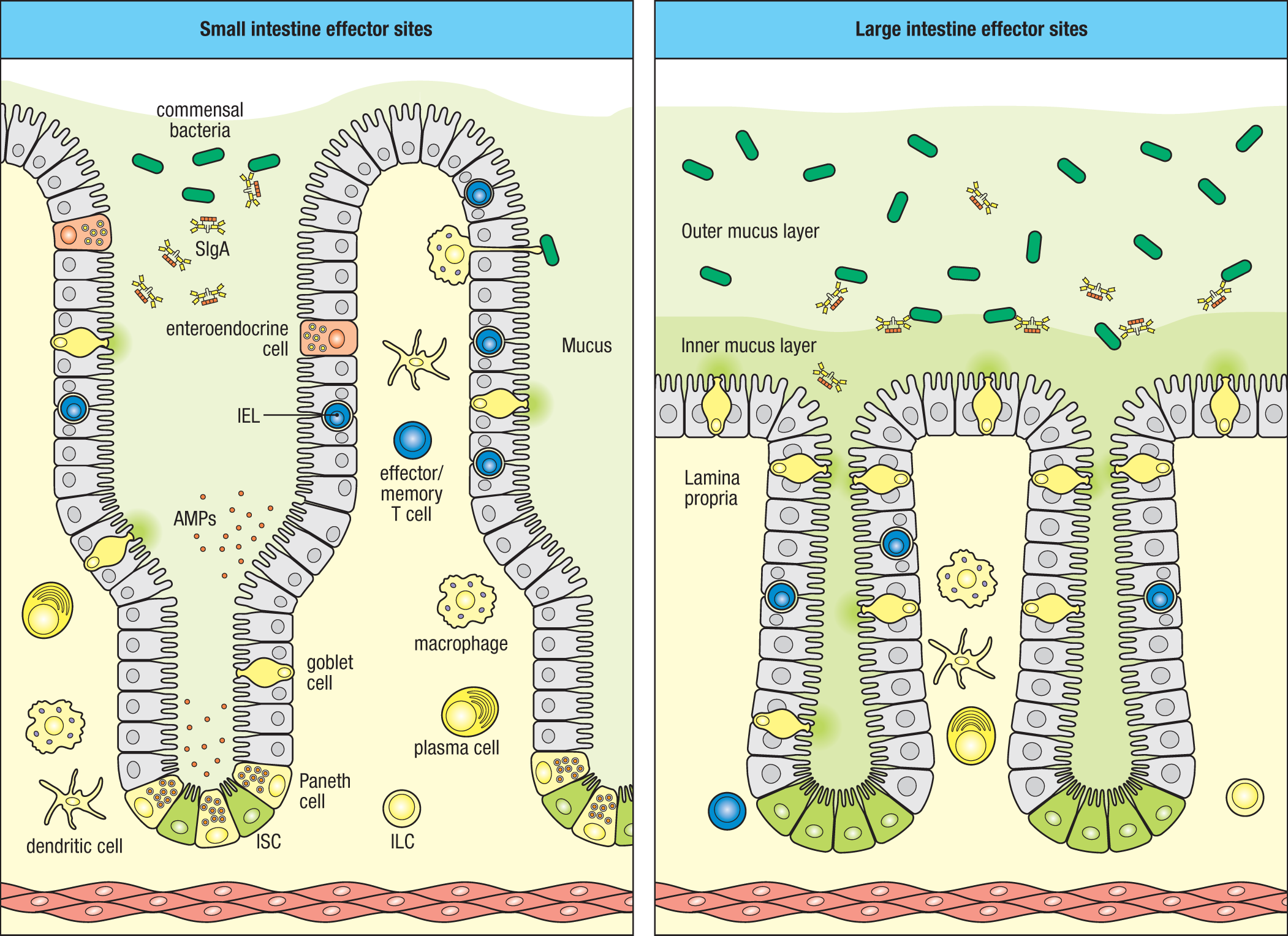
In addition to the inductive sites discussed above, the barrier function of the intestines is supported by effector sites that contain innate and adaptive immune cells that migrate through or reside within the intestinal epithelium itself or in the underlying layer of connective tissue called the intestinal lamina propria (Fig. 12.11; see also Fig. 12.8). The intestinal epithelium and lamina propria are referred to as effector sites because the preponderance of adaptive immune cells within them are effector cells that either have migrated there after recognition of antigen in inductive lymphoid tissues of the GALT or regional lymph nodes or, in the case of the epithelium, have populated the site directly from the thymus as functionally competent effector cells around the time of birth. The intestinal epithelium harbors a substantial number of conventional and unconventional T cells, so-called intraepithelial lymphocytes (IELs), as well as some innate lymphoid cells (ILCs), which will be discussed further in Section 12-5. The intestinal lamina propria is diffusely populated by macrophages and dendritic cells that are strategically located beneath the epithelium, and it also contains antigen-experienced effector and regulatory T cells, as well as antibody-producing plasma cells, which are most numerous in the small intestine. Indeed, at homeostasis the largest number of effector T cells and antibody-producing B cells is found in the intestinal mucosa. The intestinal lamina propria also contains smaller numbers of ILCs and type 2 innate effector cells, including eosinophils, basophils, and mast cells, which are also more abundant in the small intestine. In addition, a variety of innate lymphocytes and lymphoid cells on the innate-adaptive spectrum are found in gut effector sites. These are cells that express invariant antigenic receptors, such as γδ T cells, invariant NKT (iNKT) cells, and mucosal-associated invariant T cells (MAIT cells), many of which populate the lamina propria during fetal development in preparation for colonization of the gut by the microbiota.
12-3 Maturation of the gut-associated lymphoid tissue is driven by acquisition of the commensal microbiota.
The normal maturation of the intestinal mucosa and the GALT is initiated at birth by colonization of the intestines by the commensal microbiota. Lacking a commensal microbiota, the preterm infant is germ-free, but is normally exposed to the vaginal microbiota of the mother during birth, initiating the process of intestinal colonization. Although Peyer’s patches and the intestine-associated lymph nodes develop in utero, ILFs develop only after colonization by the commensal microbiota, and all of the GALT requires sensing of the intestinal microbiota to become fully populated by lymphocytes (see Fig. 12.10). Similarly, other components of the intestinal mucosa that contribute to the establishment of host–microbiota commensalism are driven by colonization of the neonate at birth. This process has been best studied in germ-free mice (or gnotobiotic mice), which are raised in a contained, microbe-free environment so that there is either no colonization of the gut by microorganisms or colonization only by microbes introduced into the gnotobiotic chamber. In the intestines of germ-free mice, Peyer’s patches do not develop normally, and isolated lymphoid follicles are absent. These mice also have severely reduced numbers of intestinal T lymphocytes and ILCs, and very few IgA-secreting plasma cells. They also have reduced mediators of intestinal immunity, such as antimicrobial peptides, retinoic acid, and the cytokines IL-7, IL-22, IL-25, IL-33, and thymic stromal lymphopoietin (TSLP).
As we learned in Chapter 9 (see Section 9-2), preparation for the adaptation to microbial colonization requires the actions of lymphoid tissue inducer (LTi) cells. This subset of ILC3 cells is instrumental in organizing development of the GALT, as well as other lymphoid tissues in the body (see Fig. 9.2). During fetal development, LTi cells disseminate to sites of future lymphoid tissue development in the gut, recruited by the chemokine CXCL13, a product of specialized stromal cells. Activation of these stromal cells by lymphotoxin (LT-α1β2) expressed on LTi cells initiates the development of structural components of the Peyer’s patches, as well as the recruitment and partitioning of B and T cells into B-cell follicles and T-cell zones. ILF development is similarly dependent on LTi cells but is initiated after birth when cells in cryptopatches are stimulated to recruit lymphocytes in response to microbial stimuli from the developing microbiota (see Fig. 12.10). Thus, ILFs develop from cryptopatches only after colonization by the intestinal microbiota. As discussed later, LTi cells continue to contribute to the intestinal response to the microbiota beyond their role in the perinatal development of the GALT, as do other subsets of ILCs that seed the intestines—and other barrier tissues—during the perinatal period.
Like the GALT, the intestinal epithelium of the newborn undergoes changes to accommodate to the microbiota. For example, expression of TLR-4, the receptor for lipopolysaccharide (LPS)—a major pathogen-associated molecular pattern (PAMP) expressed by Gram-negative bacteria—is significantly up-regulated prior to birth. Soon after exposure to the developing microbiota, however, intestinal epithelial cells (IECs) rapidly down-regulate TLR-4 and other components of its signaling apparatus to attenuate signaling at the IEC apical membrane. Nevertheless, TLR-4 remains expressed within endocytic vesicles where it can sense bacterial invasion and induce a pro-inflammatory response. An additional immune mechanism that prepares the neonate for microbial colonization is the seeding of the intestinal epithelium by intraepithelial lymphocytes (IELs) before birth. As we will see in Section 12-5, localization of IELs within the epithelium enables them to respond rapidly to help limit bacterial translocation, maintain epithelial barrier integrity, and promote repair of the epithelium after injury.
At maturity, the intestines maintain the largest number and diversity of commensal microbes in or on the body, harboring several hundred species of commensal bacteria alone. The greatest density of microbiota along the intestinal tract is in the distal ileum and large intestine, particularly in the cecum. Over the first year or so of life, however, there are marked and often erratic shifts in the intestinal microbiota as it transitions from a more limited consortium dominated by so-called pioneer bacterial species—primarily facultative anaerobes dominated by Proteobacteria, such as E. coli, which can thrive in a more aerobic microbiota-naive intestine—to a more diverse consortium dominated by strict anaerobes, such as Firmicutes (for example, Clostridium spp.) and Bacteroidetes (for example, Bacteroides fragilis), that occurs in concert with a reduction of oxygen levels in the superficial intestinal mucosa and the lumen (Fig. 12.12).
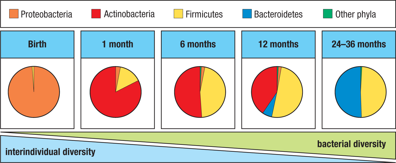
Another major driver of maturation of the intestinal microbiota is the transition from a liquid to solid diet that occurs with weaning from breast milk. Prior to the introduction of solid food, breast milk provides a specialized mix of nutrients and antimicrobial proteins that influence the ecology of the neonatal microbiota, as well as maternal secretory IgA (SIgA), the specificities of which have been shaped by the mother’s microbiota. In essence, the mother’s mucosal immune memory is transmitted to her child. Thus, in infants delivered transvaginally, the intestinal microbiota not only is seeded by maternal bacteria, but also its composition may be shaped by maternal SIgA, which is influenced, in turn, by the mother’s microbiota. The transition to solid foods introduces new substrates, such as complex carbohydrates, that serve as fuel for a transition of the microbiota to its mature composition. As different constituents of the microbiota have different nutrient and metabolic features, the foods ingested can result in marked alterations in the composition of the microbiota.
In humans, two major phyla of anaerobic bacteria—Firmicutes and Bacteroidetes—come to dominate the mature intestinal microbiota, although significant Proteobacteria, Actinobacteria, Verrucomicrobia, and Archaea are also found (see Fig. 12.5). Trillions of these microorganisms normally exist in a beneficial symbiotic relationship with the host known as mutualism, reflecting the benefits that both microbes and host derive from their sharing of resources. Microbiota–host mutualism dates back almost 500 million years to the rise of jawed vertebrates, with the result that the microbiota has coevolved with their hosts over many millennia. Indeed, it is speculated that the development of an adaptive immune system in vertebrates may have allowed them to accommodate to a complex intestinal microbiota and generate the bacterial bioreactor as a mechanism to extend the range of foods from which they derive energy, providing a competitive survival advantage. This has produced, in essence, a superorganism that includes humans and their microbiota.
The microbiota has an essential role in maintaining health. Its collective genome, or microbiome, contains more than 10 times the number of genes encoded in the human genome, including many genes that encode enzymes that metabolize dietary constituents such as cellulose and produce essential cofactors such as vitamin K1. Short-chain fatty acids (SCFAs), such as acetate, propionate, and especially butyrate, produced by anaerobic commensal bacteria during the metabolism (fermentation) of complex dietary carbohydrates, are an essential source of energy for colonic enterocytes as substrates for the tricarboxylic acid (TCA) cycle. Host benefits derived from the commensal microbiota extend beyond those related to nutrition, however. The microbiota also plays an important role in protection against pathogens by competing for colonization niches and nutrient resources, collectively referred to as colonization resistance. Indeed, the normal microbiota contains bacterial species that can cause pathology if not continually restrained by other members of the collective. These opportunistic pathogens include a number of Gram-negative and Gram-positive bacteria of the Enterobacteriaceae family, such as E. coli, Klebsiella pneumoniae, and Proteus mirabilis, which can cause serious disease if they extend beyond the confines of the intestinal lumen.
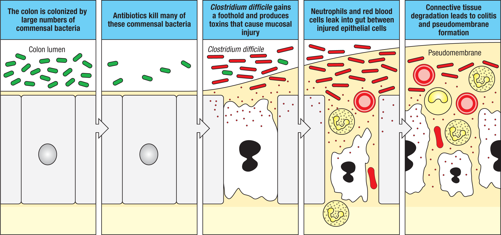
This protective role of the intestinal microbiota is dramatically illustrated by the adverse effects of broad-spectrum antibiotics, which can kill large numbers of commensal gut bacteria, thereby creating an ecological niche for bacteria that would not otherwise be able to successfully compete. The disproportionate expansion of relatively minor constituents of the microbiota that are normally restrained by the collective is called dysbiosis, which can disrupt the normal metabolic function of the intestines and even cause serious disease. One example of a bacterium that grows in the antibiotic-treated gut and can cause severe disease is the spore-forming, Gram-positive anaerobe Clostridium difficile (Fig. 12.13). This organism is an increasing problem in countries where broad-spectrum antibiotic use is prevalent, as it produces toxins that cause severe diarrhea and intestinal mucosal injury. In many cases, treatment of this infection by additional antibiotics is not effective. In these cases, it has been found that providing a normal intestinal microbiota by a transplant of fecal material from healthy individuals can cure C. difficile infection. The success of this procedure, called fecal microbiota transplantation (FMT), in treating antibiotic-resistant C. difficile infection has led to a growing consideration of its use for the treatment of other gastrointestinal and extraintestinal diseases thought to be linked to a dysbiotic microbiota, including inflammatory bowel disease (IBD), obesity, metabolic syndrome, and diabetes, and even CNS disorders such as Parkinson’s disease and multiple sclerosis.
Summary.
The barrier tissues of the body are continually exposed to an enormous diversity of antigens derived from the environment and the commensal microbiota. While in utero, the developing fetus is largely insulated from these antigens, but immediately after birth, the newborn must contend with them, beginning an interaction that affects the development and function of the mucosal immune system and persists throughout life. Specializations of the immune system at mucosal tissues facilitate free interactions with these antigens while maintaining the epithelial integrity to prevent microbes from breaching the epithelium.
The anatomy of the epithelium differs regionally, but most of the surface of mucosal tissues is covered by a single layer of epithelial cells. A unique adaptation of mucosal tissues is their deployment of inductive lymphoid tissues, such as Peyer’s patches and isolated lymphoid follicles, which lack afferent lymphatics and instead receive antigens directly across specialized epithelial cells that separate them from the lumens of the mucosae. These components of the mucosa-associated lymphoid tissue, or MALT, complete their development in response to microbes that populate the mucosae after birth and are central to the lifelong maintenance of immune tolerance to the commensal microbiota. Maintenance of the commensal microbiota confers benefits to the host, including certain nutrients and vitamins and colonization resistance against potential pathogens, but dysregulation of the microbiota, or dysbiosis, can lead to disease. The mucosal tissues are also major sites of entry of pathogens, and, as in other tissues, draining lymph nodes also serve the mucosal tissues as sites for the induction of adaptive immunity against pathogens and commensals alike. As a reflection of their important role on the front lines, barrier tissues contain the largest number of effector B and T cells in the body and produce the largest quantity of antibody.
Glossary
- enterocytes
- The cells that line the intestines. Also known as intestinal epithelial cells, or IECs. There are two general subtypes: absorptive (e.g., absorptive enterocyte) and secretory (e.g., Paneth cells), both of which differentiate from intestinal stem cells located in the base of the crypts of the small and large intestines.
- keratinocytes
- Epithelial cells that produce keratin, which are the major cell type in the skin.
- systemic immune system
- Name sometimes given to the lymph nodes and spleen to distinguish them from the mucosal immune system.
- celiac disease
- A chronic condition of the upper small intestine caused by an immune response directed at gluten, a complex of proteins present in wheat, oats, and barley. The gut wall becomes chronically inflamed, the villi are destroyed, and the gut’s ability to absorb nutrients is compromised.
- gluten
- A type of protein found in certain common grains, including wheat, barley, and rye. In individuals with certain genetic risk factors, an aberrant immune response to gluten leads to inflammatory injury in the small intestine, causing celiac disease.
- Crohn’s disease
- Chronic inflammatory bowel disease thought to result from an abnormal overresponsiveness to the commensal gut microbiota.
- mesenteric lymph nodes
- Lymph nodes located in the connective tissue (mesentery) that tethers the intestine to the rear wall of the abdomen. They drain the GALT.
- mucosa-associated lymphoid tissue (MALT)
- Generic term for all organized lymphoid tissue found at mucosal surfaces, in which an adaptive immune response can be initiated. It comprises gut-associated lymphoid tissue (GALT), nasal-associated lymphoid tissue (NALT), and bronchus-associated lymphoid tissue (BALT) (when present).
- nasal- (or nasopharynx-) associated lymphoid tissue (NALT)
- Organized lymphoid tissues found in the upper respiratory tract. In humans, NALT consists of Waldeyer’s ring, which includes the adenoids and palatine and lingual tonsils, plus other similarly organized lymphoid tissue located around the pharynx. It is part of the mucosal immune system.
- bronchus-associated lymphoid tissue (BALT)
- Organized lymphoid tissue found in the bronchi in some animals. Adult humans do not normally have such organized lymphoid tissue in the respiratory tract, but it may be present in some infants and children.
- gut-associated lymphoid tissue (GALT)
- Lymphoid tissues associated with the gastrointestinal tract, comprising Peyer’s patches, the appendix, and isolated lymphoid follicles found in the intestinal wall, where adaptive immune responses are initiated.
- Peyer’s patches
- Organized peripheral lymphoid organs under the epithelium in the small intestine, especially the ileum, and in which an adaptive immune response can be initiated. They contain lymphoid follicles and T-cell areas. They are part of the gut-associated lymphoid tissue (GALT).
- isolated lymphoid follicles (ILFs)
- A type of organized lymphoid tissue in the gut wall that is composed mainly of B cells.
- appendix
- A gut-associated lymphoid tissue located at the beginning of the colon.
- follicle-associated epithelium (FAE)
- Specialized epithelium separating the lymphoid tissues of the gut wall from the intestinal lumen. As well as enterocytes it contains microfold cells, through which antigens enter the lymphoid organs from the gut.
- microfold cells
- Specialized epithelial cell type in the intestinal epithelium over Peyer’s patches, through which antigens and pathogens enter from the gut.
- cryptopatches
- Aggregates of lymphoid tissue in the gut wall that are thought to give rise to isolated lymphoid follicles.
- lymphoid tissue inducer (LTi) cells
- Cells of the blood lineage, which arise in the fetal liver and are carried in the blood to sites where they will form lymph nodes and other peripheral lymphoid organs.
- intraepithelial lymphocytes (IELs)
- Lymphocytes present in the epithelium of mucosal surfaces such as the gut. They are predominantly T cells, and in the gut are predominantly CD8 T cells.
- germ-free mice
- Mice that are raised in the complete absence of intestinal and other microorganisms. Such mice have very depleted immune systems, but they can respond virtually normally to any specific antigen, provided it is mixed with a strong adjuvant.
- gnotobiotic mice
- Mice that are raised in the complete absence of intestinal and other microorganisms. Such mice have very depleted immune systems, but they can respond virtually normally to any specific antigen, provided it is mixed with a strong adjuvant.
- mutualism
- A symbiotic relationship between two organisms in which both benefit, such as the relationship between a human and its normal resident (commensal) gut microorganisms.
- microbiome
- The collection of genomes from all the microorganisms in an environment, such as the commensal microorganisms of the gut or skin. The term is to be contrasted with ‘microbiota,’ which refers to the collection of microorganisms themselves. See commensal microorganisms.
- short-chain fatty acids (SCFAs)
- Fatty acids with fewer than six carbon atoms that are primary metabolic products of bacterial fermentation of dietary fibers and resistant starch in the large intestine. SCFAs can act on intestinal epithelial cells as well as local immune cells to promote intestinal immune homeostasis.
- butyrate
- A short-chain fatty acid produced abundantly by anaerobic digestion of carbohydrates in the intestine by commensals and which can influence host cells in several ways, acting as an energy source for enterocytes and as an inhibitor of histone deacetylases.
- colonization resistance
- The ability of a healthy microbiota to resist incursion of new microbial species, including potential pathogens, thereby contributing to general host defense. Multiple mechanisms may be contributory, including nutrient competition and production of antimicrobial products.
- opportunistic pathogens
- Microorganisms that are unable to invade a healthy host, but can cause disease when there is compromise of host defenses, whether due to weakened immunity (e.g., chemotherapy or HIV/AIDS), alteration of the healthy microbiota (e.g., dysbiosis induced by antibiotic therapy), or breach of normal integrity of a barrier tissue (e.g., penetrating trauma).
- dysbiosis
- Altered balance of microbial species composing the microbiota resulting from a variety of causes (e.g., antibiotics, genetic disorders) and frequently associated with outgrowth of pathogenic organisms such as Clostridium difficile.
- fecal microbiota transplantation (FMT)
- The transfer of healthy donor stool (i.e., a healthy intestinal microbiota) to an individual with a damaged microbiota via colonoscopy, enema, nasoenteric tube, or capsules. FMT can be curative in treatment of some diseases, such as recurrent C. difficile infection caused by a broad-spectrum antibiotic, by engrafting a healthy microbiota that displaces a dysbiotic one.
- Clostridium difficile
- Gram-positive anaerobic toxogenic spore-forming bacterium frequently associated with severe colitis after treatment with certain broad-spectrum antibiotics.