Innate immune defenses of the intestinal immune system.
The GI tract is one of the body’s most extensive portals for the entry of microbes, whether commensals or pathogens. As such, it has evolved specialized barrier defenses with which to restrain the microbiota and minimize pathogen threats while maintaining its functions of nutrient and water uptake. The innate defenses in the gut are also tuned toward anti-inflammatory responses through regulatory networks that promote efficient removal of microbes that traverse the epithelium without untoward production of pro-inflammatory factors (for example, cytokines and chemokines). The strategy is one of a layered defense that integrates the mucus layer; an epithelium that contains resident immune cells, produces antimicrobial factors, and transports antibodies into the lumen; and a lamina propria populated by innate and adaptive immune cells positioned for rapid responses to microbial threats and poised for tissue repair should barrier breaches occur. Here we consider the innate components of this layered defense and examine their role in maintaining a defensive posture while promoting coexistence with the microbiota.
12-4 The intestines are lined by a diversity of epithelial cell types that develop from a common progenitor and play distinct roles in mucosal immunity.
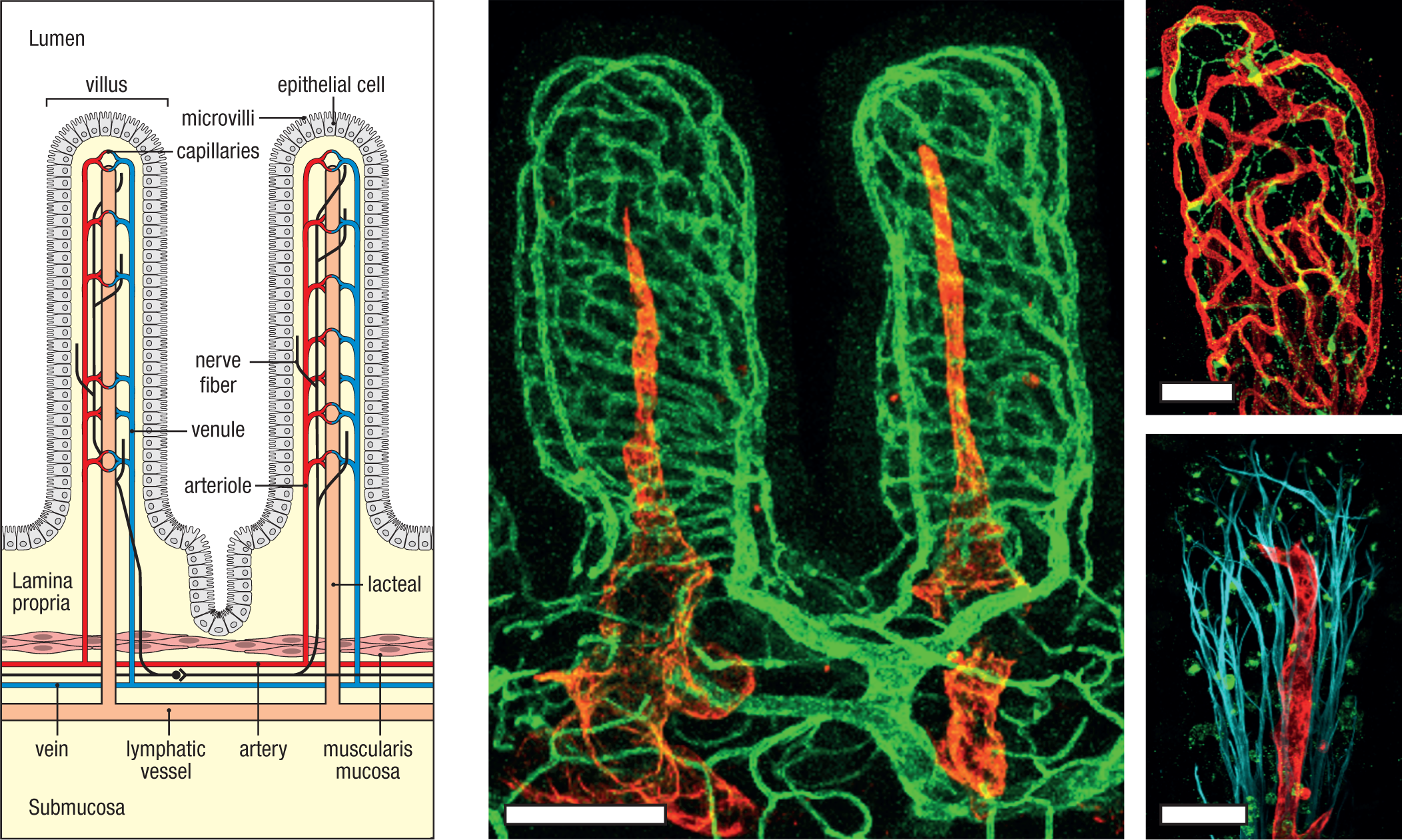
Central to intestinal host defense is the epithelium, which is an active sensor of, and conduit for, interactions between the host and the ‘external’ environment; that is, the contents of the intestinal lumen—both ingested (food) and resident (commensal microbiota). The intestinal epithelium is a simple columnar epithelium that is one cell thick and is polarized such that it distinguishes the external and internal environments. It performs a complex role in balancing the uptake of nutrients and water with its barrier function of excluding commensal and pathogenic microbes. It is composed of a diversity of cell types that, although derived from a common stem-cell precursor, play distinct roles in these varied functions. The mix of intestinal epithelial cell (IEC) types varies along the length of the intestines, as does the microanatomy of their deployment, corresponding to regional variation in the dominant functions of segments of the small and large intestines. In the small intestine, the epithelium covers fingerlike projections, or villi, which markedly increase the surface area for absorption of food (Fig. 12.14). The height of villi is greatest in the proximal small intestine and decreases progressively along the axis from the duodenum to the ileum, reflecting the greater role of the proximal and middle small intestine in nutrient uptake. Each villus is supported by a dense network of small blood vessels, nerves, and a single lacteal—a closed-ended lymphatic vessel that conducts lymph and immune cells, as well as absorbed nutrients (for example, lipids), from the lamina propria to regional lymph nodes (see Fig. 12.14). Individual villi are surrounded by multiple invaginations, called crypts, which are shared by surrounding villi and are sites for the production of new epithelial cells, as we will consider momentarily. In the large intestine, villi are scarce; most of the large intestinal mucosa is composed of tightly apposed crypts between which there is scant lamina propria at homeostasis. The absorptive surface of the large intestine is therefore reduced, reflecting its more limited role in nutrient uptake in favor of its role in water regulation and waste elimination, as well as support of the commensal microbiota.
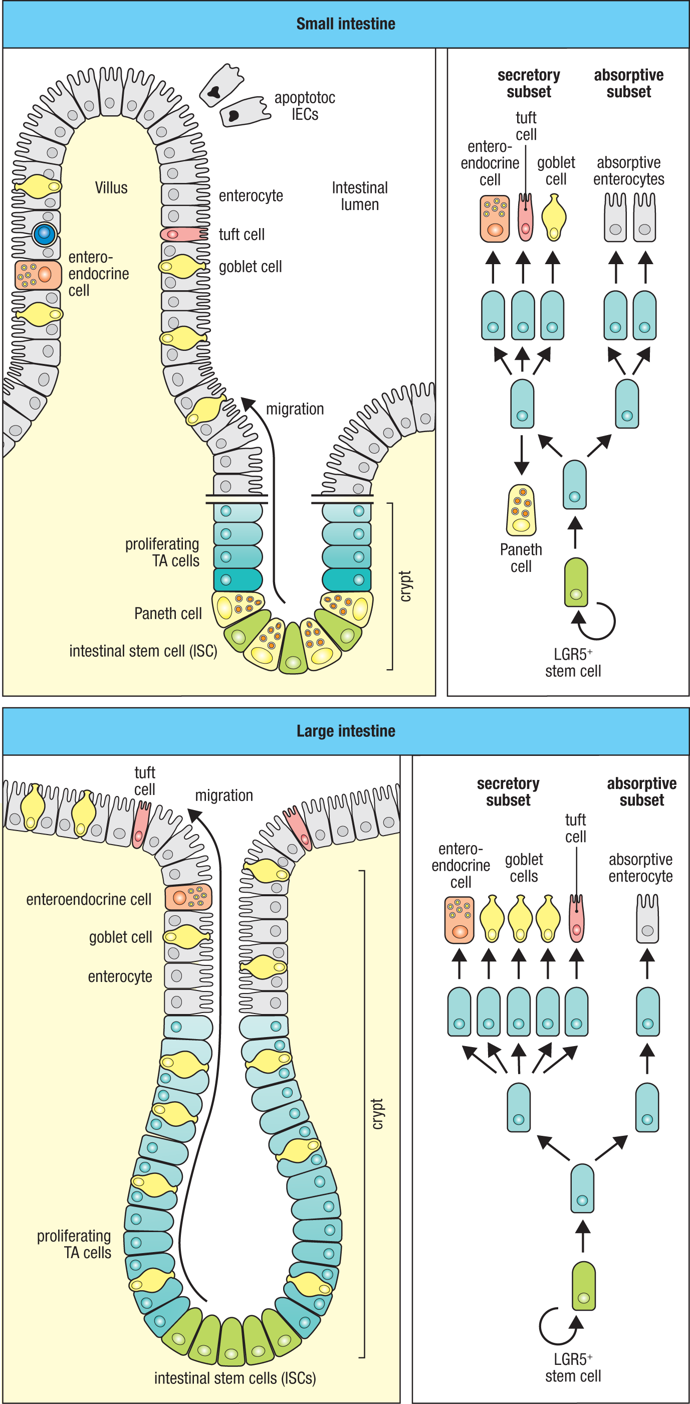
In both the small and large intestine, the crypts are the ‘nursery’ for production of new epithelial cells (Fig. 12.15; and see Fig. 12.11). Because of the substantial mechanical, chemical, and microbial stresses to which the intestinal epithelium is subjected, its turnover is among the most rapid in the body, with the lifetime of most IECs averaging 3–5 days from their generation in the depths of crypts to their shedding into the lumen from the mucosal surface. Regeneration is from a small population of continually cycling, self-renewing stem cells, called intestinal stem cells (ISCs), or crypt base columnar (CBC) cells, which reside at the bottom, or base, of the crypts and are identified by their expression of leucine-rich repeat–containing G protein–coupled receptor 5 (LGR5), which recognizes the R-spondin family of ligands. The strategic location of these cells as distant as anatomically possible from the intestinal lumen and the small opening through which the crypt connects with the intestinal lumen are thought to protect ISCs from insults and reflects their critical role in resupplying the epithelium at both homeostasis and during infection. Progeny of ISCs destined for differentiation into mature IECs are rapidly dividing cells that populate the lower and middle crypts, so-called transit-amplifying (TA) cells, which, in turn, give rise to the two major groups of IECs: absorptive IECs and secretory IECs. Within these two major subsets of differentiated IECs, at least six cell types are recognized: absorptive enterocytes and M cells (absorptive subset); and goblet cells, Paneth cells, enteroendocrine cells, and tuft cells (secretory subset) (Fig. 12.15 and Fig. 12.16).
|
Features of intestinal epithelial cell types |
||||
|
Cell |
Lineage |
Cell markers |
Tissue site(s) |
Major functions |
|
Crypt base columnar (CBC) |
Intestinal stem cell |
LGR5 |
Small intestine Large intestine |
Self-renewal Production of all intestinal epithelial cell types |
|
Absorptive enterocyte |
Absorptive |
Sucrose isomaltase Lactase |
Small intestine Large intestine |
Uptake of nutrients and fluids Microbial and metabolic sensing Transport of secretory immunoglobulins |
|
Microfold (M) cell |
Absorptive |
RANK |
Small intestine Large intestine |
Antigen uptake Bacterial translocation |
|
Goblet cell |
Secretory |
MUC2, Trefoil factor |
Small intestine Large intestine |
Production of mucins Antigen uptake |
|
Paneth cell |
Secretory |
Lysozyme |
Small intestine |
Support for intestinal stem cells Secretion of antimicrobial peptides (AMPs) |
|
Tuft cell |
Secretory |
IL-25 |
Small intestine Large intestine |
Sensing of helminthic odorants and succinate Mobilization of type 2 ILCs via IL-25 and eicosanoid release |
|
Enteroendocrine cell |
Secretory |
Chromogranin A |
Small intestine Large intestine |
Respond to multiple nutrient and microbial-secreted products to release hormones and neurotransmitters |
Fig. 12.16 Features of intestinal epithelial cell subsets.
In accord with its specialization for nutrient and fluid uptake, the absorptive enterocyte, or simply enterocyte (from the Greek enterikos, meaning ‘intestine’), is the most abundant cell type in both small and large intestines (see Figs. 12.15 and 12.16). To enhance its absorptive function, the enterocyte projects microvilli toward the intestinal lumen to increase surface area, and it expresses a range of enzymes and transporters tasked with breaking down and taking up nutrients from food. Enterocytes also are critical sensor cells of the innate immune system, reflecting their dominance on the front line separating the external from internal environments. As for all IECs, an important aspect of the barrier function is the network of intercellular junctions that bind individual cells to each other (Fig. 12.17). Organized near the luminal surface of IECs, these tight junctions are formed by complexes of a number of members of the claudin and occludin families of junctional transmembrane proteins that are linked to the epithelial-cell cytoskeleton to form a continuous seal between adjacent cells. This results in a selective, semipermeable structure that generally restricts the passage of molecules between IECs to ions, small solutes, and water. In addition to regulating the flow of these molecules across the epithelium, tight junctions organize the polarity of IECs and create an ‘outside,’ or apical, surface of enterocytes that faces the intestinal lumen and an ‘inside,’ or basolateral, surface, which rests on a basement membrane and enables communication with the underlying lamina propria.
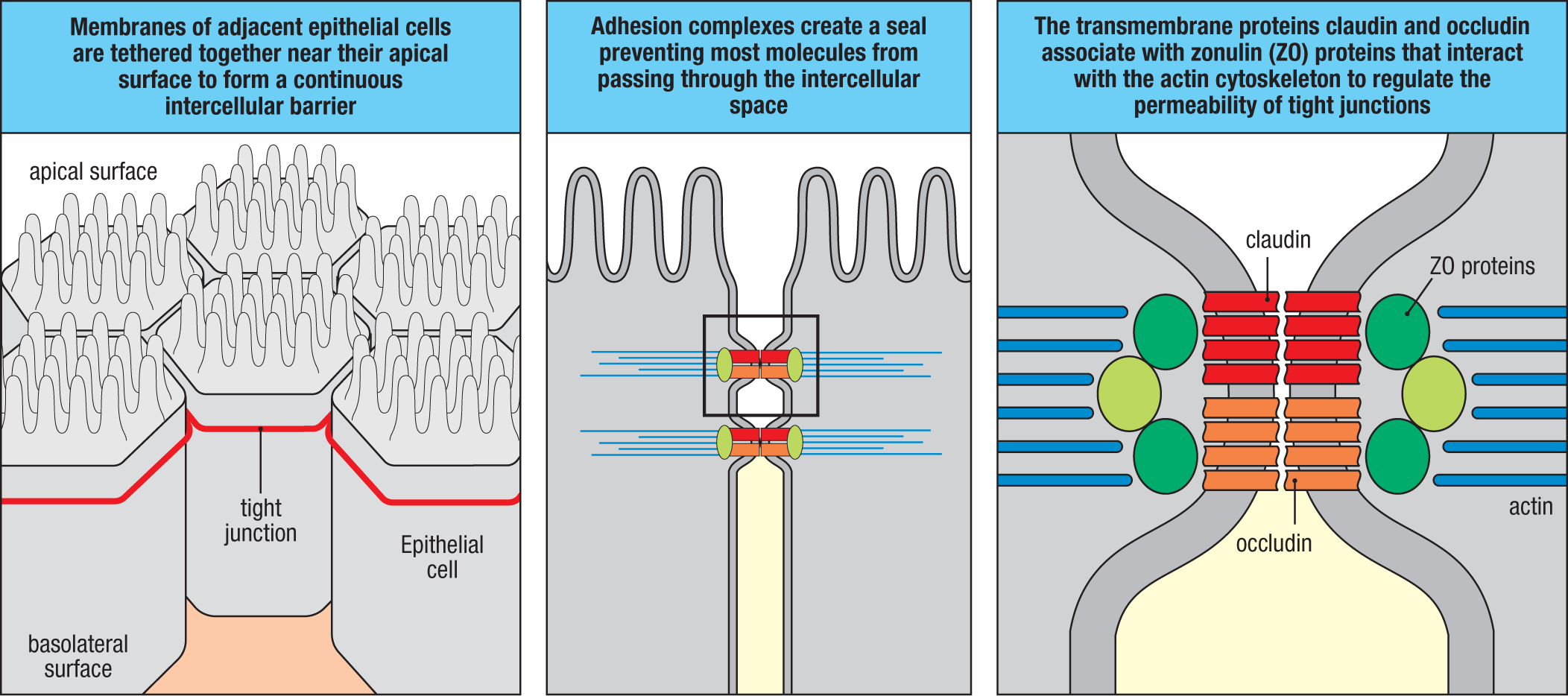
This partitioning of the cell surface of IECs is important for its responses to microbial threats, as different pattern-recognition receptors (PRRs), such as TLRs, are differentially arrayed between the apical and basolateral cell membranes. As an example, TLR-5, which senses bacterial flagellins—a component of the flagella that provide motility for bacteria and are often expressed by pathogenic strains—is preferentially arrayed on the basolateral surface of enterocytes where it can detect microbes that have breached the barrier and alert the IECs to secrete antimicrobial factors, such as antimicrobial peptides (AMPs), as well as cytokines and alarmins that activate innate and adaptive immune cells. Thus, in addition to their role in nutrient uptake, enterocytes are active participants in immune responses in the intestines, acting as critical innate sensor and effector cells.
Although the IECs that compose the follicle-associated epithelium (FAE) are a subset of enterocytes, the developmental relationship of microfold (M) cells to enterocytes is less clear. M cells represent a small fraction of all IECs (<1%). Their development is controlled by local B cells and RANK ligand (RANKL), a member of the tumor necrosis factor (TNF) superfamily (see Section 7-23). As indicated earlier (see Section 12-2), M cells are highly specialized for sampling particulate antigens from the intestinal lumen. In contrast to absorptive enterocytes, the apical surface of M cells is devoid of microvilli and lacks a glycocalyx, and because M cells reside in the mucus-depleted follicle-associated epithelium, they are in direct contact with the luminal contents so as to facilitate sampling of the molecules and microbes found there (see Fig. 12.9). M cells also express receptors that bind SIgA, facilitating the uptake of bacteria coated with SIgA (see Section 12-9). M cells have a remarkable ability to shuttle intact microbes across the epithelium in vesicles that deliver their contents to the subepithelial dome where they can be sampled by dendritic cells, a process referred to as transcytosis (meaning literally ‘across the cell’). The basolateral surfaces of M cells are highly invaginated to form large pockets typically occupied by B and T cells, as well as dendritic cells. This enables the immune system to actively interrogate the microbiota as part of the general mechanism to promote tolerance to nonharmful components of the consortium by inducing regulatory T cells (Treg cells) or SIgA to antigens of commensal microbes, as we will discuss further later (see Section 12-10). As we will also see, some pathogens utilize the M cell as a ferry to cross the epithelial barrier and gain access to the host, even though this feeds them into the heart of the intestinal adaptive immune system.
The second most abundant IEC is the goblet cell, so named because of the large secretory vacuole that occupies much of the cytoplasmic space in mature cells (see Figs. 12.15 and 12.16). The goblet cell’s primary function is the production of mucins, which are heavily charged, glycosylated, secreted or cell-associated glycoproteins that are organized into the gel-like mucus layer that coats the intestinal epithelium (Fig. 12.18); because mucus is the defining feature of mucosal tissues, the goblet cell is the defining secretory cell of the mucosae. The physicochemical properties of mucus—both slippery and sticky—make it a lubricant that reduces mechanical stress on the epithelium by the flow of material in the lumen while also trapping microbes to block their interaction with the epithelium and facilitate their removal by the normal, rhythmic muscular contractions of the intestines, termed peristalsis. Mucus also provides a scaffolding for retention of SIgA and AMPs that reinforce this physical barrier. Intestinal mucus also serves as a substrate for colonization of harmless commensal bacteria that provide a microbial buffer against pathogens.
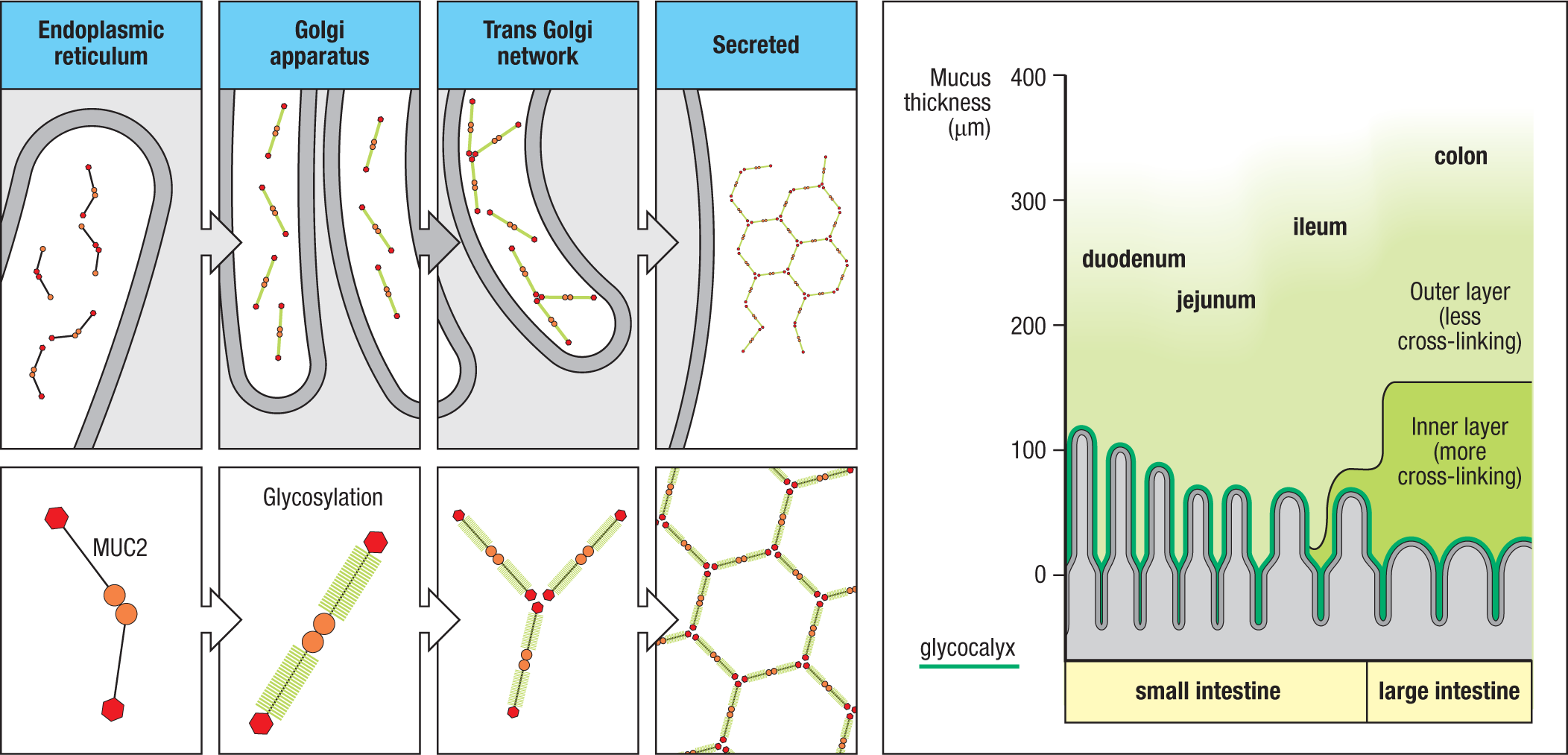
Mucus thickness varies along the duodenal–colonic axis of the intestines (Fig. 12.18 and Fig. 12.19). The mucus layer is thinner and focally discontinuous in the proximal small intestine, becoming thicker and continuous in the ileum. In the large intestine, the mucus layer becomes thicker still, several times thicker than in the proximal small intestine. This correlates with the density of goblet cells in the different regions of the intestine, which generally has an inverse relationship with the length of villi (and therefore absorptive function) and a direct relationship with the abundance of commensal bacteria; the density of both goblet cells and bacteria is greatest in the large intestine, and the density of each declines proximally. Also, in the large intestine there are two functionally distinct mucus layers: a thinner, more compact, firmly adherent inner layer that is generally devoid of microbes, and a thicker, more loosely structured, nonadherent outer layer that can be colonized by microbes. The inner, compact layer is generally lacking in the small intestine. Although bacteria can penetrate the loose, outer layer of mucus, they are normally kept away from the surface of the epithelial cells, and the cross-linking of the inner layer in the large intestine is such that the pores generated are too small for bacteria to penetrate. Defects in this structure compromise antimicrobial defense, and some enteric pathogens produce mucolytic enzymes that allow them to disrupt the mucus structure to invade.
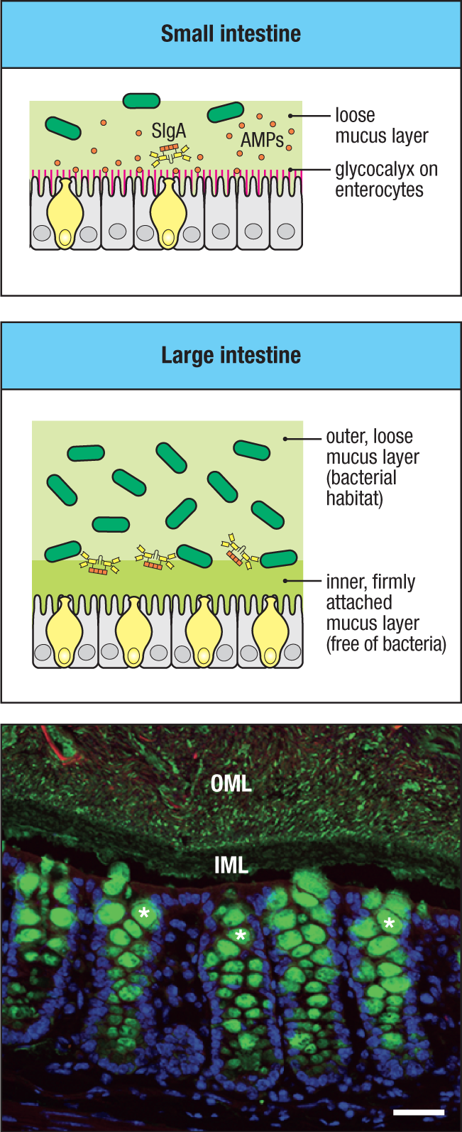
Although there are many different mucins, the dominant mucin in the intestines is the secreted mucin MUC2, which undergoes cross-linking with itself and membrane-bound mucins to form the compact inner layer (see Figs. 12.18 and 12.19). Proteolytic cleavage of the MUC2 polypeptide backbone in the outer layer results in a less compact structure that is amenable to colonization by components of the microbiota. Indeed, mucin glycans serve as nutrients for several keystone anaerobic bacterial species of the large intestinal commensal microbiota (for example, Bifidobacterium and Bacteroides spp.). The breakdown of these glycans by bacterial fermentation generates short-chain fatty acids (SCFAs), such as propionate and butyrate, which are principal fuel sources for IECs, representing a positive IEC–microbiota feedback loop that promotes retention of these protective bacterial species in the healthy gut. Other fermentation products of mucin degradation (for example, acetate and lactate) that are generated by commensal bacteria are toxic to some pathogenic bacteria, providing an indirect mechanism by which mucus participates in host defense. In contrast to most of the intestinal epithelium, the follicle-associated epithelium that is associated with Peyer’s patches and isolated lymphoid follicles is devoid of goblet cells and is not covered by mucus, thereby facilitating sampling of the luminal contents. Notably, there is functional heterogeneity among goblet cells. In the large intestine, the entrance to the colonic crypts is bordered by so-called sentinel goblet cells, which, upon sensing pathogens, stimulate a simultaneous release of mucins from other goblet cells in the crypt that is under assault, resulting in an expulsive jet of mucus that flushes the crypt to eject invading microbes. This phenomenon appears to be coordinated by cell-to-cell communication via gap junctions, which are distinct from tight junctions and are specialized structures that provide regulable pores between adjacent IECs to enable rapid cell-to-cell communication. Also, as we will detail later (see Section 12-9), goblet cells play an important role in the delivery of soluble antigens across the epithelium to underlying dendritic cells.
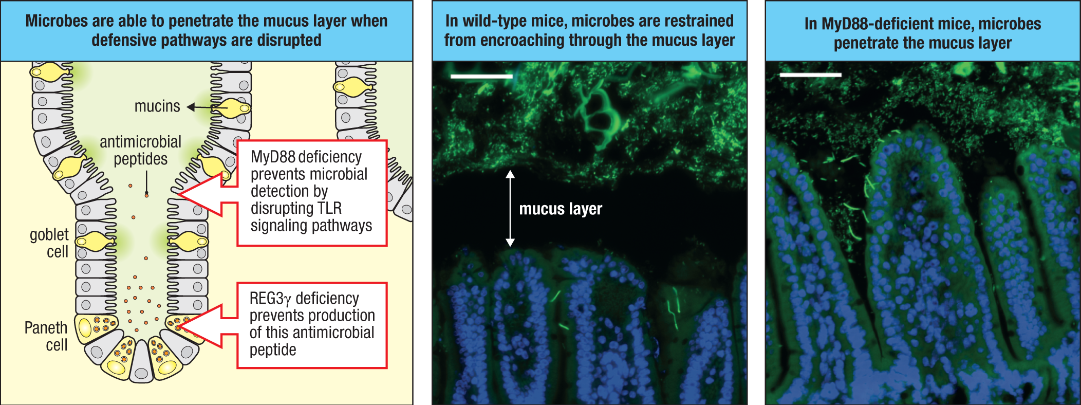
Paneth cells are a population of secretory IECs that reside in the base of crypts in the small intestine, where they are intercalated between ISCs (see Figs. 12.11, 12.15, and 12.16). They develop from early secretory cell progenitors and have two major functions: the production of antimicrobial peptides, both at homeostasis and in response to pathogen-induced activation; and the provision of maintenance signals for ISCs. Paneth cells are characterized by an abundance of intracytoplasmic secretory granules containing a diversity of antimicrobial factors (Fig. 12.20), some of which are secreted at homeostasis and appear to help in maintaining a sterile crypt and reinforcing the mucus layer to restrain bacterial incursion and protect the ISCs (Fig. 12.21). Paneth cells can also respond directly to PAMPs, as they express TLRs and NODs and they are highly autophagic. Defects in Paneth-cell function lead to reduced bacterial defense and are believed to be important in susceptibility to inflammatory bowel disease in humans due to the penetration of microbes across the mucus barrier (see Section 15-17). Notably, Paneth cells are not found in the large intestine. However, a similar cell has been identified in contact with ISCs in colonic crypts, and it appears to serve Paneth-like cell functions, although it lacks the large cytoplasmic granules characteristic of Paneth cells.
|
Antimicrobial proteins produced in the intestinal mucosa |
|||
|
Family (examples) |
Mechanism of action |
Cellular source |
Microbial targets |
|
α-Defensins (humans) Cryptidins (mice) |
Cell membrane disruption |
Paneth cells Neutrophils Macrophages |
Gram+ and Gram− bacteria, fungi, protozoa, viruses |
|
β-Defensins (e.g., BD1, BD2, BD3) |
Cell membrane disruption |
Enterocytes |
Gram+ and Gram− bacteria, fungi, protozoa, viruses |
|
Calprotectin (S100A8-S100A9) |
Metal chelation |
Enterocytes Neutrophils |
Gram+ and Gram− bacteria, fungi, viruses |
|
C-type lectins (e.g., REG3β, REG3γ) |
Cell membrane disruption |
Paneth cells Enterocytes |
Gram+ and Gram− bacteria |
|
Lysozyme |
Cleavage of bacterial cell wall peptidoglycan |
Paneth cells |
Gram+ > Gram− bacteria |
|
Phospholipase A2 |
Cleavage of bacterial cell membrane phospholipids |
Paneth cells Macrophages |
Gram+ bacteria |
Fig. 12.20 Antimicrobial proteins that contribute to innate defense of the intestinal mucosa. A variety of antimicrobial proteins are produced in the intestinal mucosa. These include small, typically cationic antimicrobial peptides (AMPs) that insert into bacterial cell walls (e.g., defensins), as well as enzymes (e.g., lysozyme) that cleave components of bacterial cell walls or cell membranes. These proteins are produced by both intestinal epithelial cells (IECs), including Paneth cells of the small intestine and non-Paneth epithelial cells in the small and large intestines, and myeloid cells, such as macrophages and neutrophils that infiltrate the intestines during inflammation.
Intestinal tuft cells, so named for the characteristic ‘tuft’ of microvilli they project into the lumen, are a chemosensory type of IEC related to odorant and taste receptors in the upper airways and tongue (see Figs. 12.15 and 12.16; see also Fig. 11.3). Tuft cells are specialized for recognition of molecules produced by helminths and are therefore key sensor IECs that promote type 2 immune responses (see Section 11-3). They are relatively rare, representing less than 1% of all IECs, although their numbers increase during helminthic infections.
Enteroendocrine cells (EECs) are a second type of chemosensory IEC that, like tuft cells, are primarily dispersed throughout the superficial, or lumen-facing, intestinal epithelium (see Figs. 12.11, 12.15, and 12.16). EECs are specialized for sensing a range of nutrients in the intestinal lumen that stimulate their release of hormones and/or neurotransmitters to regulate digestion, absorption, and satiety in the gut, as well as local and systemic metabolism. Like tuft cells, EECs compose less than 1% of all IECs, but because of the large number of cells in the intestinal epithelium, EECs represent the largest collection of endocrine cells in the body. There is considerable heterogeneity in EECs, with at least eight subtypes that are distinguished by the nutrient receptors they express and the hormones and neurotransmitters they release when stimulated. Although the roles of EECs in intestinal immunity are still being defined, the innervation of some EECs by nerve fibers that communicate with the central nervous system suggests that, in addition to their roles in regulating intestinal physiology at homeostasis, EECs may also contribute to neuroimmune interactions that regulate certain immune responses.
12-5 The intestinal epithelium contains conventional and unconventional T cells that are focused on barrier maintenance and defense.
The epithelial cells of the intestines are not alone on the front line. Compared to most other epithelia in the body, the epithelium of barrier tissues, including the intestines, is unusual in harboring a substantial number of conventional and unconventional (innate-like) T cells, or intraepithelial lymphocytes (IELs), as well as small numbers of ILC1 cells (Fig. 12.22 and Fig. 12.23). At birth, unconventional T cells are the dominant IELs, as they populate the epithelium perinatally and expand in response to colonization by the intestinal microbiota immediately after birth. As an increasing number of antigen-experienced conventional T cells take up residency in the epithelium throughout life, the balance between unconventional and conventional IELs shifts, such that conventional T cells become a greater fraction of the total with age. Because population of the epithelium by conventional T cells is controlled more by the history of antigenic exposure, the ratios of conventional to unconventional IELs can vary considerably between individuals, and in the same individual, throughout life. Normally, very few IELs are actively dividing, and there is a limited contribution of circulating cells to the IEL compartment.
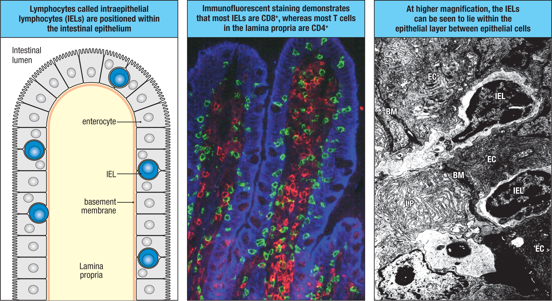
IELs are intercalated between and therefore are in immediate contact with IECs, situated beneath the tight junctions that bind IECs and above the basement membrane on which the IECs rest (see Fig. 12.22). The number and composition of IELs differs regionally along the intestinal tract; the highest density of IELs is found in the proximal small intestine and gradually decreases toward the ileum and the colon. In the healthy proximal small intestine, there is about one IEL for every 10 epithelial cells at homeostasis, the highest density of IELs among the barrier tissues. This number drops to about one IEL per 50 IECs in the colon. The difference in density largely reflects the greater proportion of non-crypt epithelium in the proximal small intestine, where the luminal or surface epithelium contains a greater density of IELs. Because of the tremendous surface area of the intestinal epithelium, IELs are one of the single largest populations of T cells in the body.
Notably, IELs are not static, rather they are highly motile within the intestinal epithelium, reflecting their surveillance function. Because of their disposition within the barrier epithelium itself and their fully mature effector status, IELs are both early sensors of changes in the epithelium resulting from environmental exposures and rapid responders that target epithelial cells for altered function or destruction so as to maintain barrier integrity. IELs participate in host defense against pathogens, in epithelial repair, and in homeostatic interactions with the epithelium, microbiota, and nutrients. They may play an important role in the removal of IECs that have sustained genotoxic stress or DNA damage that is not adequately repaired by the DNA damage response, thereby removing IECs at risk for malignant transformation. Here, we will focus on the populations of T cells that make up the large majority of IELs.
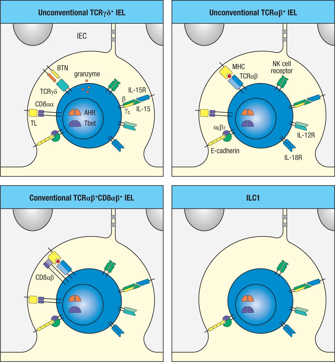
Innate-like, unconventional T cells (for example, invariant TCRγδ and oligoclonal TCRαβ T cells) take up residence within the intestinal epithelium immediately before and after birth. They are also referred to as thymic-derived, or natural, IELs (nIELs) (Fig. 12.23, upper panels, and Fig. 12.24), reflecting their development in, and immigration directly from, the thymus, without having to pass through organized lymphoid tissues to acquire their full effector functions (see Chapter 8). In contrast, conventional T cells migrate into the intestinal epithelium only after their activation by specific antigen in the GALT or gut-associated lymph nodes; these T cells are therefore referred to as induced, or peripheral, IELs (pIELs) (Fig. 12.23, lower left panel, and Fig. 12.24). (Note that this terminology resembles that of the two major subsets of Treg cells—natural, or thymic, and induced, or peripheral, Treg cells.) pIELs derive from conventional CD4 and CD8 T cells, thus their T-cell receptors (TCRs) recognize foreign peptides bound to conventional MHC I or MHC II molecules, respectively. In contrast, TCRαβ nIELs are responsive to a diversity of MHC molecules, including MHC I and MHC II molecules as well as nonclassical MHC I molecules (for example, MHC Ib), albeit through recognition mechanisms that do not appear to require specific peptides (see Chapter 6). Similarly, TCRγδ nIELs, which constitute the largest fraction of IELs in the intestines, are not activated by classical peptide:MHC recognition.
|
Characteristics of intraepithelial lymphocytes |
||||
|
IEL subset |
Co-receptor expression |
TCR repertoire |
NK receptors |
Antigenic ligands |
|
Unconventional TCRγδ nIEL |
CD4−CD8− CD8αα |
TCRVγ7 > TCRVγ4 (mouse) TCRVγ1 (human) |
NKG2 family CD94 LY49 family (mouse) KIR family (human) |
BTNLs (mouse, ? human) MULT1, H60a, Qa-1 (mouse) CD1, MICA, MICB, ULBP (human) |
|
Unconventional TCRαβ nIEL |
CD4−CD8− CD8αα |
Oligoclonal |
NKG2 family CD94 LY49 family (mouse) KIR family (human) |
MHC I, MHC II (mouse, human) Non-classical MHC I (mouse) MICA, MICB, ULBP (human) |
|
Conventional TCRαβ pIEL |
CD8+CD8αα CD4+CD8αα |
Diverse |
None |
Peptide:MHC I Peptide:MHC II |
|
ILC1 |
CD4−CD8− |
None |
NKG2 family CD94 LY49 family (mouse) KIR family (human) |
MULT1, H60a, Qa-1 (mouse) MICA, MICB, ULBP (human) |
Fig. 12.24 Characteristics of intraepithelial lymphocytes.
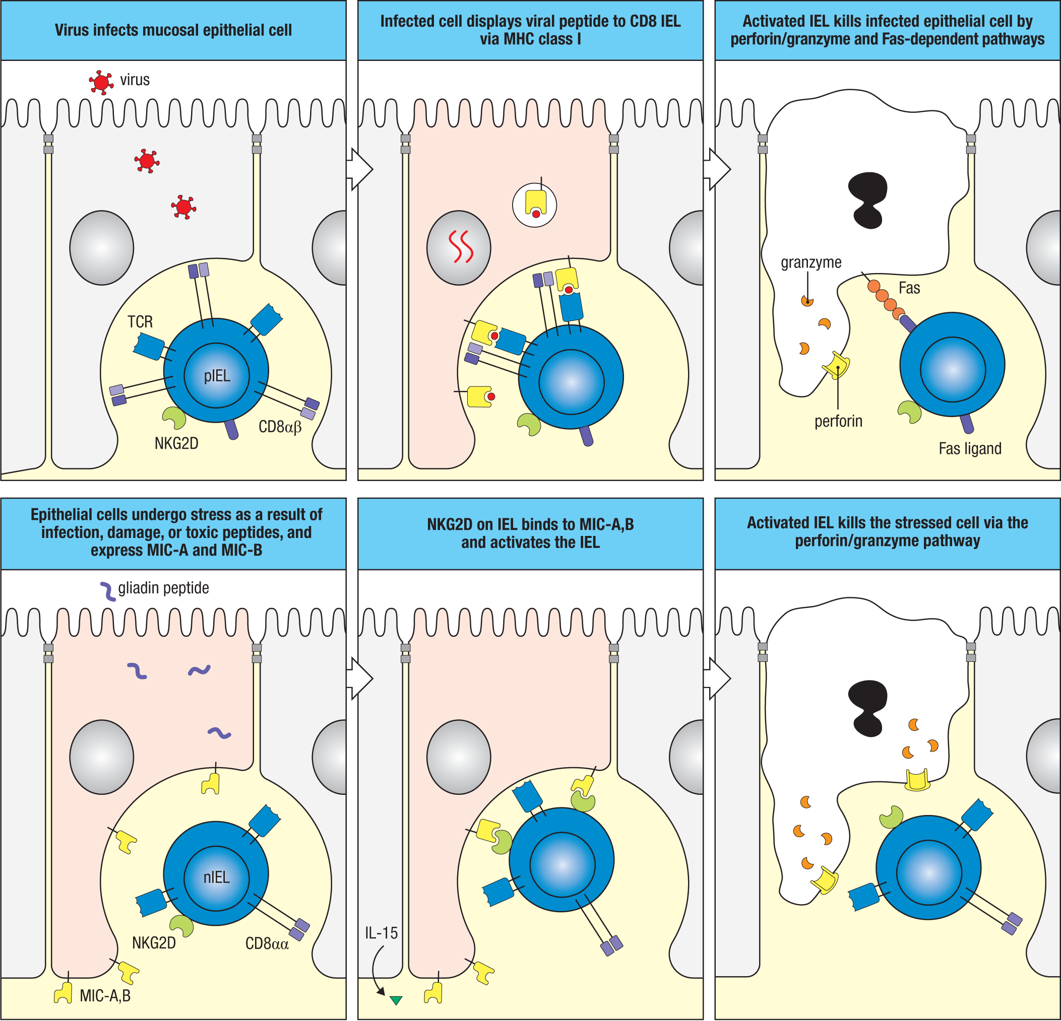
Although the variety of structures recognized by each type of nIEL is still an area of discovery, it is clear that this recognition diversity considerably expands the range of external and internal signals that can be sensed within the intestinal epithelial compartment. Nevertheless, most IEL types, both conventional and unconventional, share similar functionality in bolstering barrier defense and maintaining barrier function; most IELs are characterized by the expression of effector capabilities typical of innate and adaptive cells of the cytotoxicity immune module (see Section 11-2) (Fig. 12.25). Thus, common properties of IELs are their cytolytic capability (reflected in their expression of perforin and granzyme B) and expression of type 1 cytokines (for example, IFN-γ, TNF-α and FasL), which are activated in response to antigen recognition or to the local production of the cytokines IL-12, IL-18, and IL-15. Accordingly, a major function of nIELs and pIELs is their recognition and destruction of IECs that display properties of infection, damage, or stress. IELs can also express antimicrobial peptides, and at least a subset of nIELs is programmed for IL-17 production when activated. Additionally, IELs can express epithelial growth factors, such as epidermal growth factor (EGF), which promotes epithelial proliferation and repair in response to IEC damage.
The developmental origins of conventional and unconventional IELs are distinct. Although both subsets undergo selection in the thymus, neither TCRγδ nor TCRαβ nIELs undergo typical positive and negative selection like conventional TCRαβ T cells. Like most TCRγδ cells in the body, TCRγδ cells destined for residence in the intestinal epithelium exit the thymus as double-negative T cells; that is, prior to the double-positive stage of thymic selection (see Section 8-18). This includes TCRγδ cells with invariant use of Vγ7, which represent the majority of intestinal nIELs in mice, as well as a smaller fraction of TCRγδ cells that express invariant Vγ4. Each of these populations arise as part of the second programmed wave of TCRγδ cell development around the time of birth (see Section 8-18).
In contrast to TCRγδ nIELs, positive selection of TCRαβ nIELs requires recognition of MHC molecules. However, unlike conventional TCRαβ cells, TCRαβ nIELs appear to recognize many MHCs, including nonclassical MHC I and MHC I–like molecules (for example, CD1), rather than just a single one. Their intrathymic selection by unrestricted MHC molecules has been termed agonist selection, indicating the absence of classical TCR recognition of a specific peptide:MHC ligand. Instead, the TCRs of TCRαβ nIELs appear to be promiscuous for a range of different MHC molecules and are therefore broadly cross-reactive with MHC, including nonself MHC molecules.
Concurrent with their selection in the thymus, unconventional TCRγδ and TCRαβ cells fated for the intestinal epithelium up-regulate the integrin α4β7, which directs them to the intestines rather than to peripheral lymphoid organs. They also up-regulate CCR9 or CCR10, which directs them to the epithelium of the small or large intestine in response to CCL25 or CCL28, respectively. They remain double-negative in the intestine but are induced by the high local concentrations of TGF-β produced by IECs to express the CD8αα homodimer and αE integrin (CD103), which pairs with β7 integrin to form αEβ7, which, in turn, binds E-cadherin expressed by IECs (see Figs. 12.23 and 12.24). CD8αα binds the nonclassical MHC class Ib molecule thymus leukemia antigen (TL), which is expressed by IECs. Indeed, nearly all intraepithelial T cells, both nIELs and pIELs, express CD8αα, which appears both to down-modulate TCR signaling to prevent overreactivity of IELs to IEC antigens and to promote NK-like killing of damaged or stressed IECs. CD8αα may also serve as a survival signal to IELs.
Unlike nIELs, pIELs arise extrathymically from conventional CD4 and CD8 T cells after cognate antigen recognition in lymphoid tissues associated with the intestines. The precise signals that determine whether antigen-induced CD4 or CD8 T cells will become intraepithelial residents is not completely understood, but critical to their homing to the epithelial compartment is their expression of αEβ7 integrin (CD103) and CD8αα induced by TGF-β signaling in the intestinal mucosa (see Figs. 12.23 and 12.24). In the case of CD4 pIELs, many appear to be derived from FoxP3+ Treg cells that down-regulate FoxP3, whereas CD8 T cells retain much of their transcriptional program. Because both CD4 and CD8 pIELs retain expression of their native co-receptor, they acquire CD4+CD8αα+ and CD8+CD8αα+ phenotypes and retain responsiveness to cognate peptide:MHC II and peptide:MHC I complexes, respectively. This implies IECs must have the capacity to process and present foreign antigens on MHC I and MHC II molecules. Indeed, in response to IFN-γ signaling, IECs can up-regulate CIITA, the master transcriptional regulator of classical antigen-presenting pathways, and thus appear able to gain antigen-presenting cell (APC) functions, thereby enabling recognition by CD8 or CD4 pIELs.
As discussed in Chapter 6, the ligands recognized by unconventional T cells are heterogeneous and include both unique recognition receptors and those shared with other immune cells (see Sections 6-16 through 6-20). Both subsets of nIELs express molecules typical of the NKG2 family of NK-cell receptors, including NKG2D, an activating receptor that recognizes a subset of nonclassical MHC Ib molecules that are induced on intestinal epithelial cells in response to cellular injury, stress, or ligation of TLRs (see Section 6-16). In addition, as indicated earlier, TCRαβ nIELs express cross-reactive TCRs capable of responding to broad changes in expression of classical and nonclassical MHC molecules in a cognate peptide–independent manner. This suggests TCRαβ nIELs may be activated by alterations in MHC expression in a manner akin to NK cells.
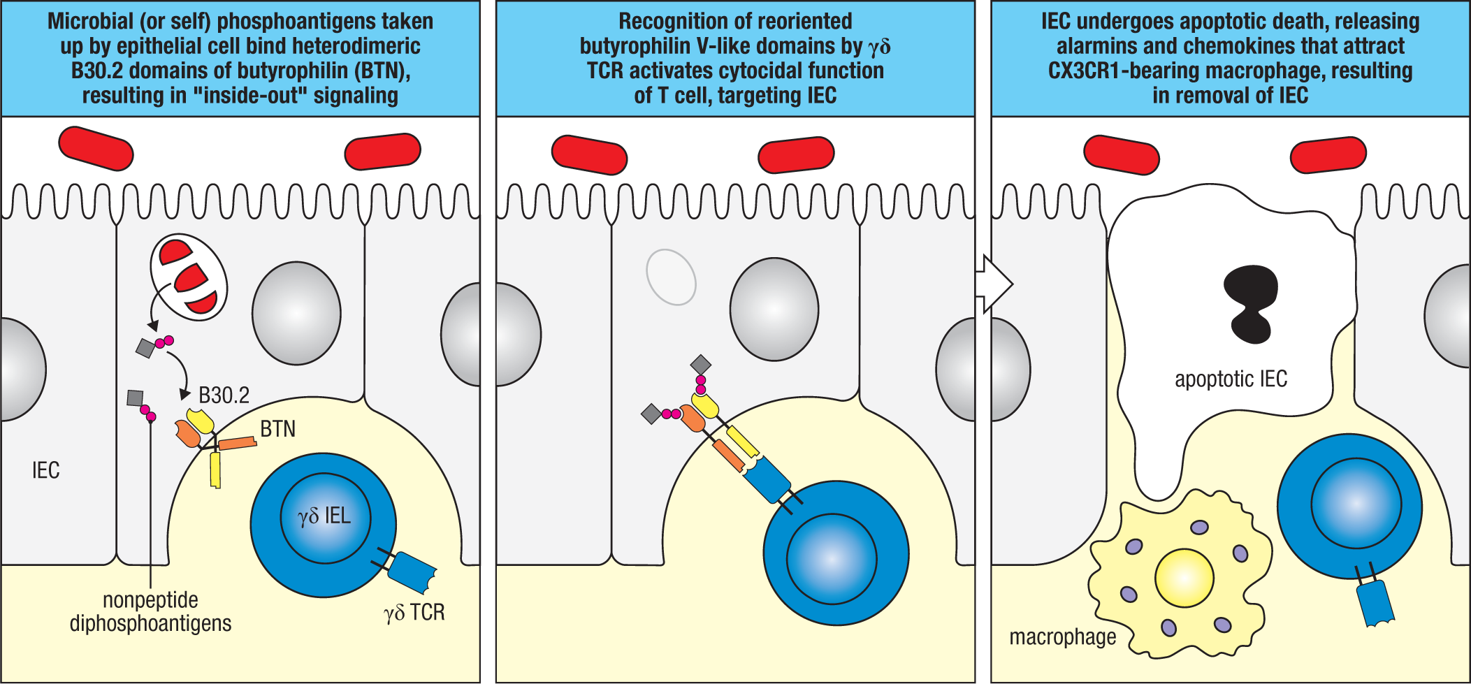
As for TCRγδ nIELs, it was recently shown that an important ligand for the dominant TCRVγ7 nIELs in mice is a heterodimer of BTNL1 and BTNL6, members of the butyrophilin (BTN) gene family. These ligands are structurally related to the B7 family of co-stimulatory molecules and are expressed on IECs but do not appear to engage receptors of the CD28 family. Rather, butyrophilins bind domains on invariant TCRs of some unconventional T cells, as has been shown for a subset of human TCRγδ cells, thereby providing an activating signal via the γδ TCR (Fig. 12.26). The TCR-liganding function of some butyrophilin family molecules is dependent on an intracellular domain, termed B30.2. This domain binds a variety of nonpeptide diphosphate (pyrophosphate)-containing molecules, or phosphoantigens, which are products of microbes or transformed epithelial cells. Butyrophilins thus appear to be a form of pattern-recognition receptor that can detect foreign or self phosphoantigens and generate an ‘inside-out’ signal; that is, the TCR recognizes metabolites inside IECs via allosteric modulation of the BTN extracellular domains. Exactly how these ligands are regulated on IECs and whether this family of ligands is generic for different TCRγδ subsets is currently unknown. However, deletion of BTNL1 leads to selective loss of TCR Vγ7 nIELs in mice, indicating that this signal is required for their maintenance.
Important for both the development and maintenance of all subsets of nIELs and pIELs is expression by the epithelium of IL-15, which is produced in response to the microbiota and is ‘trans-presented’ to IELs in a complex with the IL-15Rα chain (see Fig. 12.23). That is, IL-15 produced by IECs is bound to the IEC surface by IL-15Rα, and this complex induces signaling via the IL-2Rβ and γc subunits expressed on IELs. The necessity for IL-15 in IEL maintenance is evident from the loss of IELs in either IL-15– or IL-15R–deficient mice. IL-15 signaling induces IEL expression of the aryl hydrocarbon receptor (AHR), a transcription factor activated by various environmental ligands derived from cruciferous vegetables, microbiota metabolites, and xenobiotics that is expressed with subsets of ILC3 cells and TH17 cells. IL-15 also induces IELs to express Tbet, which is also expressed by TH1 cells and ILC1 cells. Both of these transcription factors are required for the maintenance and function of IELs. Mice that lack the AHR or Tbet have reduced numbers of IELs and show loss of nIELs, along with abnormalities in epithelial barrier repair, reinforcing the view that these unusual lymphocytes play important roles in the innate immune response to local materials in the intestine.
12-6 Innate lymphoid cells and unconventional lymphocytes are present in GALT and in the lamina propria and are rapid responders to microbes that breach the epithelium.
Just as the intestinal epithelium is populated by IELs in the perinatal period, so too are the GALT and intestinal lamina propria populated by tissue-resident innate lymphoid cells (ILCs) and innate-like lymphocytes. As discussed in Chapter 11 (see Section 11-3), in addition to cytotoxic ILCs (NK cells), three main groups of ‘helper’ ILCs are recognized (see Fig. 11.5)—ILC1, ILC2, and ILC3—each of which preferentially distributes to barrier tissues around birth. The composition of ILCs within the intestinal mucosa is dependent on the microbial exposure of the host, but in more industrialized countries where gastrointestinal worm infestation is less common, ILC3 cells are the most prominent. Most ILC3 cells are found in the GALT, but there are also ILC3 cells within the lamina propria.
Beyond the function of the LTi subset of ILC3 cells in the development of the GALT, ILCs play a central role in the early response to pathogen encroachment in the mature intestinal tissues and may also contribute to the homeostatic function of intestinal epithelial stem cells, although this is an area of ongoing study. There is an increasing appreciation of heterogeneity within ILC subsets, and at least two major mature ILC3 subsets are recognized: LTi-like ILC3 cells that lack NK receptors (NK-cell receptor negative, or NCR–) and non-LTi ILC3 cells that express NK receptors (NK-cell receptor positive, or NRC+). In humans, NCR–, LTi-like ILC3 cells are thought to preferentially express IL-17, whereas NCR+ ILC3 cells produce IL-22. In mice, there is greater overlap in cytokine expression profiles by these subsets. The development and function of both subsets is controlled by the transcription factors RORγt and AHR. Like all ILC3 cells, those in the intestines produce IL-17 and IL-22 in response to IL-23 and IL-1β, which are produced primarily by local dendritic cells and inflammatory macrophages. As we will see in Section 12-20, IL-22 produced by ILC3 cells is indispensable for host protection in certain types of bacterial infections, where it acts on the epithelium to alter IEC development and stimulate production of antimicrobial peptides that restrain bacterial growth and prevent breach of the epithelium in advance of the adaptive response. Notably, similarly to TH17 cells, with which they share many properties, ILC3 cells can be induced to transition to ILC1-type cells that lose expression of RORγt in favor of Tbet expression, resulting in a type 1 functional program. Indeed, most of the ILC1 cells that come to populate the intestinal mucosa appear to derive from ILC3 cells.
As we learned in Section 11-3, IL-5 and IL-13 produced by ILC2 cells in response to IL-33, TSLP, and IL-25 produced by enterocytes and/or tuft cells form an important layer of T cell–independent responses to helminth parasites in the intestine and may contribute to food allergies (see Chapter 14). CD1-restricted invariant NKT (iNKT) cells and mucosal-associated invariant T (MAIT) cells (see Sections 6-18 and 6-19) are also present in the lamina propria and account for 2–3% of lamina propria T cells in the human small intestine. As their names imply, both iNKT cells and MAIT cells express an invariant TCRα chain paired with a limited range of TCRβ chains. iNKT cells recognize lipids and glycolipids complexed with CD1d, whereas MAIT cells recognize metabolites of vitamin B derived mainly from the microbial riboflavin metabolism pathway complexed with MR1.
Summary.
The intestinal epithelium is composed of a diversity of cell types, each with specific functions in intestinal physiology and mucosal immunity. All intestinal epithelial cells (IECs) develop from common progenitors (intestinal stem cells, or ISCs) that reside in the base of the intestinal crypts and differentiate as they move from the base of the crypts toward the surface. There are two major developmental branches: absorptive and secretory. Absorptive enterocytes are most numerous and include absorptive enterocytes and M cells. Secretory enterocytes include goblet cells, Paneth cells (in the small intestine; not in the large intestine), tuft cells, and enteroendocrine cells. Each of these cell types contributes to innate immunity, albeit through mechanisms that reflect their specific functions. Most IECs have a high rate of turnover and must be continually replenished from the ISC pool.
A central feature of the intestinal mucosa—and other mucosal tissues—is the production of mucus, which is produced by goblet cells. Mucus plays an important role in protecting the epithelium from mechanical injury and from interactions with microbes. It provides a physical barrier but also retains molecules produced by or transported across the epithelium, including a diversity of antimicrobial peptides (AMPs) and secretory IgA (SIgA).
The integrity of the intestinal epithelium is supported by both innate and adaptive immune cells. The epithelium itself is populated by a range of natural and peripheral intraepithelial lymphocytes (nIELs and pIELs), including both conventional and unconventional TCRαβ and TCRγδ subsets. IELs actively survey the epithelium and respond by eliminating infected or damaged cells by cytolytic targeting. Beneath the epithelium there are substantial numbers of resident macrophages and dendritic cells that can take up microbes that traverse the epithelium and can eliminate them or ferry them to immune inductive sites to initiate adaptive immune responses. Intestinal phagocytes that sense pathogens can also release cytokines that activate innate lymphoid cells (ILCs) present within mucosal lymphoid tissues, such as in isolated lymphoid follicles (ILFs), as well as in the lamina propria.
Glossary
- lacteal
- A blind lymphatic capillary vessel that originates in the lamina propria of villi of the small intestine that ultimately delivers absorbed nutrients (especially lipids in the form of chylomicrons) to the draining lymph nodes. The vessels may also deliver antigens and immune cells to lymph nodes.
- intestinal stem cells (ISCs)
- Multipotent adult stem cells located in the basal region of crypts in the large and small intestine.
- TLR-5
- Cell-surface Toll-like receptor that recognizes the flagellin protein of bacterial flagella.
- transcytosis
- The active transport of molecules, such as secreted IgA, through epithelial cells from one face to the other.
- goblet cell
- Specialized epithelial cell located in many sites throughout the body and responsible for mucus production; important in protection of the epithelium.
- MUC2
- An oligomeric, mucus gel-forming protein in the colon, where it is secreted by epithelial goblet cells into the lumen of the large intestine to create a protective, lubricating barrier.
- sentinel goblet cells
- Goblet cells present at the entrance of colonic crypts and which sense TLR ligands to activate an NLRP inflammasome to alert adjacent goblet cells deeper in the crypt to secrete MUC2, producing a jet of mucus that expels invading microbes from the crypts.
- Paneth cells
- Specialized epithelial cells at the base of the crypts in the small intestine that secrete antimicrobial peptides.
- tuft cells
- Chemosensory sentinel cells present in mucosal tissues, such as the intestinal and respiratory tracts, which detect products of helminths and protozoans to activate type 2 immune responses. The ligands recognized by tuft cells share features with tastants/odorants recognized by taste-bud cells and lead to the secretion of IL-25, eicosanoids, and acetylcholine.
- enteroendocrine cells (EECs)
- A broad family of specialized intestinal epithelial cells (IECs) that produce hormones in response to a number of stimuli. EECs compose the largest collection of endocrine cells in humans. The secreted products of different types of EECs can act locally, via the bloodstream, or via the enteric nervous system to coordinate responses with other cells.
- neuroimmune interactions
- Interactions between components of the immune and nervous systems coordinated by chemical and electrophysiological signals.
- agonist selection
- A process by which T cells are positively selected in the thymus by their interaction with relatively high-affinity ligands.
- CCR9
- Chemokine receptor expressed by dendritic cells, T cells, thymocytes, and some γδ T cells that binds CCL25 and mediates recruitment of gut-homing cells.
- CCR10
- Chemokine receptor expressed by many cells that binds CCL27 and CCL28 and mediates intestinal recruitment of IgA-producing B lymphocytes.
- CCL25 (TECK)
- Chemokine made by small-intestinal epithelial cells that binds CCR9 to recruit gut-homing T and B cells.
- CCL28 (MEC, mucosal epithelial chemokine)
- Chemokine made by colonic intestinal cells, salivary gland cells, and lactating mammary gland cells that binds CCR10 to recruit B lymphocytes producing IgA into these tissues.
- thymus leukemia antigen (TL)
- Nonclassical MHC class Ib molecule expressed by intestinal epithelial cells and a ligand for CD8αα.
- butyrophilin (BTN)
- A subtype of surface membrane glycoprotein within the immunoglobulin superfamily comprising approximately 11 unique members, expressed on a variety of somatic tissues and composed of typically two extracellular immunoglobulin domains.
- aryl hydrocarbon receptor (AHR)
- A basic helix–loop–helix transcription factor that is activated by various aromatic ligands including, famously, dioxin. It functions in the normal activity of several types of immune cells including some ILCs and IELs.
- Tbet
- A transcription factor active in many immune cell types but most typically associated with ILC1 and T
- H1 function.
- invariant NKT (iNKT) cells
- A type of innate-like lymphocyte that carries a T-cell receptor with an invariant α chain and a β chain of limited diversity that recognizes glycolipid antigens presented by CD1 MHC class Ib molecules. This cell type also carries the surface marker NK1.1, which is usually associated with NK cells.
- αE integrin (CD103)
- Alpha integrin molecule (ITGAE) that pairs with β7 integrin to form the heterodimeric integrin, αEβ7, which binds the cellular adhesion molecule E-cadherin expressed by intestinal epithelial cells. Expressed by intraepithelial lymphocytes (IELs) and some dendritic cells associated with barrier tissues such as the intestines. Note that CD103 is often used to refer to the αEβ7 heterodimer despite strictly referring only to the αE molecule.
- mucosal-associated invariant T cells (MAIT cells)
- Primarily γδ T cells with limited diversity present in the mucosal immune system that respond to bacterially derived folate derivates presented by the nonclassical MHC class Ib molecule MR1.
- CD8αα
- Homodimer of CD8α molecule that is up-regulated on natural and peripheral IELs in the intestinal epithelium. Recognizes the nonclassical MHC molecule TL (thymus leukemia antigen) on intestinal epithelial cells, loss of which may trigger cytolytic NK-like activity of IELs. May also provide a survival signal for IELs.