The intestinal immune response in host defense and immune-mediated disease.
Although major functions of the intestinal immune response are the control of interactions with the commensal microbiota and barrier surveillance and repair at homeostasis, host defense against a variety of infectious agents is also a critical function. Despite the array of innate immune mechanisms in the gut and stiff competition from the indigenous microbiota, the gut is nevertheless a frequent site of infection by a wide variety of pathogenic organisms. These include many viruses; enteric bacteria such as enteropathogenic E. coli, Vibrio, Salmonella, and Shigella species, and Listeria monocytogenes; protozoans such as Entamoeba histolytica; multicellular helminth parasites such as tapeworms and pinworms; and fungi. These pathogens cause disease in different ways, and, as elsewhere in the body, the key to generating protective immunity is the activation of appropriate aspects of the innate and adaptive immune systems.
As in other tissues, the intestines must be able to generate a spectrum of immune responses tailored to individual pathogens, as discussed in the last chapter. Not surprisingly, many microbes have evolved means of adapting to and subverting these host responses. To ensure an adequate response to pathogens, the mucosal immune system needs to be able to recognize and respond to any foreign antigen, but it must not produce the same effector response to harmless antigens (from food or commensals). A major role of the intestinal immune system is to balance these competing demands. As we have highlighted previously, both innate and adaptive responses to intestinal infection are initiated through, and integrated with, the intestinal epithelium. In the last chapter, we highlighted features of type 2 immunity in the intestines (see Section 11-10). In this part of the chapter, we will consider several types of enteric bacterial infections to highlight some of the different strategies taken by pathogens to penetrate the intestinal defenses, along with mechanisms available to resist these pathogens. In the final part of the chapter, we will consider viral infections of the respiratory tract and fungal infection of the skin to highlight features of barrier defense against these types of pathogens.
12-17 Enteric pathogens elicit ‘danger signals’ by activating pattern-recognition receptors that are sequestered in the intestinal epithelium.
Central to the response of innate and adaptive immune cells in the intestinal tissues is the assessment of relative threat of those microbes that interact with and traverse the epithelial barrier. As we have seen, at homeostasis there is continual, albeit low-level transcytosis of commensal microbes. However, because these microbes enter in an orderly fashion, transported in large part across M cells, their recognition generally favors the induction of tolerance. Moreover, the large majority of commensal bacteria pose little risk to the host because they are strict anaerobes that cannot persist outside of the low-oxygen environment of the intestinal lumen, especially in the large intestine, where most reside. Although the surface epithelium of the intestines is relatively hypoxic—due to high oxygen consumption induced by the metabolism of short-chain fatty acids produced by commensal anaerobes—the more aerobic environment of host tissues, including the deeper lamina propria, prevents the growth of these anaerobes. However, a small but persistent fraction of the intestinal microbiota includes facultative anaerobes, particularly Enterobacteriaceae family members from the phylum Proteobacteria, which can thrive outside the intestinal lumen and retain the potential for serious disease should they traverse the epithelium in substantial numbers and disseminate as opportunistic pathogens. Similarly, most bacterial pathogens are facultative anaerobes that can thrive outside the gut lumen. The sensing of these organisms by the intestinal mucosa is therefore of paramount importance to host defense.
A central mechanism for discriminating friend from foe at the intestinal barrier is, as in most tissues, the activation of pattern-recognition receptors (PRRs), such as Toll-like receptors (TLRs). However, because TLRs that sense bacteria are down-modulated in the intestines to accommodate to the commensal microbiota, there are special adaptations of the intestinal epithelium that allow bugs to reach the border, but not cross it, except via M cells that direct them directly to the GALT. Central to this balancing act is the partitioning of PRRs within the polarized epithelium. Thus, while some receptors that sense microbes at the apical surface of IECs are down-modulated, these same receptors are retained within intracellular vacuoles, as well as in the IEC cytosol and at basolateral surfaces, such that penetration of microbes into or across the epithelial barrier elicits pro-inflammatory responses, ignited by signals from the epithelium itself (Fig. 12.38). TLRs in intracellular vacuoles or at the basolateral surface of IECs detect intracellular pathogens or extracellular pathogens and their products that have been internalized by endocytosis or have penetrated the epithelium. Moreover, IECs express a variety of cytosolic sensors, as described in Chapter 3, which are activated when microorganisms or their products enter the cytoplasm. These include the nucleotide-binding and oligomerization domain (NOD) proteins NOD1 and NOD2, as well as cGAS and STING, among others (see Sections 3-9 and 3-10; Figs. 3.18 and 3.19). NOD1 recognizes a diaminopimelic acid–containing peptide that is found only in the cell walls of Gram-negative bacteria. NOD2 recognizes a muramyl dipeptide found in the peptidoglycans of most bacteria, as well as structures in certain viruses and mycobacteria. Epithelial cells defective in NOD2 are less resistant to infection by intracellular bacteria. Mice lacking NOD2 also show increased translocation of bacteria across the epithelium. A defect in recognition of the commensal microbiota by NOD2 also seems to be important in some cases of Crohn’s disease, as up to 15% of Caucasian individuals carry a mutation in the NOD2 gene that renders the NOD2 protein nonfunctional.
 Case Study: Crohn’s Disease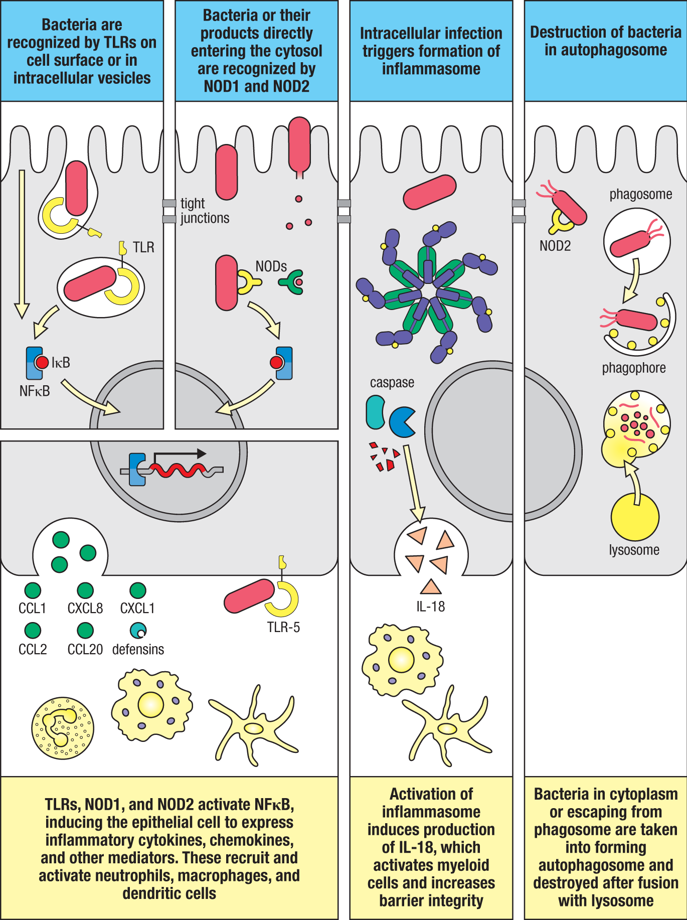
Ligation of TLRs or NOD proteins in IECs stimulates the production of cytokines, such as IL-18, and the production of chemokines. It can also modulate the production of antimicrobial peptides, mucins, and tight junctions, whether through direct actions or by indirect actions of cytokines induced by local immune cells that act, in turn, on the epithelium. Epithelial cells also express members of the intracellular NOD-like receptor (NLR) family, including NLRC4 and NLRP6, which can form inflammasomes (see Fig. 12.38). As described in Section 3-11, the formation of an inflammasome leads to activation of caspase 1, which cleaves pro-IL-18 to produce the active cytokine (see Fig. 3.20). While the cell targets activated by IL-18 (for example, macrophages) contribute to epithelial defense against bacterial invasion by promoting barrier integrity, these cells can cause tissue damage if activated for long periods, as in IBD.
One mechanism recently recognized as important for epithelial defense against infection is autophagy, which we discussed in Section 6-6 in regard to its relationship to antigen processing. In this process, a crescent-shaped double-membrane fragment in the cytoplasm, called the isolation membrane, or phagophore, engulfs various cytoplasmic contents to form a complete vesicle, the autophagosome, which fuses with lysosomes to degrade the contents (see Fig. 12.38). When autophagy is disrupted, bacteria cannot be contained effectively, and epithelial cells become stressed. This can lead to increased penetration of bacteria into the body and to NFΚB-mediated inflammation. Autophagy is promoted by the NOD1 and NOD2 intracellular bacterial sensors. As with NOD2, mutations in the autophagy-related genes ATG16L1 and IRGM1 are associated with susceptibility to Crohn’s disease in humans.
12-18 Pathogens induce inflammatory adaptive immune responses when innate defenses have been breached.
If pathogens gain access to the subepithelial space, they interact with innate immune cells in the underlying tissue. Although resident macrophages are inflammation-anergic at homeostasis (see Section 12-8), and therefore resistant to activation of pro-inflammatory responses, in the context of pro-inflammatory signals from the epithelium and products from pathogens themselves, these cells can become activated to release pro-inflammatory cytokines and chemokines. This response, in turn, results in the rapid recruitment of monocytes from the circulating blood, which are not inflammation-anergic, and amplifies the pro-inflammatory cascade in a feed-forward manner. Together with the cascade of inflammatory mediators released by epithelial cells, this process dramatically alters the environment of the mucosa and changes the behavior of local antigen-presenting cells, including dendritic cells.
As described in Section 9-8, activated dendritic cells will traffic to local T-cell zones and express high levels of co-stimulatory molecules, as well as polarizing cytokines such as IL-6, IL-23, IL-12, or IL-4, which promote development of the appropriate subset of effector T cells. Dendritic cells activated in Peyer’s patches migrate to the T cell–dependent areas of the patch, whereas dendritic cells that encounter antigen in the lamina propria migrate to the mesenteric lymph node. The effector T cells activated in these ways acquire gut-homing molecules (see Section 12-11), ensuring that they return to the gut wall to encounter the invading organisms. Similarly, SIgA-producing B lymphocytes are generated in Peyer’s patches and mesenteric lymph nodes, giving rise to plasma cells that accumulate in the lamina propria. IgA secretion into the lumen is enhanced in response to infection because pIgR expression is enhanced by TLR ligands and pro-inflammatory cytokines. In a number of infections, B cells undergo class switching to IgG, generating intestinal plasma cells that produce complement-fixing IgG isotypes that enhance the eradication of the invading microbes, as we discuss later in Section 12-20.
The activated myeloid cells found in the inflamed mucosa may also contribute to amplifying and sustaining the functions of effector T and B cells that arrive in the mucosa. For example, IL-1 and IL-23 produced by recently arrived monocytes appear to be important for the survival and function of local TH17 cells. Pro-inflammatory myeloid cells also produce mediators such as IL-6, TNF-α, and nitric oxide, which help drive IgA switching and secondary expansion of mucosal B cells.
12-19 Effector T-cell responses in the intestine protect the function of the epithelium.
Once activated, the effector T cells that accumulate in the intestine behave much like their counterparts elsewhere in the body, producing cytokines and generating cytolytic activity as appropriate to the pathogen. What is different is that a major aim of the protective immune response in the intestine is tailored to preserving the integrity and function of the epithelial barrier. This is achieved in a number of ways, depending on the nature of the pathogen.
In viral infections, CD8 cytotoxic T cells among intraepithelial lymphocytes kill infected epithelial cells (see Fig. 12.25), triggering their replacement by uninfected cells derived from the rapidly dividing stem cells in the crypts. A similar process can occur during other forms of protective immune responses, with cytokines from CD4 effector T cells directly stimulating stem cells in the colonic crypts to accelerate epithelial-cell generation and modulate the developmental trajectory of IECs toward either the secretory pathway (for example, in response to helminths) or the absorptive pathway (for example, in response to extracellular bacteria). This forces the replacement of infected cells and generates a moving target for organisms that are attempting to attach to the surface of the epithelium. It also expands the types of cells best suited to combat particular types of pathogens.
As an example, the production of IL-13 by ILC2 cells and TH2 cells during parasitic infections increases the number of goblet cells and enhances their production of mucus, and also expands the number of tuft cells that contribute to a positive feedback loop by producing IL-25, which enhances production of IL-13 from ILC2 cells and TH2 cells. Conversely, IL-22 produced by TH17 cells contributes to defense against extracellular bacteria and fungi by promoting a shift in the type of mucin produced by goblet cells, promoting the cell death and sloughing of infected IECs and stimulating the production of antimicrobial peptides by Paneth cells and other IECs, as we discuss in the next section. Finally, these mediators and others can enhance the peristaltic action of the intestine and its outward secretion of fluid, washing out pathogens within the lumen of the intestine. Together these processes aim to generate a hostile and unstable environment for the pathogen, reducing its ability to invade and damage the epithelial barrier.
12-20 Noninvasive and invasive enteric bacterial pathogens use different strategies to colonize the intestines.
Bacterial pathogens elicit different host defenses contingent on their pattern of intestinal infection. Some bacterial pathogens are invasive; that is, they typically penetrate the intestinal epithelial barrier and are able to disseminate beyond the mucosa. Other bacterial pathogens are noninvasive and neither traverse the intestinal epithelium nor disseminate. Enteric bacterial pathogens can also differ in the region of the intestinal tract they colonize. In any case, in order for enteropathogens to gain access to the host, they must first adhere to the epithelium, whether or not they subsequently traverse it. In the small intestine, which lacks the thick, two-layer mucus defense present in the large intestine, the opportunity for directly engaging the epithelium is greater, and, in the case of the proximal small intestine or follicle-associated epithelium overlying Peyer’s patches and ILFs, there is scant mucus to prevent it. Indeed, as we shall see, like bacterial constituents of the commensal microbiota, several enteric pathogens specifically target M cells as an entry point to cross the epithelium. Thus, the epithelium can serve as both barrier and gateway for enteropathogens.
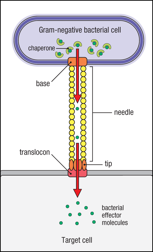
Enteropathogenic bacteria have evolved a variety of strategies for colonization and/or invasion of the intestinal epithelium, most of which are organism-specific. The bacterial molecules that mediate these properties are typically encoded by a cluster of genes called a pathogenicity island, which is either present on an extrachromosomal plasmid or integrated into the bacterial genome flanked by genetic mobility elements, enabling transfer between bacteria. The factors encoded within these elements that confer pathogenicity are referred to as virulence factors. Although each pathogen has developed distinctive ways to colonize the intestine, the expression of molecules that promote their attachment to, and/or internalization by, intestinal epithelial cells is a common feature. These include fimbriae (that is, specialized pili) and/or adhesins that bind host-membrane molecules. Many Gram-negative bacteria also assemble a needle-like protein structure, or injectisome, such as a type III secretion system (T3SS) or a type IV secretion system (T4SS) (Fig. 12.39), through which a wide spectrum of bacterial proteins, or effectors, is injected into host cells, including receptors for enhanced binding of the bacterium. In addition to their role in bacterial adhesion, these bacterial effectors can also target cellular pathways in IECs to modulate host-cell signaling pathways to facilitate pathogen replication and spread. However, as discussed earlier (see Section 12-17), these bacterial effectors can also activate intracellular pattern-recognition receptors that initiate host immunity. Some bacteria also use injectisomes, particularly type VI secretion systems (T6SSs), to deliver effectors to other bacteria and kill them in a type of interbacterial warfare that allows the pathogen to outcompete commensals.
Here we consider two noninvasive bacterial pathogens that attach to the intestinal epithelium, but do not invade. Vibrio cholerae, a Gram-negative, flagellum-expressing (motile) bacterium, is the causative agent of cholera, a prominent diarrheal disease that affects more than 2 million people and causes more than 75,000 deaths annually worldwide, particularly in underdeveloped regions with poor sanitation. Although most strains of V. cholerae are nonpathogenic free-living aquatic bacteria, pathogenic strains express at least two virulence factors that result in human disease: cholera toxin (CT) and the adhesin toxin-coregulating pilus (TCP). Both are required for virulence; TCP mediates initial attachment of V. cholerae to IECs of the small intestine, and CT mediates the pathogenic effect of cholera. Cholera toxin is a multi-subunit exotoxin composed of a pentameric binding subunit (CTB) and a monomeric active subunit (CTA). CTB binds to the cell-surface ganglioside GM1, which is widely expressed. The binding of CTB to the surface of an IEC stimulates endocytosis of the toxin and cleavage of CTB from the CTA subunit. The CTA subunit is delivered into the host cell where it activates adenylate cyclase, resulting in accumulation of intracellular cyclic AMP (cAMP). This stimulates the robust secretion of chloride ions and blocks the uptake of sodium chloide, causing a massive efflux of water. The resulting efflux of water and electrolytes results in severe diarrhea and dehydration that can be lethal, particularly for infants and small children. Infection by V. cholerae stimulates long-lasting immunity that is thought to be mediated primarily by neutralizing SIgA antibodies that block the bacteria’s attachment to the small-intestinal epithelium. Because of its potency in delivering antigens to the intestinal mucosa, the CTB subunit of cholera toxin has been explored as a mucosal adjuvant for antigens linked to it for delivery to the intestines.
Another noninvasive bacterial pathogen that causes diarrheal disease is enteropathogenic E. coli (EPEC). Although E. coli strains are normal constituents of the commensal microbiota, enteropathogenic E. coli strains contain a pathogenicity island called the locus of enterocyte effacement (LEE), so-named because it comprises the genes responsible for attaching and effacing lesions, a feature of EPEC characterized by tight adherence of the bacterium to enterocytes that results in characteristic destruction of microvilli (Fig. 12.40). The LEE pathogenicity island encodes multiple virulence factors, including components of a T3SS, which is used to deliver bacterial effectors into enterocytes. LEE also encodes two genes required for the attachment of EPEC to enterocytes: intimin and a translocated intimin receptor (Tir) to which intimin binds to establish firm adhesion to the cell surface of IECs. Unlike V. cholerae, which stimulates remarkably little inflammation in the involved small-intestinal mucosa, EPEC, which colonizes the large intestine, induces colonic inflammation (colitis) that resolves only when the infection is cleared.
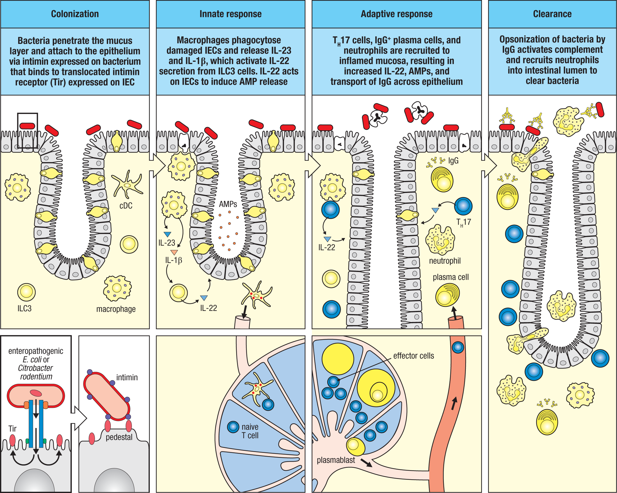
The immune response to EPEC has been modeled in mice using the highly related rodent pathogen, Citrobacter rodentium. Host defense against C. rodentium infection is dominated by a type 3 response that is initiated when the bacterium penetrates the mucus layer in the distal colon and interacts with colonic enterocytes (see Fig. 12.40). C. rodentium does not express a flagellum and is thus nonmotile, so how it is able to traverse the thick, two-layer colonic mucus barrier is unknown. However, during the process of traversing the mucus layers, the bacterium assembles its T3SS from proteins encoded in the LEE, allowing it to dock to an epithelial cell. Adhesion is stabilized by injection of the Tir protein, which inserts into the apical plasma membrane of the IEC where it can bind intimin on the bacterial surface. This adhesion is strengthened by creation of a pedestal formed from disruption and effacement of host-cell microvilli by other bacterial effectors injected into the IEC, enabling a broad area of contact between pedestal and bacterium—hence the term ‘attaching and effacing’ bacterial pathogen. Detection of bacterial effectors injected into the cytosol of the IECs is thought to initiate the IEC’s programmed death, resulting in the phagocytosis of dying bacteria-laden cells by subepithelial CX3CR1+ macrophages and cDC2 cells. TEDs projected across the infected epithelium by these phagocytes may also contribute by direct uptake of the pathogen. The local production of IL-1β and IL-23 by activated mononuclear phagocytes activates ILC3s within the mucosa, which, in turn, release type 3 cytokines, including IL-17 and IL-22, as described in Chapter 11 (see Section 11-3). The induction of IL-22 by ILC3 cells is particularly important to the innate immune response to C. rodentium, as mice deficient for either IL-22 or IL-23 rapidly succumb to infection prior to induction of an adaptive immune response. However, the adaptive response is also indispensable for host protection, as mice deficient for CD4 T cells or B cells succumb to infection, albeit later, after the earlier innate phase of the response.
The adaptive immune response against C. rodentium is dependent on the development of pathogen-specific TH17 cells that produce IL-22, which are recruited to the lamina propria of the infected segment of the large intestine and are required to fully amplify the protection of the epithelium initiated by ILC3 cells. Indeed, as the host response progresses, CD4 T cells become the major source of IL-22. The adaptive response also contributes Citrobacter-specific IgG antibodies, which are transported across the epithelium to bind the bacterium in the intestinal lumen. This IgG, which is dependent on FcRn for transepithelial transport from the lamina propria into the lumen, opsonizes bacteria and leads to their clearance by neutrophils recruited to the infection site by CCR2-activating chemokines and complement fragments (see Fig. 12.40). In normal mice, the infection is completely cleared within 3–4 weeks, and IgG-dependent protective immunity is generated.
In contrast to noninvasive bacterial pathogens, invasive bacteria can penetrate the intestinal epithelium to cause local injury or disseminate from the intestinal mucosa to cause systemic disease. Examples include Listeria, Shigella, and Salmonella spp., among others. Although we have previously considered Listeria monocytogenes in the context of systemic immunity (see Chapter 11), it is normally transmitted via the intestinal tract. L. monocytogenes is a food-borne, aerobic, Gram-positive rod, an intracellular pathogen that has evolved mechanisms to escape killing within phagocytes (see Section 11-6). It uses the virulence factor internalin A to initially establish a foothold within IECs of the small intestine. Internalin A binds to E-cadherin, which is normally expressed beneath tight junctions and therefore inaccessible to the bacterium. However, E-cadherin becomes luminally accessible around goblet cells during their exocytosis of mucins, and L. monocytogenes is then able to bind and be internalized into endocytic vesicles. L. monocytogenes may take advantage of the goblet cell–associated passage (GAP) pathway to be transcytosed to the basolateral surface of the epithelium, from which it can spread to other epithelial cells and to macrophages. Another L. monocytogenes virulence factor, listeriolysin O (LLO), is a pore-forming toxin that allows L. monocytogenes to escape phagolysosomes within macrophages to enter the cytosol, where it avoids intracellular killing and replicates. Once in macrophages, L. monocytogenes can be transported out of the gut to systemic tissues, where it can spread cell-to-cell without exposure to the extracellular environment (see Section 13-22), rendering antibody responses ineffectual. Cell-to-cell transmission is achieved by L. monocytogenes–induced polymerization of actin filaments within the host-cell cytoplasm that propel the bacterium to the cell membrane, where it forms protrusions into neighboring cells that can be internalized, and from which the bacterium can again escape, initiating another cycle of cell-to-cell dissemination. The host response to L. monocytogenes, an obligate intracellular pathogen, bridges type 1 and cytotoxic immunity (see Section 11-6).
 11.2 Listeria Parasites
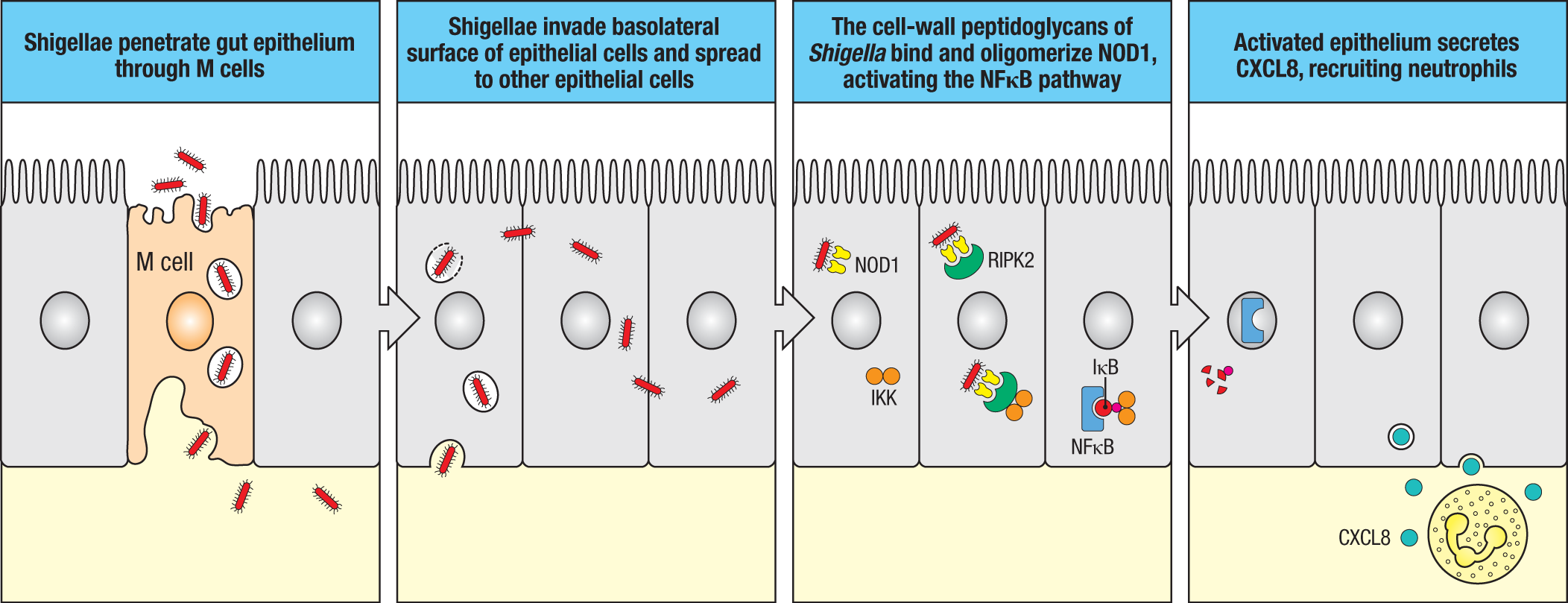
Shigella dysenteriae is a nonmotile, Gram-negative rod that causes shigellosis, or bacillary dysentery, which is transmitted via the fecal–oral route. Shigellosis is a major enteric infection in developing countries, where it causes as many as 150 million cases and 100,000 deaths each year. Like Listeria, Shigella spp. are intracellular pathogens that have evolved mechanisms for escape from phagolysosomes and use a similar actin-mediated propulsion to spread cell-to-cell and evade host immunity. However, Shigella uses the M cell for transport across the intestinal epithelium of the ileum and colon (Fig. 12.41), where it is delivered to subepithelial dome macrophages, which it is able to infect and kill. The release of the bacterium from macrophages allows it to spread locally to intestinal epithelial cells via their basolateral surfaces. By activating intracellular sensors of IECs such as NOD1 as it migrates from cell to cell, Shigella stimulates a robust innate immune response that, along with direct bacterial damage, causes extensive damage to the epithelium. Shigella also releases Shiga toxin, an exotoxin that injures local endothelial cells, resulting in bloody diarrhea; it can also affect the microvasculature in other organs, such as the kidney, to produce hemolytic uremic syndrome (HUS), so named for its intravascular destruction of red blood cells (hemolysis) and damage to kidney function (uremia). Although the more virulent Shigella spp. such as Shigella dysenteriae can cause considerable local injury of the intestinal mucosa, they do not typically disseminate beyond the intestines. As for L. monocytogenes, eradication of Shigella is dependent on type 1 and cytotoxic immunity; antibody responses are poorly induced and nonprotective.
Like Shigella, Salmonella enterica serovar Typhi, the causative agent of typhoid fever in humans, targets the M cells of Peyer’s patches to traverse the intestinal epithelium, although it is also able to directly invade other enterocytes and can be taken up by TEDs of subepithelial mononuclear phagocytes (Fig. 12.42). Salmonella enterica serovar Typhi is a food-borne, Gram-negative, rod-shaped facultative anaerobe that infects only humans. It is equipped with multiple flagella and is highly motile. Salmonella uses both type III and type VI secretion systems to cause disease. Expression of the T6SS is induced first to destroy commensal bacteria to clear a niche for infection. This is down-regulated as the T3SS is expressed. As for EPEC, the T3SS is important in the attachment of the bacterium to host cells, although Salmonella can also enter IECs after attachment. As we learned in Chapter 11 (see Section 11-14), Salmonella are facultative intracellular pathogens that can shift from an extracellular lifestyle to an intracellular lifestyle as they try to evade first a type 3 and then a type 1 host immune response, respectively. By adapting to survival within migratory dendritic cells, Salmonella can be transported with their phagocyte host to regional lymph nodes and, if not contained there, can disseminate widely.
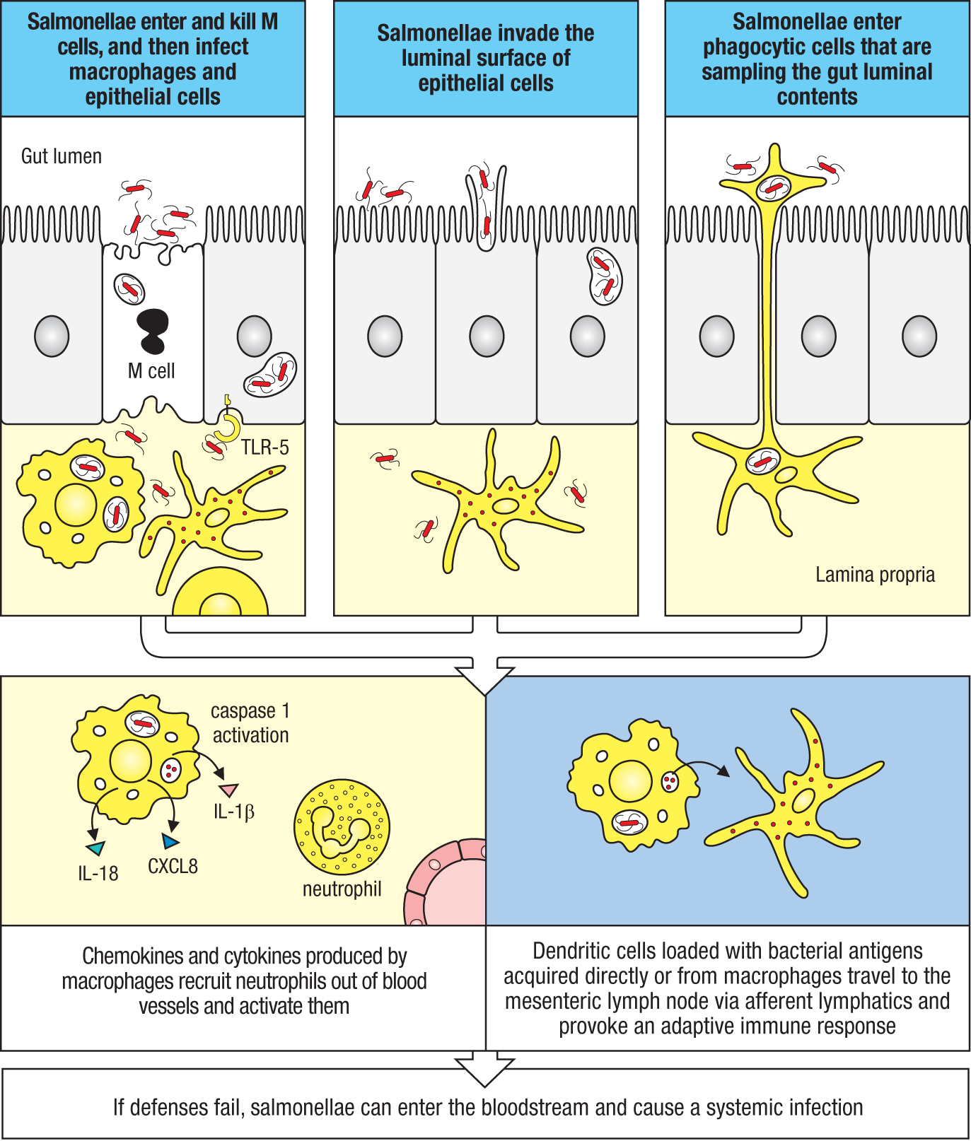
12-21 Dysregulated immune responses to commensal bacteria provoke intestinal disease.
Because of their antigenic memory and longevity, effector CD4 T cells are a liability when inappropriately directed against self antigens or, in the case of the microbiota, the ‘extended’ self that includes antigens of the commensal microbiota. Indeed, in view of the enormous number of antigens encoded in the collective genome (microbiome) of the intestinal microbiota, it is remarkable that dysregulated effector responses against the microbiota are the exception. When they do occur, the result is inflammatory bowel disease (IBD), including Crohn’s disease (CD) and ulcerative colitis (UC). Although these two types of IBD have distinct clinical and histopathologic features, both are characterized by aberrant immune responses to antigens of the intestinal microbiota. In all experimental models of IBD, the intestinal damage depends on the presence of commensal bacteria; it can be prevented by administration of broad-spectrum antibiotics and does not occur in germ-free animals. As in autoimmunity directed against host self antigens (see Chapter 15), dysregulated CD4 T-cell responses to antigens of the microbiota lead to chronic, typically relapsing and remitting disease that reflects the immune system’s inability to eliminate the antigens that drive the abnormal response.
Many genes that are associated with susceptibility to Crohn’s disease in humans encode proteins that regulate innate immunity. When these regulatory processes fail, systemic immune responses are generated against antigens from commensal bacterial, such as flagellin, the monomer that polymerizes to form bacterial flagella. Studies of both mouse models and humans with CD reveal a remarkably similar reactivity to specific flagellin epitopes expressed by Clostridium spp. that appear to be immunodominant despite their minor representation within the commensal flora. Why these commensals are common targets in a large subset of individuals with CD—but not UC—is unknown, but it likely reflects unique properties that place them in specific geographic niches in the microbiota and/or in intimate contact with the immune system, where their expression of flagella, which is atypical for commensal bacteria, makes them unusually proficient at inducing effector responses rather than regulatory responses.
In view of their prominent role in adaptive immune responses to the intestinal microbiota and propensity for generating inflammation promoted by both IL-17 and IFN-γ, it is not surprising that TH17 cells have emerged as leading contributors to IBD pathogenesis. Prior to the discovery of TH17, CD and UC were viewed in the context of TH1- and TH2-centric mechanisms, respectively, but increasingly, data from genome-wide association studies (GWASs) implicate the contributions of the TH17 pathway in both disorders. Indeed, it is fitting that IL-23, discovery of which revolutionized views on the immunopathogenesis of autoimmunity and led to discovery of TH17 cells, was the first cytokine linked to IBD by GWASs, as variants of its receptor (IL-23R) have been found to confer both protection and susceptibility to CD and UC. As the number of GWASs and next-generation sequencing studies has proliferated, so too has the number of genes linked to the TH17 pathway in IBD (see Section 15-14). This has been supplemented by genes of innate and adaptive immune pathways that are integrated with, and control, TH17 responses, spanning the gamut from epithelial barrier integrity maintenance and restitution to microbial sensing to immunomodulatory cytokines.
Summary.
Despite robust mechanisms that favor immune tolerance at homeostasis, as a major portal for the entry of pathogens the intestinal mucosa must be able to respond rapidly to all classes of pathogens. Thus, cytotoxic, type 1, type 2, or type 3 responses are each efficiently induced in response to enteric pathogens. How this is achieved in the face of a dominant tolerogenic tone is not completely understood, but a major contributor is the generation of pro-inflammatory signals generated by the intestinal epithelium upon sensing pathogen invasion. Polarization of the epithelium into apical (lumen-facing) and basolateral (internal tissue–facing) surfaces by the tight-junctional apparatus allows distribution of pattern-recognition receptors into endosomal/cytosolic and basolateral compartments where they sense microbes that have violated epithelial integrity but remain relatively insensate with respect to microbes in the mucus layer or lumen.
For invasive pathogens that directly violate the integrity of the intestinal epithelium, they must first transit or avoid the mucus barrier. A number of pathogens have evolved mechanisms that direct them to target mucosal sites that are relatively mucus-free, such as the small-intestinal villi or the follicle-associated epithelium and M cells of Peyer’s patches and ILFs. Others have evolved mechanisms, which are not fully understood, by which they can penetrate the mucus layers to gain access to epithelial cells. Whatever the mechanism, the result is the production by the activated epithelium of pro-inflammatory signals that override the tolerogenic tone of the homeostatic mucosa to activate local ILCs and recruit circulating monocytes and granulocytes that further amplify inflammatory signals and promote adaptive immune effector responses.
Glossary
- nucleotide-binding and oligomerization domain (NOD)
- A type of conserved domain originally recognized in ATP-binding cassette (ABC) transporters present in a large number of proteins, but which also mediates protein homo-oligomerization.
- inflammasome
- A pro-inflammatory protein complex that is formed after stimulation of the intracellular NOD-like receptors. Production of an active caspase in the complex processes cytokine proproteins into active cytokines.
- autophagy
- The digestion and breakdown by a cell of its own organelles and proteins in lysosomes. It may be one route by which cytosolic proteins can be processed for presentation on MHC class II molecules.
- isolation membrane
- A crescent-shaped double-membrane cytoplasmic structure.
- phagophore
- A crescent-shaped double-membrane cytoplasmic structure.
- autophagosome
- A double bilayer membrane structure that functions in macroautophagy by engulfing cytoplasmic contents and fusing with lysosomes.
- injectisome
- Needle-like molecular complex employed by pathogenic bacteria to inject bacterial effector molecules into eukaryotic cells or other bacteria.
- listeriolysin O (LLO)
- A hemolysin secreted by the bacterium Listeria monocytogenes to disrupt endosomal membranes and allow bacterial entry into the cytosol of an infected cell.
- ulcerative colitis (UC)
- One of the two major types of inflammatory bowel disease thought to result from an abnormal overresponsiveness to the commensal gut microbiota. See also Crohn’s disease.
- flagellin
- A protein that is the major constituent of the flagellum, the tail-like structure used in bacterial locomotion. TLR-5 recognizes intact flagellin protein that has dissociated from the flagellum.
- NLRP
- A group of 14 NOD-like receptor (NLR) proteins that contain a pyrin domain and function in the formation of a signaling complex called the inflammasome.