 3.1 Phagocytosis
Pathogen recognition by cells of the innate immune system.
The basis of the adaptive immune system’s enormous capacity for antigen recognition has long been appreciated. In contrast, the basis of recognition of microorganisms by innate immune receptors was discovered only in the late 1990s. Overall, the diversity of features recognized by innate immune receptors is quite limited compared with the diversity of features recognized by antigen receptors of the adaptive immune system. Nevertheless, the innate immune system has the capacity to recognize essentially any microorganism because innate immune receptors recognize conserved molecular features shared by related groups of microbes. These features are often referred to as pathogen-associated molecular patterns (PAMPs), and the receptors that recognize them are called pattern-recognition receptors (PRRs).
We will discuss specific examples of PAMPs and PRRs later in this chapter, but before doing so it is useful to establish some key principles that shape our understanding of innate immunity and distinguish it from adaptive immunity. The general characteristics of PRRs are contrasted with those of the antigen-specific receptors of adaptive immunity in Fig. 3.1. One key feature of PRRs is that their specificities are genetically encoded and inherited through the germline, in contrast to the random generation of antigen receptors of the adaptive immune system. Therefore, the specificities of PRRs have been shaped through evolution to recognize PAMPs, which are typically molecules that cannot be mutated without a significant fitness cost to the microbe. This selection of receptors that recognize conserved, largely invariant microbial features is how the innate immune system can recognize the broad diversity of potential pathogens with a limited number of receptors. In general, ligands for PRRs do not occur in healthy host cells, which ensures that immune responses are only directed against microbes; however, we will cover some interesting exceptions to this rule later in the chapter. Another critical feature of innate immune recognition is that cells express multiple PRRs, which we refer to as nonclonal expression, to distinguish it from the clonal expression of antigen receptors of the adaptive immune system. Nonclonal expression enables a given cell to respond to a range of pathogens so that large numbers of cells can rapidly mobilize when an infection is first detected. For example, macrophages, neutrophils, and dendritic cells express a variety of PRRs and can therefore respond to many distinct pathogens.
|
Receptor characteristic |
Innate immunity |
Adaptive immunity |
|
Specificity inherited in the genome |
Yes |
No |
|
Triggers immediate response |
Yes |
No |
|
Recognizes broad classes of pathogens |
Yes |
No |
|
Encoded in multiple gene segments |
No |
Yes |
|
Requires gene rearrangement |
No |
Yes |
|
Clonal expression |
No |
Yes |
|
Able to discriminate between even closely related molecular structures |
Yes |
Yes |
Fig. 3.1 Comparison of the characteristics of recognition molecules of the innate and adaptive immune systems. The innate immune system uses germline-encoded receptors while the adaptive immune system uses antigen receptors of unique specificity assembled from incomplete gene segments during lymphocyte development. Antigen receptors of the adaptive immune system are clonally expressed on individual lymphocytes and their progeny. Typically, receptors of the innate immune system are expressed nonclonally; that is, they are expressed on many, if not all, the cells of a given cell type.
In addition to direct PAMP recognition by PRRs, a more recently appreciated concept is that certain innate immune receptors respond to the consequences of infection. Pathogens often disrupt specific cellular functions while establishing infection, and some innate receptors detect these perturbations. The actions of pathogens can also induce the release or production of self-derived host molecules, which in turn activate innate immune receptors. Such molecules have been termed damage-associated molecular patterns (DAMPs). While the cellular perturbations and DAMPs are often the result of infection, they can also be induced in other contexts, such as tissue damage or neoplastic transformation.
Coordination of the innate immune response relies on the information provided by many types of receptors. Innate immune receptors can be classified into four main groups on the basis of their cellular localization and their function: free receptors in the serum, such as ficolins and histatins (discussed in Chapter 2); membrane-bound phagocytic receptors; membrane-bound signaling receptors; and cytoplasmic signaling receptors. Phagocytic receptors primarily signal for phagocytosis of the microbes they recognize. A diverse group of chemotactic receptors helps to guide cells to sites of infection; other receptors, including PRRs and cytokine receptors, help to control the activity of effector molecules at those sites.
In this part of the chapter we first look at the recognition properties of phagocytic receptors and of signaling receptors that activate phagocytic microbial killing mechanisms. Next, we describe the main PRR families that detect infection, including the Toll-like receptors (TLRs), the first of the innate sensor systems to be discovered, and several families of receptors that detect intracellular infections.
3-1 After entering tissues, many microbes are recognized, ingested, and killed by phagocytes.
If a microorganism crosses an epithelial barrier and begins to replicate in the tissues of the host, in most cases it is immediately recognized by resident phagocytic cells. The main classes of phagocytic cells in the innate immune system are macrophages and monocytes, granulocytes, and dendritic cells. Macrophages are the major phagocyte population resident in most normal tissues at homeostasis. They can arise either from progenitor cells that enter the tissues during embryonic development, and then self-renew at steady state during life, or from circulating monocytes. Studies suggest that the embryonic progenitors arise from either the fetal liver, the yolk sac, or an embryonic region near the dorsal aorta called the aorta–gonad–mesonephros (AGM), although the relative contribution of these origins is still debated. Macrophages are found in especially large numbers in connective tissue: for example, in the submucosal layer of the gastrointestinal tract; in the submucosal layer of the bronchi and in the lung interstitium—the tissue and intercellular spaces around the air sacs (alveoli)—and in the alveoli themselves; along some blood vessels in the liver; and throughout the spleen, where they remove senescent blood cells. Macrophages in different tissues were historically given different names; for example, microglial cells in neural tissue and Kupffer cells in the liver. The self-renewal of these two types of cells is dependent on a cytokine called interleukin-34 (IL-34) that is produced in these tissues and acts on the same receptor as macrophage-colony stimulating factor (M-CSF).
During infection or inflammation, macrophages can also arise from monocytes that leave from the circulation to enter into tissues. Monocytes in both mouse and human develop in the bone marrow and circulate in the blood as two main populations. In humans, 90% of circulating monocytes are ‘classical’ monocytes that express CD14, a co-receptor for a PRR described later, and function during infection by entering tissues and differentiating into activated inflammatory monocytes or macrophages. In mice, this monocyte population expresses high levels of the surface marker Ly6C. A smaller population of ‘patrolling’ monocytes roll along the endothelium rather than circulating freely in the blood. In humans, they express CD14 and CD16, a type of Fc receptor (FcγRIII; see Section 10-21), and are thought to survey for injury to the endothelium, but they do not differentiate into tissue macrophages. In mice, they express low levels of Ly6C.
3.2 Patrolling Monocytes
The second major class of phagocytes comprises the granulocytes, which include neutrophils, eosinophils, and basophils. Of these, neutrophils have the greatest phagocytic activity and are the cells most immediately involved in innate immunity against infectious agents. Also called polymorphonuclear neutrophilic leukocytes (PMNs, or polys), they are short-lived cells that are abundant in the blood but are not present in healthy tissues. Macrophages and granulocytes have an important role in innate immunity because they can recognize, ingest, and destroy many pathogens without the aid of an adaptive immune response. Phagocytic cells that scavenge incoming pathogens represent an ancient mechanism of innate immunity, as they are found in both invertebrates and vertebrates.
The third class of phagocytes in the immune system is the immature dendritic cells that reside in lymphoid organs and in peripheral tissues. There are two main functional types of dendritic cells: conventional (or classical) dendritic cells (cDCs) and plasmacytoid dendritic cells (pDCs). Both types of cells arise from progenitors within the bone marrow, and they migrate via the blood to tissues throughout the body and to peripheral lymphoid organs. Dendritic cells ingest and break down microbes, but, unlike macrophages and neutrophils, their primary role in immune defense is not the frontline, large-scale direct killing of microbes. A major role of cDCs is to process ingested microbes in order to generate peptide antigens that can activate T cells and induce an adaptive immune response. They also produce cytokines in response to microbial recognition (via PRRs) that activate other types of cells against infection. cDCs are thus considered to act as a bridge between innate and adaptive immune responses. pDCs can produce large amounts of a class of cytokines known as type I interferons, or antiviral interferons, and are considered to be part of innate immunity; they are discussed in detail later in the chapter.
Because most microorganisms enter the body through the mucosa of the gut and respiratory system, the skin, or the urogenital tract, macrophages in the submucosal tissues are the first cells to encounter most pathogens, but they are soon reinforced by the recruitment of large numbers of neutrophils to sites of infection. Macrophages and neutrophils recognize pathogens by means of cell-surface PRRs that bind PAMPs on the surface of pathogens, as discussed at the beginning of this chapter. Although they are both phagocytic, macrophages and neutrophils have distinct properties and functions in innate immunity.
The process of phagocytosis is initiated when certain receptors on the surface of the cell—typically a macrophage, neutrophil, or dendritic cell—interact with the microbial surface. The bound pathogen is first surrounded by the phagocyte plasma membrane and then internalized in a large membrane-enclosed endocytic vesicle known as a phagosome. The phagosome fuses with one or more lysosomes to generate a phagolysosome, in which the lysosomal contents are released. The phagolysosome also becomes acidified, acquires antimicrobial peptides and enzymes, and undergoes enzymatic processes that produce highly reactive superoxide radicals and nitric oxide radicals, which together kill the microbe (Fig. 3.2). Neutrophils are highly specialized for the intracellular killing of microbes and contain different types of cytoplasmic granules—the primary granules and secondary granules described in Section 2-4. These granules fuse with phagosomes, releasing additional enzymes and antimicrobial peptides that attack the microbe. Another pathway by which extracellular material, including microbial material, can be taken up into the endosomal compartment of cells and degraded is receptor-mediated endocytosis, which is not restricted to phagocytes. Dendritic cells and other phagocytes can also take up pathogens by a nonspecific process called macropinocytosis, in which large amounts of extracellular fluid and its contents are ingested.
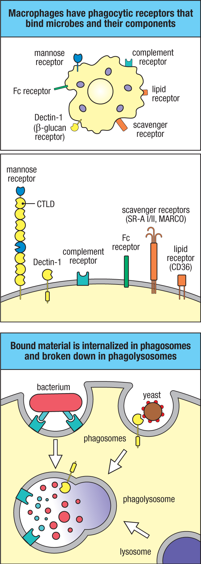
Macrophages and neutrophils constitutively express a number of cell-surface PRRs that stimulate the phagocytosis and intracellular killing of microbes bound to them, although some also signal through other pathways to trigger responses such as cytokine production. This set of phagocytic PRRs includes several members of the C-type lectin–like family (see Fig. 3.2). For example, Dectin-1 is strongly expressed by macrophages and neutrophils and recognizes β-(1,3)–linked glucans (polymers of glucose), which are common components of fungal cell walls in particular. Dendritic cells also express Dectin-1, as well as several other C-type lectin–like phagocytic receptors. Another C-type lectin, the mannose receptor expressed by macrophages and dendritic cells, recognizes various mannosylated ligands, including some present on fungi, bacteria, and viruses; it was once suspected of having an important role in resistance to microbes. However, experiments with mice that lack this receptor do not support this idea. The macrophage mannose receptor is now thought to function mainly as a clearance receptor for host glycoproteins such as β-glucuronidase and lysosomal hydrolases, which have mannose-containing carbohydrate side chains and whose extracellular concentrations are raised during inflammation.
Another set of phagocytic PRRs on macrophages, called scavenger receptors, recognize various anionic polymers and acetylated low-density lipoproteins. These receptors are structurally heterogeneous, consisting of at least six different molecular families. Class A scavenger receptors are membrane proteins composed of trimers of collagen domains (see Fig. 3.2). They include SR-A I, SR-A II, and MARCO (macrophage receptor with a collagenous structure), all of which bind various bacterial cell-wall components and help to internalize bacteria, although the basis of their specificity is poorly understood. Class B scavenger receptors bind high-density lipoproteins, and they internalize lipids. One of these receptors is CD36, which binds many ligands, including long-chain fatty acids.
A third set of receptors of crucial importance in macrophage and neutrophil phagocytosis is the complement receptors and Fc receptors introduced in Chapters 1 and 2. These receptors bind to complement-coated microbes or to antibodies that have bound to the surface of microbes and facilitate the phagocytosis of a wide range of microorganisms.
3-2 G protein–coupled receptors on phagocytes link microbe recognition with increased efficiency of intracellular killing.
Phagocytosis of microbes by macrophages and neutrophils is generally followed by the death of the microbe inside the phagocyte. In addition to the phagocytic receptors described above, macrophages and neutrophils have other receptors that signal to stimulate antimicrobial killing. These receptors belong to the evolutionarily ancient family of G protein–coupled receptors (GPCRs), which are characterized by seven membrane-spanning segments. Members of this family are crucial to immune-system function because they also direct responses to anaphylatoxins such as the complement fragment C5a (see Section 2-14), leukotrienes, and to many chemokines, recruiting phagocytes to sites of infection and promoting inflammation.
3.3 Chemokine Signaling
The fMet-Leu-Phe (fMLF) receptor is a G protein–coupled receptor that senses the presence of bacteria by recognizing a unique feature of bacterial polypeptides. Protein synthesis in bacteria is typically initiated with an N-formylmethionine (fMet) residue, an amino acid present in prokaryotes but not in eukaryotes. The fMLF receptor is named after a tripeptide, formyl-methionyl-leucyl phenylalanine, for which it has a high affinity, although it also binds other peptide motifs. Bacterial polypeptides binding to this receptor activate intracellular signaling pathways that direct the cell to move toward the most concentrated source of the ligand. Signaling through the fMLF receptor also induces the production of microbicidal reactive oxygen species (ROS) in the phagolysosome. The C5a receptor 1 recognizes the small fragment of C5 generated when complement is activated, usually by the presence of microbes (see Section 2-13), and signals by a similar pathway as the fMLF receptor. Thus, stimulation of these receptors both guides monocytes and neutrophils toward a site of infection and leads to increased antimicrobial activity; these cell responses can be activated by directly sensing unique bacterial products or by messengers such as C5a that indicate previous recognition ofa microbe.
The G protein–coupled receptors are so named because ligand binding activates a member of a class of intracellular GTP-binding proteins called G proteins, sometimes referred to as heterotrimeric G proteins to distinguish them from the family of ‘small’ GTPases typified by Ras. Heterotrimeric G proteins are composed of three subunits: Gα, Gβ, and Gγ, of which the α subunit is similar to the small GTPases (Fig. 3.3). In the resting state, the G protein is inactive, not associated with the receptor, and a molecule of GDP is bound to the α subunit. Ligand binding induces conformational changes in the receptor that allow it to bind the G protein, which results in the displacement of the GDP from the G protein and its replacement with GTP. The active G protein dissociates into two components, the Gα subunit and a complex consisting of a Gβ and a Gγ subunit. Each of these components can interact with other intracellular signaling molecules to transmit and amplify the signal. G proteins can activate a wide variety of downstream enzymatic targets, such as adenylate cyclase, which produces the second messenger cyclic AMP, and phospholipase C, whose activation gives rise to the second messenger inositol 1,4,5-trisphosphate (IP3) and the release of Ca2+ from stores in the endoplasmic reticulum. The subsequent increase in cytosolic Ca2+ can induce several signaling pathways, including the activation of protein kinase C (PKC) (see Chapter 7 for a more detailed discussion of these and other signaling pathways).
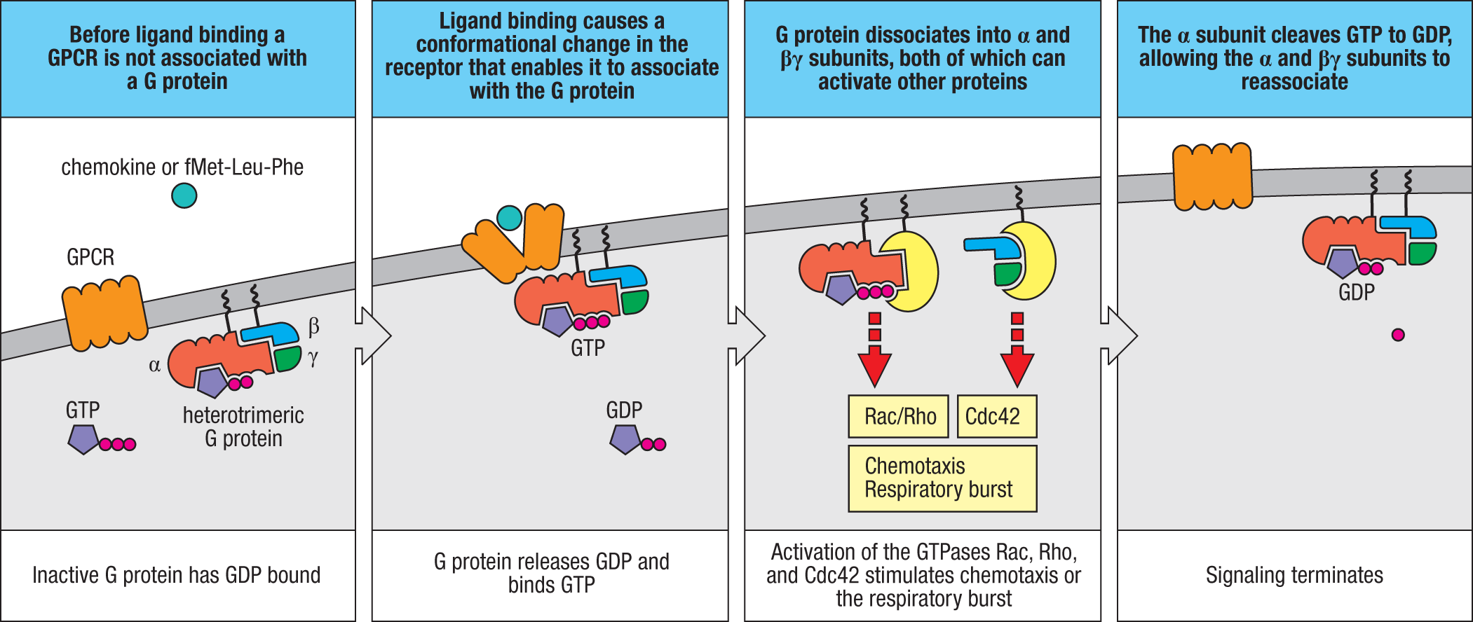
Signaling by fMLF and C5a receptors influences cell motility, metabolism, gene expression, and cell division through activation of several Rho family small GTPase proteins. The α subunit of the activated G protein indirectly activates Rac and Rho, while the βγ subunit indirectly activates the small GTPase Cdc42 (see Fig. 3.3). Activation of these GTPases is controlled by guanine nucleotide exchange factors (GEFs) (see Fig. 7.5), which exchange GTP for GDP bound to the GTPase. The G proteins activated by fMLF activate the GEF protein PREX1 (phosphatidylinositol 3,4,5-trisphosphate–dependent Rac exchanger 1 protein), which can directly activate Rac. Other GEFs, including members of the Vav family that are controlled by other types of receptors (see Section 7-19), can also activate Rac activity, and their activity synergizes with the actions of fMLF and C5a.
The activation of Rac and Rho helps to increase the microbicidal capacity of macrophages and neutrophils that have ingested pathogens. Upon phagocytosing microbes, macrophages and neutrophils produce a variety of toxic products that help to kill the engulfed microorganism (Fig. 3.4). The most important of these are the antimicrobial peptides described in Section 2-4, reactive nitrogen species such as nitric oxide (NO), and ROS such as the superoxide anion (O2–) and hydrogen peroxide (H2O2). Nitric oxide is produced by a high-output form of nitric oxide synthase, inducible NOS2 (iNOS2), whose expression is induced by a variety of stimuli, including fMLF.
|
Antimicrobial mechanisms of phagocytes |
||
|
Class of mechanism |
Macrophage products |
Neutrophil products |
|
Acidification |
pH ≈ 3.5-4.0, bacteriostatic or bactericidal |
|
|
Toxic oxygen–derived products |
Superoxide O2−, hydrogen peroxide H2O2, singlet oxygen 1O2•, hydroxyl radical •OH, hypochlorite OCl− |
|
|
Toxic nitrogen oxides |
Nitric oxide NO |
|
|
Antimicrobial peptides |
Cathelicidin, macrophage elastase–derived peptide |
α-Defensins (HNP1–4), β-defensin HBD4, cathelicidin, azurocidin, bacterial permeability inducing protein (BPI), lactoferricin |
|
Enzymes |
Lysozyme: digests cell walls of some Gram-positive bacteria Acid hydrolases (e.g., elastase and other proteases): break down ingested microbes |
|
|
Competitors |
Lactoferrin (sequesters Fe2+), vitamin B12–binding protein |
|
Fig. 3.4 Bactericidal agents produced or released by phagocytes after uptake of microorganisms. Most of the agents listed are directly toxic to microbes and can act directly in the phagolysosome. They can also be secreted into the extracellular environment, and many of these substances are toxic to host cells. Other phagocyte products sequester essential nutrients in the extracellular environment, rendering them inaccessible to microbes and hindering microbial growth. Besides being directly bacteriostatic or bactericidal, acidification of lysosomes also activates the many acid hydrolases that degrade the contents of the vacuole.
Activation of phagocytic receptors, including the fMLF and C5a receptors, can promote ROS generation by inducing assembly of a multicomponent, membrane-associated NADPH oxidase, also called phagocyte oxidase. In unstimulated phagocytes, this enzyme is inactive because it is not fully assembled. One set of subunits, the cytochrome b558 complex (composed of p22 and gp91), is localized in the plasma membranes of resting macrophages and neutrophils, and it appears in lysosomes after the maturation of phagolysosomes. The other components, p40, p47, and p67, are in the cytosol. Activation of phagocytes induces the cytosolic subunits to join with the membrane-associated cytochrome b558 to form a complete, functional NADPH oxidase in the phagolysosome membrane (Fig. 3.5). Activation of Rac promotes the movement of the cytosolic components to the membrane to assemble the active NADPH oxidase. Phosphorylation of certain residues of the NADPH oxidase complex also plays a critical role in activation and is mediated by PKC activation downstream of GPCRs. PKC activation can also occur downstream of phagocytic receptors, such as Dectin-1 and Fc receptors, which contain immunoreceptor tyrosine-based activation motifs (ITAMs) in their cytosolic tails. The signaling mediated by ITAMs is discussed in greater detail later in this chapter (Section 3-26) as well as in later chapters.
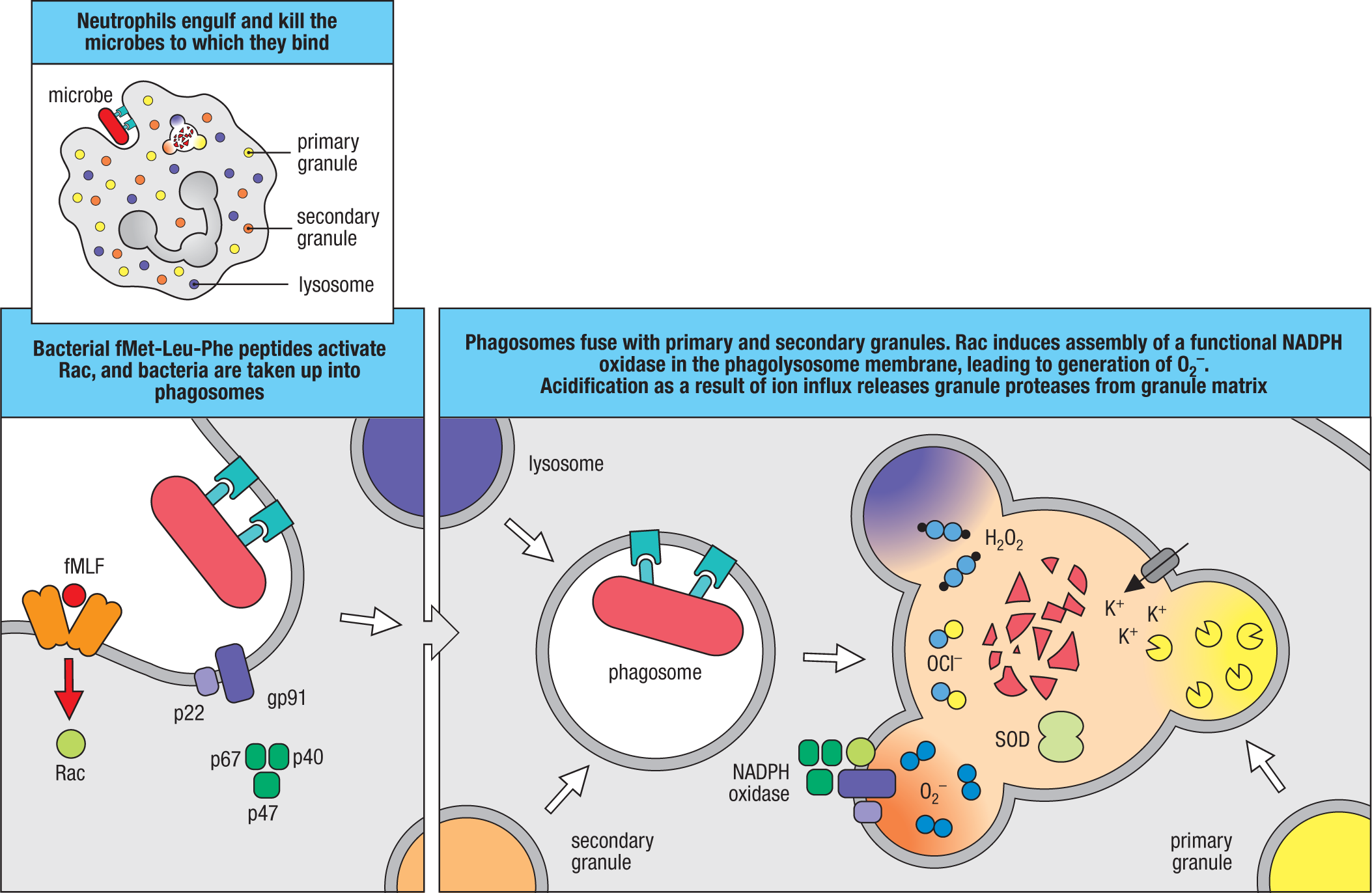
The NADPH oxidase reaction results in a transient increase in oxygen consumption by the cell, which is known as the respiratory burst. It generates superoxide anion within the lumen of the phagolysosome, and this is converted by the enzyme superoxide dismutase (SOD) into H2O2. Further chemical and enzymatic reactions produce a range of toxic ROS from H2O2, including the hydroxyl radical (•OH), hypochlorite (OCl–), and hypobromite (OBr–). In this way, the direct recognition of bacterially derived polypeptides or previous pathogen recognition by the complement system activates a potent killing mechanism within macrophages and neutrophils that have ingested microbes via their phagocytic receptors. However, phagocyte activation can also cause extensive tissue damage because hydrolytic enzymes, membrane-disrupting peptides, and reactive oxygen species can be released into the extracellular environment and are toxic to host cells.
Neutrophils use the respiratory burst described above in their role as an early responder to infection. Neutrophils are not tissue-resident cells, and they need to be recruited to a site of infection from the bloodstream. Their main function is to ingest and kill microorganisms, but they also produce cytokines and lipid mediators that promote and orchestrate the inflammatory response. Although neutrophils are eventually present in much larger numbers than macrophages in some types of acute infection, they are short-lived, dying soon after they have accomplished a round of phagocytosis and used up their primary and secondary granules. Dead and dying neutrophils are a major component of the pus that forms in abscesses and in wounds infected by certain extracellular capsulated bacteria such as streptococci and staphylococci, which are thus known as pus-forming, or pyogenic, bacteria. Macrophages, in contrast, are long-lived cells and continue to generate new lysosomes.
Individuals with a disease called chronic granulomatous disease (CGD) have a genetic deficiency of NADPH oxidase, which means their phagocytes do not produce the toxic oxygen derivatives characteristic of the respiratory burst and so are less able to kill ingested microorganisms and clear an infection. The most common form of CGD is an X-linked heritable disease that arises from inactivating mutations in the gene encoding the gp91 subunit of cytochrome b558. People with this defect are unusually susceptible to bacterial and fungal infections, especially in infancy, though they remain susceptible for life. One autosomal recessive form of NADPH oxidase deficiency, p47phox deficiency, has very low but detectable activity and causes a milder form of CGD.
3.4 Neutrophil Extracellular Traps
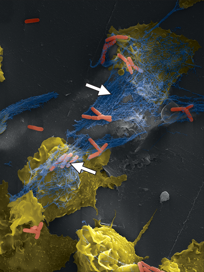
In addition to killing microbes engulfed by phagocytosis, neutrophils use another rather novel mechanism of destruction that is directed at extracellular pathogens. During infection, some activated neutrophils undergo a unique form of cell death in which the nuclear chromatin, rather than being degraded as occurs during apoptosis, is released into the extracellular space and forms a fibril matrix known as neutrophil extracellular traps, or NETs (Fig. 3.6). It has been proposed that NETs act to capture microorganisms, which may then be more efficiently phagocytosed by other neutrophils or macrophages, but the importance of this mechanism during infection remains rather unclear.
Macrophages can phagocytose pathogens and produce the respiratory burst immediately upon encountering an infecting microorganism, and this can be sufficient to prevent an infection from becoming established. In the 19th century, the immunologist Élie Metchnikoff believed that the innate response of macrophages encompassed all host defenses; indeed, invertebrates such as the sea star that he was studying rely entirely on innate immunity to overcome infection. Although this is not the case in humans and other vertebrates, the innate response of macrophages still provides an important front line of defense that must be overcome if a microorganism is to establish an infection that can be passed on to a new host.
Pathogens have, however, developed a variety of strategies to avoid immediate destruction by macrophages and neutrophils. Many extracellular pathogenic bacteria coat themselves with a thick polysaccharide capsule that is not recognized by any phagocytic receptor. In such cases, however, the complement system can recognize microbial surfaces and coat them with C3b, thereby flagging them for phagocytosis via complement receptors, as described in Chapter 2. Other pathogens, for example, mycobacteria, have evolved ways to grow inside macrophage phagosomes by inhibiting their acidification and fusion with lysosomes. Without such strategies, a microorganism must enter the body in sufficient numbers to overwhelm the immediate innate host defenses and to establish a focus of infection.
3-3 Microbial recognition and tissue damage initiate an inflammatory response.
An important effect of the interaction between microbes and tissue macrophages is the activation of macrophages and other immune cells to release small proteins called cytokines and chemokines, and other chemical mediators, such as lipid mediators. Collectively, these signals induce a state of inflammation in the tissue, attract monocytes and neutrophils to the infection, and allow plasma proteins to enter the tissue from the blood. An inflammatory response is usually initiated within minutes to hours of infection or wounding. Macrophages are stimulated to secrete pro-inflammatory cytokines, such as TNF-α, and chemokines by interactions between microbes or microbial products and specific receptors expressed by macrophages. We will examine how the cytokines interact with pathogens later in the chapter, but first we describe some general aspects of inflammation and how it contributes to host defense.
Inflammation has three essential roles in combating infection. The first is to deliver additional effector molecules and cells from the blood into sites of infection, and so increase the destruction of invading microorganisms. The second is to induce local blood clotting, which provides a physical barrier to the spread of the infection in the bloodstream. The third is to promote the repair of injured tissue, which occurs during the resolution phase, after the infection has been cleared.
Inflammatory responses are characterized by heat, pain, redness, and swelling at the site of an infection, reflecting four types of change in the local blood vessels, as shown in Fig. 3.7. The first is an increase in vascular diameter, leading to increased local blood flow—hence the heat and redness—and a reduction in the velocity of blood flow, especially along the inner walls of small blood vessels. The second change is the activation of endothelial cells lining the blood vessel to express cell-adhesion molecules that promote the binding of circulating leukocytes. The combination of slowed blood flow and adhesion molecules allows leukocytes to attach to the endothelium and migrate into the tissues, a process known as extravasation. All these changes are initiated by the pro-inflammatory cytokines and chemokines produced by activated macrophages and other cells in the tissue.
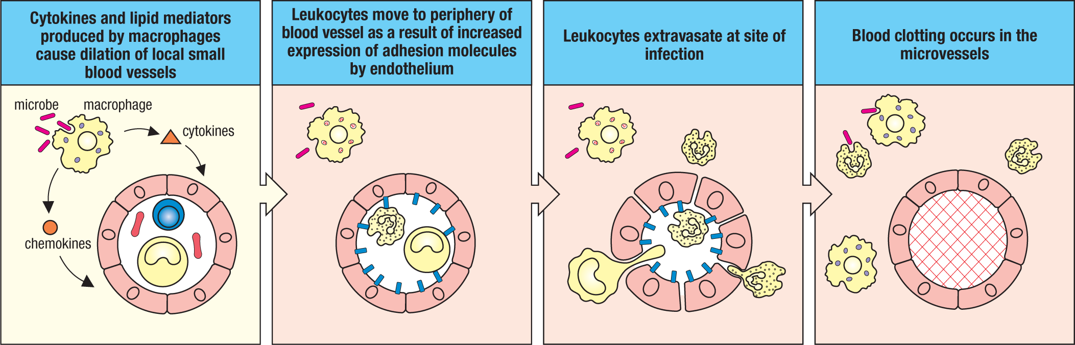
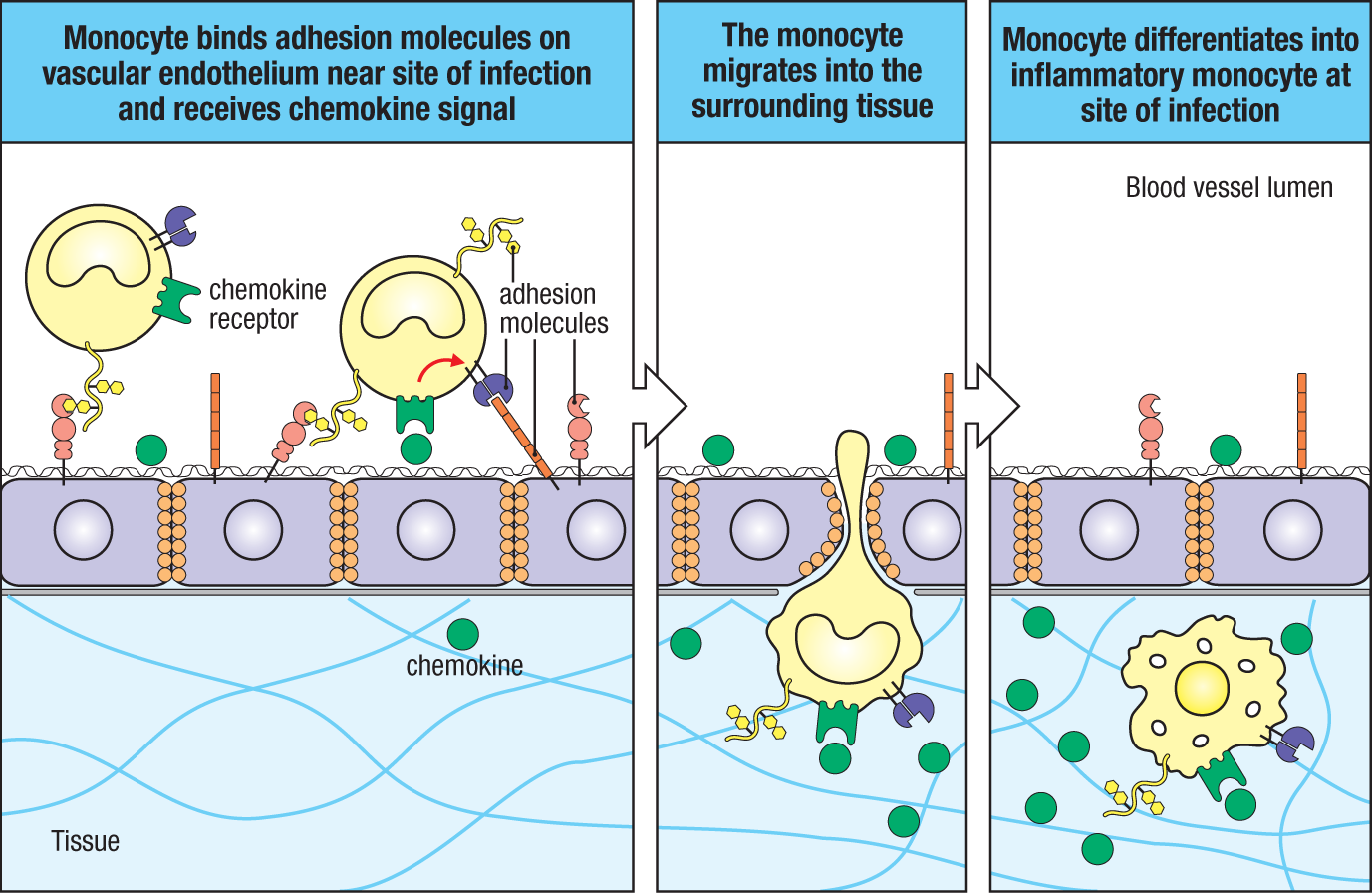
Once inflammation has begun, the first white blood cells attracted to the site are neutrophils. These are followed by monocytes (Fig. 3.8), which upon activation are called inflammatory monocytes. Monocytes can produce various pro-inflammatory cytokines but are distinguishable from macrophages by their lack of expression of the adhesion G protein–coupled receptor E1, commonly called F4/80. Monocytes are also able to give rise to dendritic cells in the tissues, depending on signals that they receive from their environment. In the later stages of inflammation, other leukocytes such as eosinophils and lymphocytes also enter the infected site.
The third major change in local blood vessels is an increase in vascular permeability. Thus, instead of being tightly joined together, the endothelial cells lining the blood vessel walls become separated, leading to an exit of fluid and proteins from the blood and their local accumulation in the tissue. This accounts for the swelling, or edema, and pain—as well as the accumulation in tissues of plasma proteins such as complement and mannose-binding lectin (MBL) that aid in host defense. The changes that occur in endothelium as a result of inflammation are known generally as endothelial activation. The fourth change, clotting in microvessels in the site of infection, prevents the spread of the pathogen via the blood.
These changes are induced by a variety of inflammatory mediators released as a consequence of the recognition of pathogens by macrophages, and later by neutrophils and other white blood cells. Macrophages and neutrophils secrete lipid mediators of inflammation—prostaglandins, leukotrienes, and platelet-activating factor (PAF)—which are rapidly produced by enzymatic pathways that degrade membrane phospholipids. Their actions are followed by those of the chemokines and cytokines that are synthesized and secreted by macrophages and inflammatory monocytes in response to pathogens. The cytokine tumor necrosis factor-α (TNF-α, also known simply as TNF), for example, is a potent activator of endothelial cells. We describe TNF-α and related cytokines in more detail in Section 3-15.
Besides stimulating the respiratory burst in phagocytes and acting as a chemoattractant for neutrophils and monocytes, C5a also promotes inflammation by increasing vascular permeability and inducing the expression of certain adhesion molecules on endothelium. C5a also activates local mast cells (see Section 1-4), which are stimulated to release their granules containing the small inflammatory molecule histamine as well as TNF-α and cathelicidins.
If wounding has occurred, the injury to blood vessels immediately triggers two protective enzyme cascades. One is the kinin system of plasma proteases that is triggered by tissue damage to generate several polypeptides that regulate blood pressure, coagulation, and pain. Although we will not fully describe its components here, one inflammatory mediator produced is the vasoactive peptide bradykinin, which increases vascular permeability to promote the influx of plasma proteins to the site of tissue injury. It also causes pain. Although unpleasant to the victim, pain draws attention to the problem and leads to immobilization of the affected part of the body, which helps to limit the spread of the infection.
The coagulation system is another protease cascade that is triggered in the blood after damage to blood vessels, although its full description is also outside our present scope. Its activation leads to the formation of a fibrin clot, whose normal role is to prevent blood loss. With regard to innate immunity, however, the clot physically encases the infectious microorganisms and prevents their entry into the bloodstream. The kinin and the coagulation cascades are also triggered by activated endothelial cells, and so they can have important roles in the inflammatory response to pathogens even if wounding or gross tissue injury has not occurred. Thus, within minutes of the penetration of tissues by a pathogen, the inflammatory response causes an influx of proteins and cells that may control the infection.
Damage to tissues can occur in the absence of infection by microbes; for example, as a result of physical trauma, ischemia, or metabolic or autoimmune disorders. In such sterile injury, many of the changes associated with infection, such as neutrophil recruitment, can occur, in addition to activation of the kinin system and clot formation.
3-4 Toll-like receptors represent an ancient pathogen-recognition system.
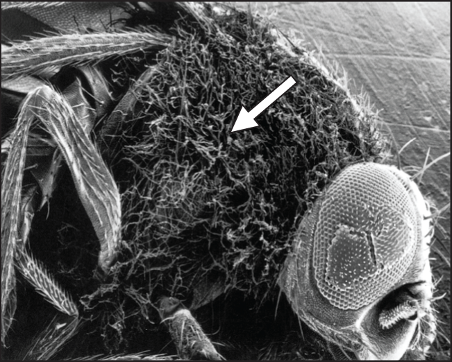
Section 1-5 introduced pattern-recognition receptors (PRRs), which function as sensors for pathogen-associated molecular patterns (PAMPs). The existence of these receptors was predicted by Charles Janeway, Jr., before mechanisms of innate recognition of PAMPs were known, to explain how complex mixtures of microbial molecules, called adjuvants, were able to stimulate adaptive immune responses to purified antigens. The first example of such a receptor was the Toll protein, discovered in the fruit fly Drosophila melanogaster. Although Toll was identified earlier as a gene controlling the dorso-ventral patterning during development of Drosophila, in 1996 Jules Hoffmann discovered that Toll signaling induces the expression of several host-defense mechanisms. The importance of this mechanism was illustrated by the decreased production of antimicrobial peptides and the enhanced susceptibility to infection of adult flies with mutations in the Toll pathway (Fig 3.9). For this work, Hoffmann was awarded part of the 2011 Nobel Prize in Physiology or Medicine.
In 1997, Janeway and Ruslan Medzhitov discovered a human homolog of Toll, subsequently termed Toll-like receptor (TLR), and showed that signaling by this receptor activates innate and adaptive immune responses. Subsequently, numerous TLRs associated with resistance to infection by pathogens have been found in humans and other animals. In plants, proteins with domains resembling the ligand-binding regions of TLR proteins are involved in the production of antimicrobial peptides, indicating the ancient association of these domains with host defense.
3.5 Pattern-Recognition Receptors
3-5 Mammalian Toll-like receptors are activated by many different pathogen-associated molecular patterns.
There are 10 expressed TLR genes in humans and 12 in mice. Each TLR recognizes one or more distinct molecular ‘patterns’ present on microbes, which are referred to as pathogen-associated molecular patterns (PAMPs), as discussed at the beginning of this chapter. These molecules are general components of both pathogenic and nonpathogenic microorganisms and are sometimes called microbial-associated molecular patterns, or MAMPs, to reflect this fact. Collectively, the mammalian TLRs recognize molecules from bacteria, fungi, viruses, and protozoa. The mammalian TLRs and their known microbial ligands are listed in Fig. 3.10. Note that in almost all cases the ligands represent highly conserved features that are characteristic of these microbial groups. For example, lipoteichoic acids of Gram-positive bacterial cell walls and lipopolysaccharide (LPS) of the outer membrane of Gram-negative bacteria (see Fig. 2.9) are recognized by TLRs. In general, these microbial molecules are not found in vertebrate cells, which prevents inappropriate immune recognition of host cells. The major exceptions to this rule, however, are the DNA and RNA ligands for certain TLRs. As discussed later in this section, additional mechanisms are used to distinguish between microbial and host DNA and RNA.
|
Innate immune recognition by mammalian Toll-like receptors |
|
|
Toll-like receptor |
Ligand |
|
TLR-1:TLR-2 heterodimer |
Lipomannans (mycobacteria) Diacyl and triacyl lipopeptides (bacteria) Lipoteichoic acids (Gram-positive bacteria) Cell-wall β-glucans (fungi) |
|
TLR-2:TLR-6 heterodimer |
|
|
TLR-3 |
Double-stranded RNA (viruses), poly I:C |
|
TLR-4 |
LPS (Gram-negative bacteria) |
|
TLR-5 |
Flagellin (bacteria) |
|
TLR-7 |
Single-stranded RNA (viruses) |
|
TLR-8 |
Single-stranded RNA (viruses) |
|
TLR-9 |
DNA with unmethylated CpG (bacteria and DNA viruses) |
|
TLR-10 (human only) |
Unknown |
|
TLR-11 (mouse only) |
Profilin and profilin-like proteins (Toxoplasma gondii, uropathogenic bacteria) |
|
TLR-12 (mouse only) |
Profilin (Toxoplasma gondii) |
|
TLR-13 (mouse only) |
Single-stranded RNA (bacterial ribosomal RNA) |
Fig. 3.10 Innate immune recognition by Toll-like receptors. Each of the human or mouse TLRs recognizes one or more microbial molecular patterns, generally by direct interaction with molecules on the pathogen surface. Some Toll-like receptor proteins form heterodimers (for example, TLR-1:TLR-2 and TLR-2:TLR-6). LPS, lipopolysaccharide.
TLRs are sensors for microbes present in extracellular spaces. Some mammalian TLRs are cell-surface receptors, but others are located intracellularly in the membranes of endosomes, where they detect pathogens or their components that have been taken into cells by phagocytosis, receptor-mediated endocytosis, or macropinocytosis (Fig. 3.11). TLRs localized to the plasma membrane can also be incorporated into phagosomes, which further enables detection of phagocytosed microbes. TLRs are transmembrane proteins in which the extracellular region of the monomer is composed of 18–25 copies of a leucine-rich repeat (LRR). Each LRR of a TLR protein is composed of approximately 20–25 amino acids, and multiple LRRs create a horseshoe-shaped protein scaffold that is adaptable for ligand binding and recognition on both the outer (convex) and inner (concave) surfaces. Signaling by mammalian TLRs is activated when binding of a ligand induces formation of a dimer or induces conformational changes in a preformed TLR dimer. All mammalian TLR proteins have in their cytoplasmic tails a TIR (for Toll–IL-1 receptor) domain, which is also found in the cytoplasmic tail of the receptor for the cytokines interleukin-1α (IL-1α) and interleukin-1β (IL-1β). The TLR TIR domains mediate interaction with other TIR domain–containing signaling molecules, discussed in greater detail in Section 3-7. For years after the discovery of the mammalian TLRs it was not known whether they made direct contact with microbial products or whether they sensed the presence of microbes by some indirect means. Drosophila Toll, for example, does not recognize pathogen products directly, but instead is activated when it binds a cleaved version of a self protein, Spätzle. Drosophila has other direct pathogen-recognition molecules, and these trigger the proteolytic cascade that ends in the cleavage of Spätzle. In this sense, Toll is not a classical pattern-recognition receptor. However, X-ray crystal structures of several mammalian dimeric TLRs bound to their ligands show that mammalian TLRs make direct contact with microbial ligands.
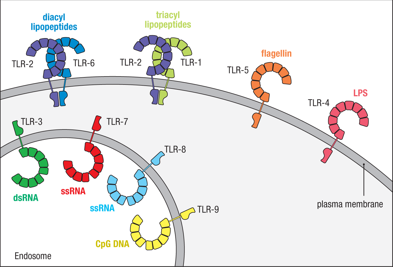
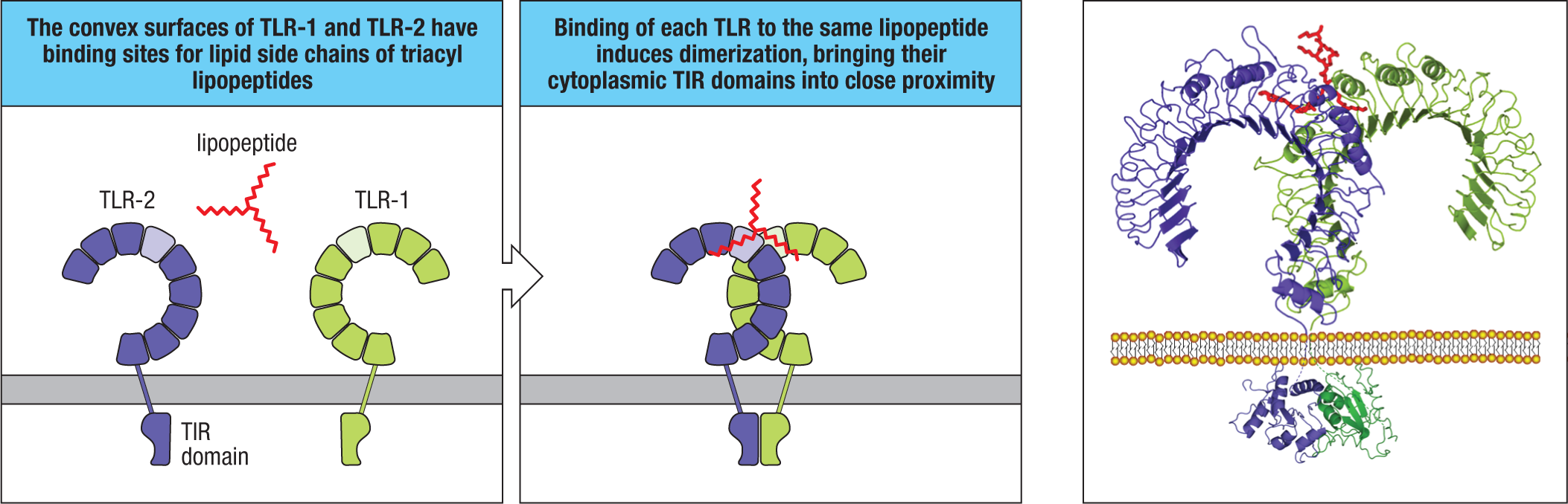
TLRs are expressed on many cell types, including macrophages, dendritic cells, B cells, stromal cells, and certain epithelial cells, enabling the initiation of antimicrobial responses in many tissues. Mammalian TLR-1, TLR-2, and TLR-6 are cell-surface receptors that are activated by various ligands, including lipoteichoic acid from Gram-positive bacteria and diacyl and triacyl lipoproteins present in both Gram-positive and Gram-negative bacteria. Ligand binding induces the formation of heterodimers of TLR-2 and TLR-1 or of TLR-2 and TLR-6. The X-ray crystal structure of a synthetic triacyl lipopeptide ligand bound to TLR-1 and TLR-2 shows exactly how ligand binding induces dimerization (Fig. 3.12). Two of the three lipid chains bind to the convex surface of TLR-2, while the third binds to the convex surface of TLR-1. Dimerization brings the cytoplasmic TIR domains of the TLR chains into close proximity with each other to initiate signaling. Similar interactions are presumed to occur with the diacyl lipopeptide ligands that induce the dimerization of TLR-2 and TLR-6.
TLR-5 is expressed on the cell surface of certain macrophages and dendritic cells and also in intestinal epithelial cells; it recognizes flagellin, a protein subunit of bacterial flagella. TLR-5 recognizes a highly conserved site on flagellin that is buried and inaccessible in the assembled flagellar filament. This means the receptor is activated only by monomeric flagellin, which can be shed during flagellar assembly or when flagella are degraded.
Mice, but not humans, express TLR-11 and TLR-12. TLR-11–deficient mice develop urinary infections caused by uropathogenic strains of Escherichia coli, although the bacterial ligand for TLR-11 has not yet been identified. TLR-11 and TLR-12 also both recognize protozoan parasites such as Toxoplasma gondii and Plasmodium falciparum. They bind to motifs that are present in the protozoan actin-binding protein profilin but absent in mammalian profilins. TLR-11 and TLR-12 are both required in macrophages and conventional dendritic cells for activation by T. gondii profilin, but TLR-12 plays a more dominant role. Mice lacking TLR-11 develop more severe tissue injury than normal mice on infection with Toxoplasma, whereas mice lacking TLR-12 die rapidly after infection.
Five of the mammalian TLRs (TLR3, TLR7, TLR8, TLR9, and TLR13) recognize nucleic acids. These receptors are all localized to endosomes. The endosome lumen is acidic and contains many proteases, which degrade internalized microbes and release their nucleic acids for recognition by TLRs. All microbes possess some form of nucleic acid and so can be recognized by one or more of the nucleic acid–sensing TLRs; however, analysis of mice and humans with defects in one or more of these TLRs indicates that these receptors are most important for detection of viruses. In general, viruses lack other conserved features that could serve as PAMPs.
TLR-7 and TLR-8 share very similar amino acid sequences, and both receptors are activated by degradation products of single-stranded RNA (ssRNA). Many viruses (for example, orthomyxoviruses such as influenza; flaviviruses such as West Nile virus; and coronaviruses such as SARS-CoV-2) utilize ssRNA genomes, and TLR-7 and TLR-8 bind to small RNA fragments released when viruses are degraded in endosomes. Recent X-ray crystal structures of TLR-7 and TLR-8 have revealed two pairs of binding sites for distinct RNA fragments: guanosine and a short RNA fragment bind TLR-7, while uridine and a short RNA fragment bind TLR-8. Whether differences in the RNA sequences that activate TLR-7 and TLR-8 have consequences for antiviral immunity remains unclear. In mice, TLR-7 plays a dominant role over TLR-8 for detection of ssRNA viruses. For example, mice lacking TLR-7 have impaired immune responses to influenza. A viral detection function for TLR-8 in mice has not been established as clearly. The difference between mice and humans may be based, at least in part, on which cell types express the receptors in each species. In mice, TLR-7 is expressed broadly in many innate immune cell types, but in humans TLR-7 expression is limited to plasmacytoid dendritic cells and B cells, while TLR-8 is expressed more broadly. TLR-13 also binds RNA but has specificity for certain bacterial ribosomal RNA sequences instead of viral RNA. TLR-13 is present in mice but not humans.
TLR-9 recognizes single-stranded DNA sequences that contain unmethylated CpG dinucleotides. In mammals, CpG dinucleotides in genomic DNA are often methylated on the cytosine by DNA methyltransferases. In the genomes of bacteria and many viruses, CpG dinucleotides remain unmethylated, so these sequences serve as ligands for TLR-9. The requirement for unmethylated CpG sequences is thought to limit the potential for recognition of self DNA by TLR-9. The structure of the TLR-9 dimer bound to DNA reveals a ligand-binding modality similar to what was previously described for TLR-7 and TLR-8: two pairs of binding sites within the dimer bind distinct DNA sequences. While the DNA genomes of viruses and bacteria are typically double-stranded, TLR-9 recognizes single-stranded DNA, so the DNA strands of pathogen genomes must be melted apart within endosomes prior to TLR-9 binding.
TLR-3 is expressed by macrophages, conventional dendritic cells, and intestinal epithelial cells; it recognizes double-stranded RNA (dsRNA). Some viruses have genomes composed of dsRNA, but dsRNA is also generated when ssRNA viruses replicate or express genes. dsRNA can be recognized by TLR-3 after direct endocytosis of viruses with double-stranded RNA genomes, such as rotavirus, or by the phagocytosis of dying cells in which viruses are replicating. Crystallographic analysis shows that TLR-3 binds directly to dsRNA. TLR-3 has two contact sites for dsRNA: one on the amino terminus and a second near the membrane-proximal carboxyl terminus. Mutations in the ectodomain of human TLR-3, which produce a dominantly acting loss-of-function mutant receptor, have been associated with encephalitis that is caused by a failure to control the herpes simplex virus. This increased susceptibility is somewhat surprising because herpes simplex virus is a DNA virus; the most likely explanation for sensing by TLR-3 is generation of dsRNA after transcription of overlapping viral genes encoded on opposing strands of DNA.
To reach endosomes, TLR-3, TLR-7, TLR-8, TLR-9, TLR-11, TLR-12, and TLR-13 must interact with a specific protein, UNC93B1. UNC93B1, which is composed of 12 transmembrane domains, binds TLRs in the endoplasmic reticulum and shuttles them to endosomes. Mice lacking this protein have defects in signaling by these endosomal TLRs because the receptors cannot exit the endoplasmic reticulum. Rare human mutations in UNC93B1 have been identified as causing susceptibility to herpes simplex encephalitis, similarly to TLR-3 deficiency, but they do not impair immunity to many other viral pathogens, presumably because of the existence of other viral sensors, which are discussed later in this chapter.
Sensing of nucleic acids enables broad recognition of diverse pathogens, but, in abnormal settings, TLRs may be activated by self-derived nucleic acids, leading to autoimmunity. In healthy cells, RNA and DNA are normally confined to the nucleus and cytoplasm and are not present in endosomes; however, nucleic acids can be released into the extracellular space during tissue damage or inflammation (for example, NETs). The localization of nucleic acid–sensing TLRs to endosomes reduces the likelihood that these receptors encounter such extracellular RNA and DNA. RNases and DNases in the extracellular space degrade RNA and DNA before they are taken up by endocytosis and delivered to TLRs. Another mechanism that limits TLR activation to endosomes is that the ectodomains of each of the nucleic acid–sensing TLRs must undergo proteolytic cleavage before the receptors can be activated; this cleavage occurs when TLRs arrive in endosomes, which prevents receptors from responding to ligands in other subcellular compartments, such as the cell surface (see Fig. 3.11). The two halves of the cleaved TLR remain associated and both contribute to ligand binding. Without this proteolytic event, the receptor cannot undergo the necessary conformational change required to initiate signaling.
In mouse models of systemic lupus erythematosus (SLE), an autoimmune disease associated with responses to self nucleic acids, TLR-7 and TLR-9 both contribute to disease. Several studies have identified polymorphisms in the human TLR-7 gene that are associated with increased risk of SLE, suggesting a potential role in this disease.
3-6 TLR-4 recognizes bacterial lipopolysaccharide in association with the host accessory proteins MD-2 and CD14.
Not all mammalian TLRs bind their ligands so directly. TLR-4 is expressed by several types of immune-system cells, including dendritic cells and macrophages, and is important in sensing and responding to numerous bacterial infections. TLR-4 recognizes the LPS of Gram-negative bacteria by a mechanism that is partly direct and partly indirect. LPS varies in composition among different bacteria but essentially consists of a polysaccharide core attached to an amphipathic lipid, lipid A, with a variable number of fatty-acid chains per molecule. To recognize LPS, the ectodomain of TLR-4 uses an accessory protein, MD-2. MD-2 initially binds to TLR-4 within the cell and is necessary both for the correct trafficking of TLR-4 to the cell surface and for the recognition of LPS. MD-2 associates with the central section of the curved ectodomain of TLR-4, binding off to one side as shown in Fig. 3.13. When the TLR-4:MD-2 complex encounters LPS, five lipid chains of LPS bind to a deep hydrophobic pocket of MD-2, but not directly to TLR-4, while a sixth lipid chain remains exposed on the surface of MD-2. This last lipid chain and parts of the LPS polysaccharide backbone can then bind to the convex side of a second TLR-4 ectodomain, inducing TLR-4 dimerization, which activates intracellular signaling pathways.
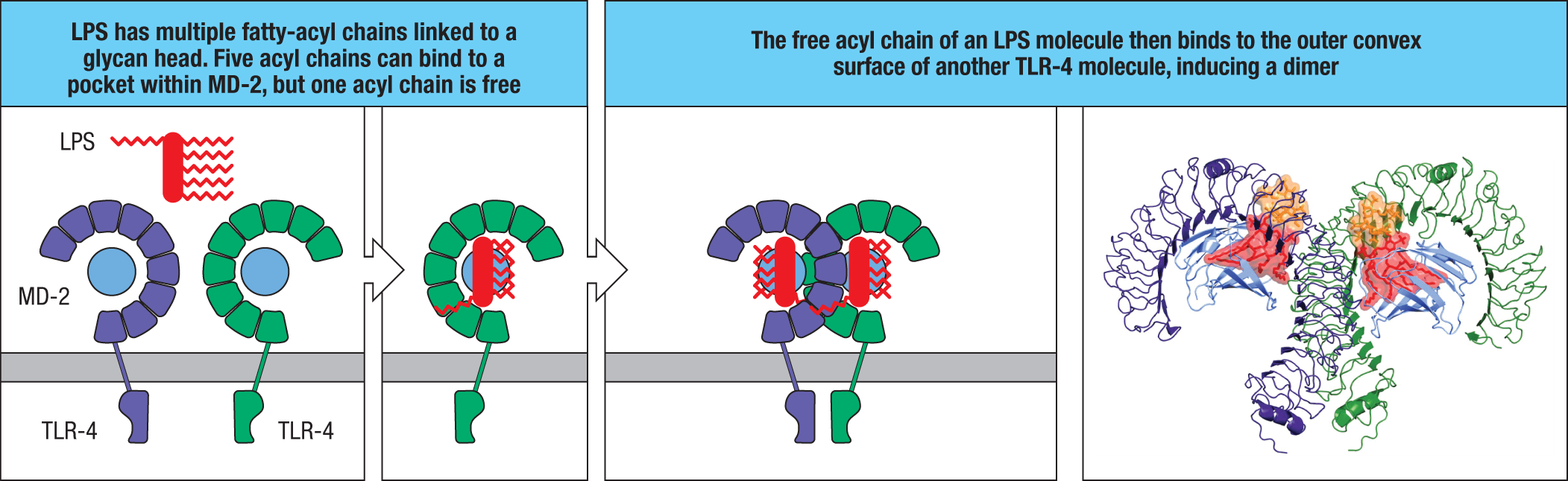
TLR-4 activation by LPS involves two other accessory proteins besides MD-2. While LPS is normally an integral component of the outer membrane of Gram-negative bacteria, during infections it can become detached from the membrane and be picked up by the host LPS-binding protein present in the blood and in extracellular fluid in tissues. LPS is transferred from LPS-binding protein to a second protein, CD14, which is present on the surface of macrophages, neutrophils, and dendritic cells. On its own, CD14 can act as a phagocytic receptor, but on macrophages and dendritic cells it also acts as an accessory protein for TLR-4.
The systemic injection of LPS causes a collapse of the circulatory and respiratory systems, a condition known as shock. These dramatic effects of LPS are seen in humans as septic shock, which results from an uncontrolled systemic bacterial infection, or sepsis. In this case, LPS induces an overwhelming secretion of cytokines, including IL-1β and TNF-α, causing systemic vascular permeability, an undesirable effect of its normal role in containing local infections. Mutant mice lacking TLR-4 function are resistant to LPS-induced septic shock but are highly sensitive to LPS-bearing pathogens such as Salmonella enterica ssp. typhimurium. In fact, TLR-4 (the first human homolog of Toll discovered by Medzhitov and Janeway, as discussed earlier) was identified as a receptor for LPS by positional cloning of its gene from the LPS-resistant C3H/HeJ mouse strain, which harbors a naturally occurring mutation in the cytoplasmic tail of TLR-4 that interferes with the receptor’s ability to signal. For this discovery, the 2011 Nobel Prize in Physiology or Medicine was partly awarded to Bruce Beutler.
3-7 TLRs activate NFκB, AP-1, and IRF transcription factors to induce the expression of inflammatory cytokines and type I interferons.
Signaling by mammalian TLRs in various cell types induces a diverse range of intracellular responses that together result in the production of inflammatory cytokines, chemotactic factors, antimicrobial peptides, and the antiviral cytokines interferon-α (IFN-α) and interferon-β (IFN-β), the type I interferons. TLR signaling achieves this by activating several signaling pathways that each activate different transcription factors. As mentioned earlier, ligand-induced dimerization of two TLR ectodomains brings the cytoplasmic TIR domains together, allowing them to interact with the TIR domains of cytoplasmic adaptor molecules that initiate intracellular signaling. There are five such adaptors used by mammalian TLRs: MyD88, TRIF, MAL (also known as TIRAP), TRAM, and BCAP. It is significant that the TIR domains of the different TLRs interact with different combinations of these adaptors because the choice of adaptor influences which of several downstream signals will be activated by the TLR. Recruitment of MyD88 and/or TRIF controls most of the signaling downstream of TLR activation, while MAL and TRAM facilitate the recruitment of MyD88 and TRIF, respectively, but do not directly participate in signal transduction. Most TLRs interact with MyD88 and not TRIF. TLR-3 interacts only with TRIF, and TLR-4 interacts with both MyD88 and TRIF. BCAP links TLR activation to distinct signaling pathways, but the details of this signaling are less clear because BCAP was only recently discovered as a TLR adaptor.
Signaling by most TLRs activates the transcription factor NFΚB (Fig. 3.14). Mammalian TLRs also activate several members of the interferon regulatory factor (IRF) transcription factor family through a second pathway, and they activate members of the activator protein 1 (AP-1) family, such as c-Jun, through yet another signaling pathway involving mitogen-activated protein kinases (MAPKs). NFΚB and AP-1 act primarily to induce the expression of pro-inflammatory cytokines and chemotactic factors. The IRF factors IRF3 and IRF7 are particularly important for inducing antiviral type I interferons, whereas a related factor, IRF5, is involved in the production of pro-inflammatory cytokines. Here we will describe how TLR signaling induces the transcription of various cytokine genes; later in the chapter, we will explain how those cytokines exert their various actions.
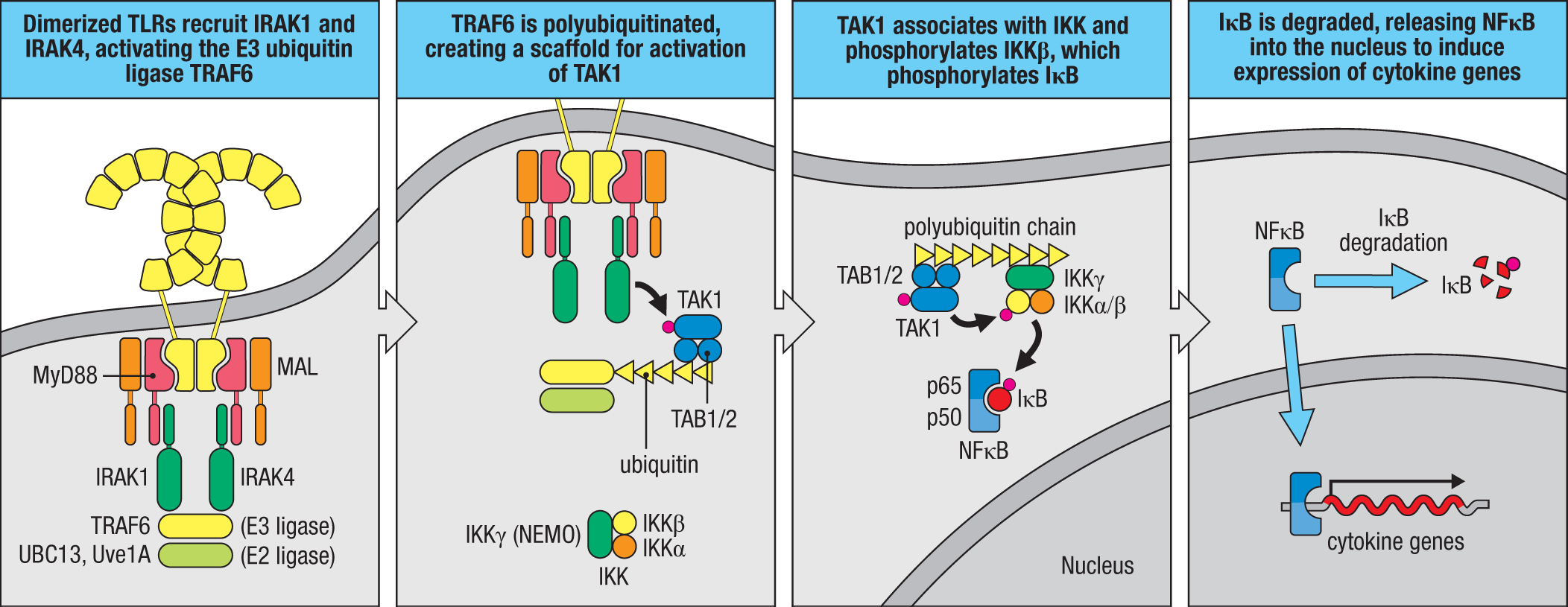
We consider first the signaling pathway triggered by TLRs that use MyD88. Two protein domains of MyD88 are responsible for its function as an adaptor. MyD88 has a TIR domain at its carboxyl terminus that associates with the TIR domains in the TLR cytoplasmic tails. At its amino terminus, MyD88 has a death domain, so named because it was first identified in signaling proteins involved in apoptosis, a type of programmed cell death. The MyD88 death domain associates with a similar death domain present in other intracellular signaling proteins. Both MyD88 domains are required for signaling, and rare mutations in either domain are associated with immunodeficiency characterized by recurrent bacterial infections in humans. The MyD88 death domain recruits and activates two serine/threonine protein kinases—IRAK4 (IL-1 receptor–associated kinase 4) and IRAK1—via their death domains. This IRAK complex performs two functions: it recruits enzymes that produce a signaling scaffold, and it uses this scaffold to recruit other molecules that are then phosphorylated by the IRAKs.
 Case Study: Interleukin-1 Receptor–Associated Kinase 4 Deficiency
To form a signaling scaffold, the IRAK complex recruits the enzyme TRAF6 (tumor necrosis factor receptor–associated factor 6), which is an E3 ubiquitin ligase that acts in cooperation with UBC13, an E2 ubiquitin ligase, and its cofactor Uve1A (together called TRIKA1) (see Fig. 3.14). The combined activity of TRAF6 and UBC13 is to ligate (unite with a chemical bond) one ubiquitin molecule to another protein, which can be another ubiquitin molecule, and thereby generate protein polymers. The polyubiquitin involved in signaling contains linkages between the lysine 63 residue on one ubiquitin and the carboxyl terminus of the next, forming K63 linkages. This polyubiquitin polymer can be initiated on other proteins, including TRAF6 itself, or produced as free linear ubiquitin polymers, and can be extended to produce polyubiquitin chains that act as a platform—or scaffold—to which other signaling molecules bind. Next, the scaffold recruits a signaling complex consisting of the polyubiquitin-binding adaptor proteins TAB1, TAB2, and the serine/threonine kinase TAK1 (see Fig. 3.14). By being brought onto the scaffold, TAK1 is phosphorylated by the IRAK complex, and activated TAK1 propagates signaling by activating certain MAPKs, such as c-Jun terminal kinase (JNK) and MAPK14 (p38 MAPK). These then activate AP-1 family transcription factors that transcribe cytokine genes.
TAK1 also phosphorylates and activates the IΚB kinase (IKK) complex, which is composed of three proteins: IKKα, IKKβ, and IKKγ (the latter also known as NEMO, for NFΚB essential modifier). IKKγ functions by binding to polyubiquitin chains, which brings the IKK complex into proximity with TAK1. TAK1 phosphorylates and activates IKKβ. IKKβ then phosphorylates IΚB (inhibitor of ΚB), which is a distinct molecule whose name should not be confused with IKKβ. IΚB is a cytoplasmic protein that constitutively binds to the transcription factor NFΚB, which is a family of transcription factors. One version of NFΚB is composed of the subunits p50 and p65, but other heterodimers and homodimers also exist. The binding of IΚB traps the NFΚB proteins in the cytoplasm. Phosphorylation by IKK induces the degradation of IΚB, and this releases NFΚB into the nucleus, where it can drive transcription of genes for pro-inflammatory cytokines such as TNF-α, IL-1β, and IL-6. The actions of these cytokines in the innate immune response are described in the second half of this chapter. The outcome of TLR activation can also vary depending on the cell type in which it occurs.
The ability of TLRs to activate NFΚB is crucial to their role of alerting the immune system to the presence of bacterial pathogens. Rare instances of inactivating mutations in IRAK4 in humans cause an immunodeficiency, IRAK4 deficiency, which, like MyD88 deficiency, is characterized by recurrent bacterial infections. Mutations in human IKKγ(NEMO) produce a syndrome known as X-linked hypohidrotic ectodermal dysplasia and immunodeficiency or NEMO deficiency, which is characterized by both immunodeficiency and developmental defects.
TLR signaling also activates several members of the IRF family. IRF proteins reside in the cytoplasm and are inactive until they become phosphorylated on serine and threonine residues in their carboxyl termini. They then move to the nucleus as active transcription factors. Of the nine IRF family members, IRF3 and IRF7 are particularly important for TLR signaling and expression of antiviral type I interferons. For TLR-3, the cytoplasmic TIR domain interacts with the adaptor protein TRIF. TRIF interacts with the E3 ubiquitin ligase TRAF3, which, like TRAF6, generates a polyubiquitin scaffold. In TLR-3 signaling, this scaffold recruits a multiprotein complex containing the kinases IKKϵ and TBK1, which phosphorylate IRF3 (Fig. 3.15). TLR-4 also triggers this pathway by binding TRIF, but the IRF3 response induced by TLR-4 is relatively weak compared with that induced by TLR-3, and its functional role in vivo remains elusive. In contrast to TLR-3, the TLRs TLR-7, TLR-8, and TLR-9 use MyD88-dependent signaling to activate IRFs and induce expression of type I interferons. This signaling pathway is particularly relevant in plasmacytoid dendritic cells and represents a good example of how the outcome of TLR signaling can vary depending on the cell type in which it occurs. Upon TLR-7 and TLR-9 signaling in plasmacytoid dendritic cells, the MyD88 TIR domain recruits the IRAK1:IRAK4 complex as described earlier. However, the IRAK complex carries out a distinct function beyond recruiting TRAFs that generate a signaling scaffold. In these cells, IRAK1 can also physically associate with IRF7, which is highly expressed by plasmacytoid dendritic cells. This allows IRF7 to become phosphorylated by IRAK1, leading to induction of type I interferons (see Fig. 3.15). Not all IRF factors regulate type I interferon genes; IRF5, for example, plays a role in the induction of pro-inflammatory cytokines.
Case Study: X-linked Hypohidrotic Ectodermal Dysplasia and Immunodeficiency
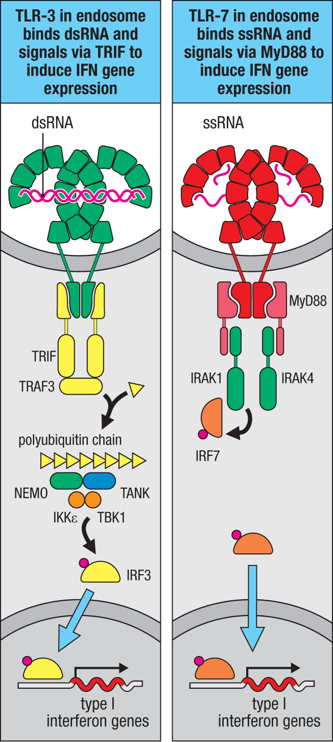
The collective ability of TLRs to activate both IRFs and NFΚB means that they can stimulate either antiviral or antibacterial responses as needed. TLRs are expressed by different types of cells involved in innate immunity and by some stromal and epithelial cells, and the responses generated will differ in some respects depending on what type of cell is being activated.
3-8 The RIG-I–like receptors detect cytoplasmic viral RNAs and activate MAVS to induce type I interferon production and pro-inflammatory cytokines.
The TLRs, being expressed on the cell’s plasma membrane or endocytic vesicles, are primarily sensors of extracellular microbial products. Since the discovery of the mammalian TLRs, additional families of innate sensors have been identified that detect microbial products in the cytosol (Fig. 3.16). Certain TLR family members detect extracellular viral RNAs and DNAs that enter the cell from the endocytic pathway. By contrast, viral RNAs produced within a cell are sensed by a separate family of proteins called the RIG-I–like receptors (RLRs). These proteins serve as viral sensors by binding to viral dsRNAs using an RNA helicase–like domain. The first of these sensors to be discovered was RIG-I (retinoic acid–inducible gene I). RIG-I is widely expressed across tissues and cell types. Mice deficient in RIG-I are highly susceptible to infection by several kinds of single-stranded RNA viruses, including paramyxoviruses, rhabdoviruses, orthomyxoviruses, and flaviviruses, but not picornaviruses.
|
Ligand |
Recognition strategies |
|
RIG-I |
Triphosphate dsRNA |
|
MDA-5 |
dsRNA |
|
cGAS |
DNA |
|
NOD1 |
γ-Glutamyl diaminopimelic acid (iE-DAP) |
|
NOD2 |
Muramyl dipeptide (MDP) |
|
NLRP1 |
Pathogen protease activity |
|
NLRP3 |
Reduced intracellular potassium, ROS, disruption of lysosomes |
|
NAIP1 with NLRC4 (mouse) |
Needle subunit of bacterial T3SS |
|
NAIP2 with NLRC4 (mouse) |
Rod subunit of bacterial T3SS |
|
NAIP5 with NLRC4 (mouse) |
Flagellin |
|
NAIP with NLRC4 (human) |
Flagellin |
|
Pyrin |
Inactivation of Rho GTPases |
|
AIM2 |
DNA |
Fig. 3.16 Ligands and recognition strategies for cytosolic innate immune receptors. Some cytosolic innate immune receptors detect pathogens directly, while others are activated by the activities of pathogens. This figure lists the major receptors for which the ligands or mechanisms of activation are known. ROS, reactive oxygen species; T3SS, type 3 secretion system.
The RLR proteins also contain two amino-terminal caspase recruitment domains (CARDs). CARD was initially recognized in a family of proteases called caspases (for cysteine–aspartic acid proteases), which are important in many intracellular pathways, including those leading to cell death by apoptosis. CARD is structurally related to the death domain in MyD88 and can dimerize with CARDs on other proteins to activate signaling to produce type I interferons when viral RNAs are bound (Fig. 3.17).
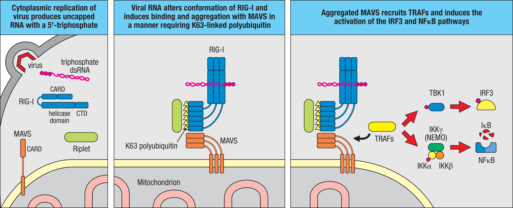
RIG-I discriminates between host and viral RNA by sensing differences at the 5′ end of RNA transcripts. Eukaryotic RNA is transcribed in the nucleus and contains a 5′-triphosphate group on its initial nucleotide that undergoes subsequent enzymatic modification called capping by the addition of a 7-methylguanosine to the 5′-triphosphate. Most RNA viruses do not replicate in the nucleus, where capping normally occurs, so their RNA genomes do not undergo this modification. Biochemical and structural studies have determined that RIG-I senses dsRNAs with the unmodified 5′-triphosphate. Many viruses produce dsRNAs with 5′-triphosphate that are detected by RIG-I, either during viral replication or due to hairpins that form when base pairing occurs within the viral RNA. In contrast, the picornaviruses, which include poliovirus and hepatitis A, replicate by a mechanism that involves the covalent attachment of a viral protein to the 5′ end of the viral RNA, so that the 5′-triphosphate is masked, which explains why RIG-I is not involved in sensing them.
MDA-5 (melanoma differentiation–associated 5) is similar in structure to RIG-I, but it senses longer dsRNAs and does not require the 5′-triphosphate for recognition. In contrast to RIG-I–deficient mice, mice deficient in MDA-5 are susceptible to picornaviruses, indicating that these two sensors of viral RNAs have crucial but distinct roles in host defense. The RLR family member LGP2 (encoded by DHX58) contains a helicase domain but lacks CARDs. LGP2 appears to cooperate with RIG-I and MDA-5 in the recognition of viral RNA, as mice lacking LGP2 have impaired antiviral responses normally mediated by RIG-I or MDA-5. This cooperative viral recognition by LGP2 appears to depend on its helicase domain, because in mice, mutations that disrupt the helicase ATPase activity result in impaired IFN-β production in response to various RNA viruses.
Sensing of viral RNAs by RIG-I and MDA-5 activates signaling that leads to type I interferon production appropriate for defense against viral infection (see Fig. 3.17). Upon binding dsRNA, both RIG-I and MDA-5 assemble into filaments along the dsRNA, which promotes the aggregation of CARDs from multiple RIG-I or MDA-5 molecules. For RIG-I, aggregation of the CARDs is facilitated by recruitment of E3 ligases Riplet (encoded by RNF153) and TRIM25, which generate K63-linked polyubiquitin scaffolds (see Section 3-7) that stabilize interactions between the CARDs. The initiation of MDA-5 signaling does not require Riplet or TRIM25, most likely because MDA-5 more readily assembles into filaments along dsRNA, which enables aggregation of the CARDs. The aggregated CARDs interact with a downstream adaptor protein called MAVS (mitochondrial antiviral signaling protein). MAVS is attached to the outer mitochondrial membrane and contains its own CARD that mediates interaction with RIG-I and MDA-5 assembled on dsRNAs. Activated MAVS also forms polymers. In this state, MAVS propagates signals by recruiting various TRAF family E3 ubiquitin ligases, including TRAF2, TRAF3, TRAF5, and TRAF6. The relative importance of each E3 ligase may differ between cell types, but their further production of K63-linked polyubiquitin leads to activation of TBK1 and IRF3 and production of type I interferons, as described for TLR-3 signaling (see Fig. 3.15), and also to activation of NFΚB.
The recognition of specific features of dsRNA (5′-triphosphate for RIG-I and longer dsRNAs for MDA-5) reduces the chance that cellular RNAs will trigger activation of RLRs. However, humans have been identified with mutations in RIG-I and MDA-5 that result in inappropriate activation of the receptors, which manifests as severe diseases such as SLE, Aicardi–Goutières syndrome, and Singleton–Merten syndrome. How these mutations facilitate RIG-I or MDA-5 activation is an area of active investigation, but in some cases they appear to enable the inappropriate activation of the receptors by endogenous RNAs.
3-9 Cytosolic DNA activates the cGAS–STING pathway to induce production of type I interferons.
Innate sensors that recognize cytosolic RNA use specific modifications, such as the 5′ cap, to discriminate between host and viral origin. Host DNA is generally restricted to the nucleus, but viral, microbial, or protozoan DNA may become located in the cytosol during various stages of infection. The fact that introduction of DNA into the cytosol induces the production of type I interferons was known for a number of years before the sensor or signaling proteins that mediate the response were identified. It is now known that the main sensor of cytosolic DNA is an enzyme called cGAS, for cyclic GAMP synthase. cGAS binds directly to cytosolic DNA, which stimulates its enzymatic activity to produce a second messenger molecule called cyclic guanosine monophosphate–adenosine monophosphate (cyclic GMP–AMP), or cGAMP. cGAMP binds to and activates STING (stimulator of interferon genes), which is a protein encoded by TMEM173 and localized within the endoplasmic reticulum membrane by an amino-terminal tetraspan transmembrane domain (Fig. 3.18). STING exists as an inactive homodimer until cGAMP binding, which induces a conformation change, recruitment of TBK1, activation of IRF3, and production of type I interferons, similarly to signaling by TLR-3 and RLRs (see Figs. 3.15 and 3.17).
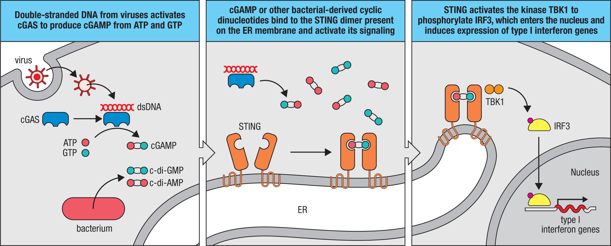
cGAMP is a member of a larger family of cyclic dinucleotides (CDNs) produced by diverse organisms. In fact, the importance of CDNs was first appreciated in bacteria, where CDNs such as cyclic diguanylate monophosphate (c-di-GMP) and cyclic diadenylate monophosphate (c-di-AMP) are synthesized by bacterial enzymes and function as signaling molecules. Before the discovery of mammalian cGAS and cGAMP, these bacterial CDNs were shown to bind and activate STING directly, suggesting that STING may itself serve as a direct sensor of intracellular bacterial infection.
cGAS and STING are expressed in most immune-cell types and many nonimmune cells, and there is good evidence that detection of DNA by the cGAS–STING pathway is important for the immune response to infection, especially viral infection. Mice harboring inactivated cGAS or STING genes show increased susceptibility to a number of DNA viruses. The cGAS–STING pathway can also be activated during infections with certain intracellular bacterial pathogens, either by bacterial DNA released during infection or by bacterial CDNs that bind STING directly, as discussed above. However, the relevance of cGAS–STING signaling for the host response to bacterial infections remains rather unclear, as the type I interferon response induced by cGAS–STING signaling is predominantly antiviral. In some contexts, the production of type I interferons can impair the response to bacteria, so it is possible that activation of cGAS–STING by some pathogens represents a pathogen-evolved mechanism of immune evasion. It is also possible that STING activation can induce other host-defense mechanisms besides production of type I interferons, such as autophagy, which may help to degrade intracellular bacteria.
As with many of the nucleic acid–sensing PRRs discussed earlier, inappropriate activation of the cGAS–STING pathway can lead to inflammatory diseases. An enzyme called Trex1 limits activation of cGAS by degrading DNA present in the cytosol. Mice and humans with defective Trex1 develop autoimmune diseases similar to those described earlier for humans with gain-of-function mutations in MDA-5 and RIG-I (see Section 3-8). DNA accumulates in the cytosol of Trex1-deficient cells, where it activates cGAS. Accordingly, loss of cGAS reverses disease symptoms seen in Trex1-mutant mice. The origin of the cytosolic DNA in Trex1-mutant cells is an area of active investigation, but one source appears to be endogenous retroelements, mobile genetic elements present within mammalian genomes. Other individuals have been identified with mutations in STING that result in constitutive signaling, leading to an autoimmune disorder called STING-associated vasculopathy with onset in infancy (SAVI).
3-10 NLRs comprise a large family of intracellular sensors with diverse functions.
One large group of cytosolic innate sensors is collectively called NOD-like receptors (NLRs), and their family members contain a nucleotide-binding and oligomerization domain (NOD) and leucine-rich repeats (LRRs) in their structure. Each NLR possesses additional domains that result in diverse functions and, in some instances, enable detection of infection. Some NLRs activate NFΚB to initiate the same inflammatory responses as the TLRs, while other NLRs trigger a very different response that leads to cell death and the release of different pro-inflammatory cytokines. We will discuss this second type of response in the next section. Not all NLRs are involved in pathogen detection. For example, NLRC5 and CIITA regulate the transcription of MHC class I and class II genes, respectively.
NOD1 and NOD2 are NLRs that recognize fragments of bacterial cell-wall peptidoglycans, although it is not known whether this occurs through direct binding or via accessory proteins. NOD1 senses γ-glutamyl diaminopimelic acid (iE-DAP), a breakdown product of peptidoglycans of Gram-negative bacteria such as Salmonella and some Gram-positive bacteria such as Listeria, whereas NOD2 recognizes muramyl dipeptide (MDP), which is present in the peptidoglycans of most bacteria. NOD ligands may enter the cytoplasm as a result of intracellular infection, but they may also be transported from materials captured by endocytosis, as mice lacking certain oligopeptide transporters have reduced responses to NOD ligands.
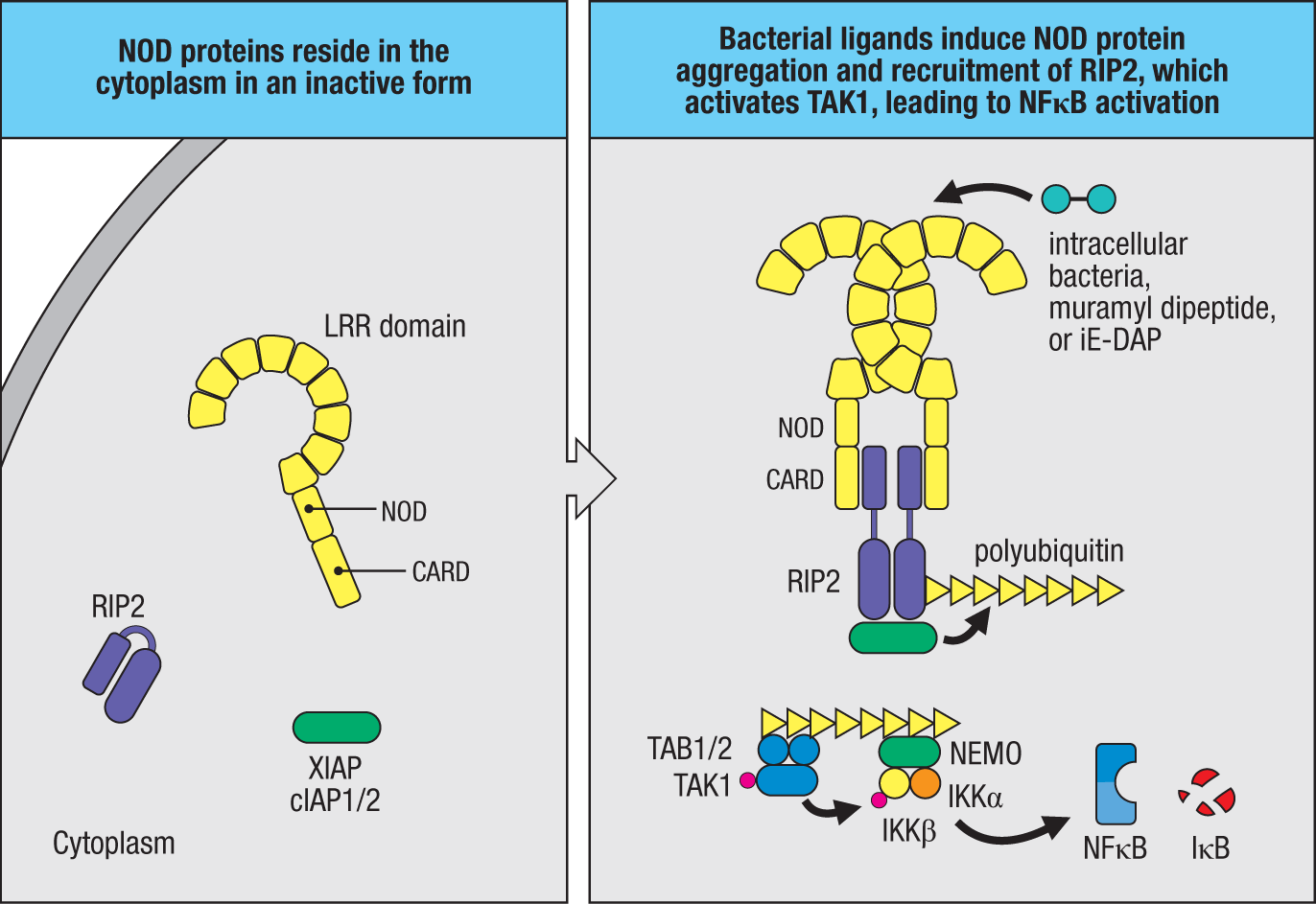
Both NOD1 and NOD2 contain CARDs. When NOD1 or NOD2 is activated, it recruits the CARD-containing serine/threonine kinase RIP2 (also known as RICK and RIPK2) (Fig. 3.19). RIP2 associates with the E3 ligases cIAP1, cIAP2, and XIAP, whose activity generates a polyubiquitin scaffold similar to the scaffold already discussed in the context of TLR signaling. This scaffold recruits TAK1 and IKK and results in activation of NFΚB as shown in Fig. 3.14. NFΚB then induces the expression of genes for inflammatory cytokines and for enzymes involved in the production of nitric oxide (NO), which is toxic to bacteria and intracellular parasites. In keeping with their role as sensors of bacterial components, NOD proteins are expressed in cells that are routinely exposed to bacteria. These include epithelial cells forming the barrier that bacteria must cross to establish an infection in the body and the macrophages and dendritic cells that ingest bacteria that have succeeded in entering the body. Macrophages and dendritic cells express TLRs as well as NOD1 and NOD2 and are activated by both pathways.
NOD2 seems to have a more specialized role, being strongly expressed in the Paneth cells of the gut, where it regulates the expression of potent antimicrobial peptides such as the α- and β-defensins (see Chapter 2). Loss-of-function mutations in NOD2 in humans are associated with the inflammatory bowel condition known as Crohn’s disease (discussed in Chapter 15). Lack of NOD2 function is thought to diminish the production of defensins and other antimicrobial peptides, thereby weakening the natural barrier function of the intestinal epithelium and leading to the inflammation characteristic of this disease. Gain-of-function mutations in human NOD2 are associated with the inflammatory disorders early-onset sarcoidosis and Blau syndrome, which are characterized by spontaneous inflammation in tissues such as the liver or in the joints, eyes, and skin. Activating mutations in the NOD domain seem to promote the signaling cascade in the absence of ligand, leading to an inappropriate inflammatory response in the absence of pathogens.
3-11 Certain NLR proteins react to infection or cellular damage by forming an inflammasome that induces cell death and secretion of inflammatory cytokines.
Some NLR proteins respond to infection or, in some cases, cellular damage in the absence of infection by inducing the formation of a multiprotein complex known as an inflammasome. Inflammasomes serve as platforms for the activation of inflammatory caspases such as caspase 1, a protease that when activated induces the proteolytic cleavage and release of pro-inflammatory cytokines IL-1β and IL-18. Cleavage of these cytokines converts them into their active forms. Activated caspase 1 also cleaves gasdermin D, a cytosolic protein that, once cleaved, inserts into the plasma membrane and assembles into a pore (Fig. 3.20). This pore is required for exit of active IL-1β and IL-18 from the cell, as these cytokines lack signal peptides and are otherwise confined to the cytosol. The gasdermin D pore also induces a lytic form of cell death called pyroptosis (‘fiery death’). While the induction of host-cell death in response to infection may seem counterproductive at first glance, pyroptosis exposes intracellular pathogens that otherwise hide within host cells (for example, macrophages). These pathogens are now visible and thus susceptible to killing by neutrophils and activated macrophages recruited by the inflammatory cytokines produced by inflammasome activation.
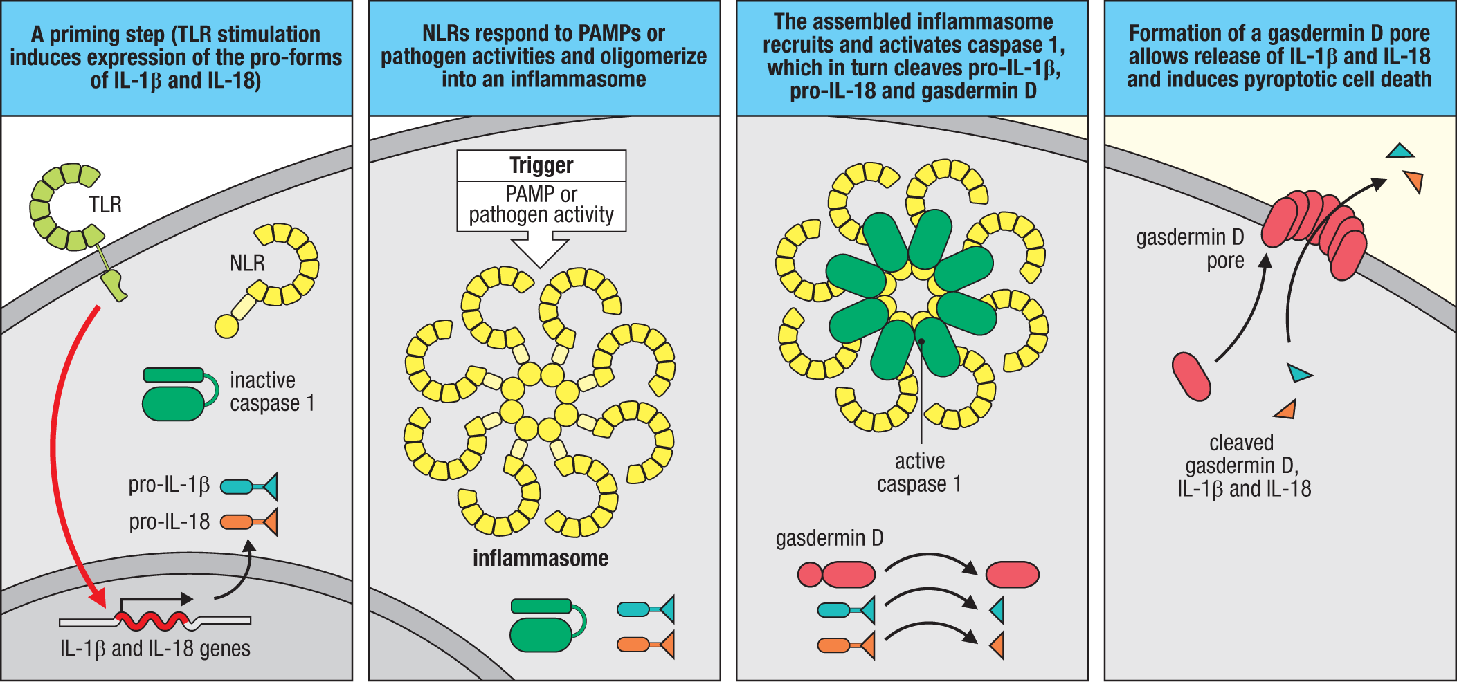
Certain NLR proteins function as PRRs by inducing inflammasome assembly after directly binding to molecules produced by pathogens. For example, NAIPs (NLR family, apoptosis inhibitory proteins) assemble into inflammasomes with NLRC4 after recognizing certain bacterial proteins that access the host cytosol. Mice encode a small family of NAIP proteins, each of which recognizes a distinct ligand. For example, NAIP5 recognizes bacterial flagellin, whereas NAIP1 and NAIP2 recognize distinct protein subunits of the type III secretion system (T3SS), a needle-like macromolecular machine that bacterial pathogens use to inject proteins into host cells. Humans encode a single NAIP protein that has a broad specificity for flagellin and T3SS proteins. NAIP5 recognizes the highly conserved D0 domain of flagellin, which is distinct from the region of flagellin recognized by TLR-5. As a cell-surface receptor, TLR-5 can detect flagellin from potentially any extracellular flagellated bacterium, including harmless commensal species, whereas cytosolic NAIP5 will respond only to bacteria that have the capacity to invade or access the cytosol—typically only pathogenic species.
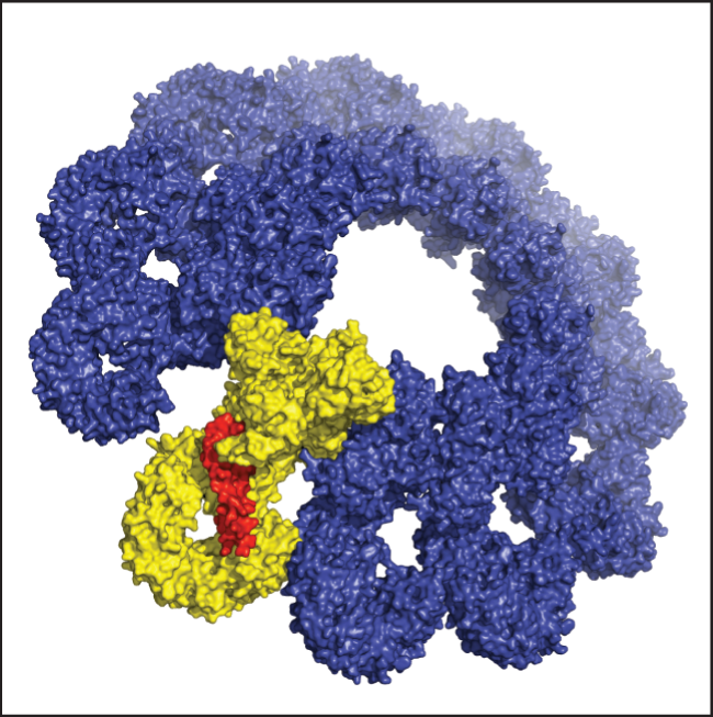
Structural analyses of the NAIP5–NLRC4 inflammasome bound to flagellin have confirmed that NAIP5 binds flagellin directly. This binding induces a conformational change in NAIP5, promoting interaction with NLRC4 and formation of an inflammasome (Fig. 3.21). NLRC4 contains an N-terminal CARD that, upon inflammasome assembly, interacts with the CARD present in caspase 1 to mediate its dimerization and activation. Active caspase 1 then cleaves multiple substrates, including the pro-forms of IL-1β, IL-18, and gasdermin D, as discussed earlier.
The importance of NAIP–NLRC4 inflammasomes for the host response to infection has been established by studies of cells or mice with disrupted NAIP or NLRC4 genes. Mice lacking NLRC4 are more susceptible to infection by numerous intracellular bacterial pathogens, including Salmonella enterica ssp. typhimurium and Legionella pneumophila. NAIP and NLRC4 genes are highly expressed in intestinal epithelial cells. In these cells, inflammasome activation leads to pyroptotic cell death, which is accompanied by expulsion of the infected cell back into the intestinal lumen, which limits the ability of bacteria to establish infection.
While the NAIPs represent classic PRRs, other NLRs do not recognize pathogens directly but rather assemble inflammasomes in response to the consequences of infection, such as pathogen-induced disruptions of cellular structures. The most studied member of this subgroup of NLRs is NLRP3 (also known as NALP3 or cryopyrin). In contrast to NLRC4, NLRP3 has a pyrin domain at its N terminus, hence its NLRP designation (NLRs with CARDs are designated NLRC). Pyrin domains are structurally related to CARDs and interact with other pyrin domains. Several seemingly unrelated triggers can induce NLRP3 activation: reduced intracellular potassium, the generation of reactive oxygen species (ROS), or the disruption of lysosomes (for example, via phagocytosis of crystalline material). It remains unclear how such diverse stimuli are sensed by this NLR, but each represents a potential consequence of infection. The loss of intracellular potassium through efflux can occur during infection with, for example, intracellular bacteria such as Staphylococcus aureus that produce pore-forming toxins. Also, the death of nearby cells during infection can release ATP into the extracellular space; this ATP can activate the purinergic receptor P2X7, which itself is a potassium channel, and allow K+ ion efflux. As discussed earlier in this chapter, ROS are produced by phagocytes upon infection. Finally, a number of pathogens are known to disrupt or permeabilize the phagosomal membrane as a mechanism of escape or to manipulate the host cell.
While it is still not understood how NLRP3 senses such diverse changes to cellular homeostasis, structural and biochemical approaches have delineated the steps of NLRP3 inflammasome assembly. As mentioned earlier, NLRP3 lacks a CARD, so it cannot recruit caspase 1 directly. Instead, upon activation, NLRP3 oligomerizes, and this aggregation induces the pyrin domains of NLRP3 to interact with pyrin domains of another protein named ASC (also called PYCARD). ASC is an adaptor protein composed of an amino-terminal pyrin domain and a carboxyl-terminal CARD. Recruitment of ASC creates a platform for caspase 1 recruitment and activation, via CARD–CARD interactions, as described earlier for the NAIP–NLRC4 inflammasomes. Once caspase 1 is activated, the downstream events are similar for all inflammasomes.
NLRP1 is another NLR that assembles into an inflammasome in response to the activity of pathogens. Specifically, NLRP1 is partially degraded by several different bacterial enzymes; however, instead of inactivating NLRP1, this partial degradation releases a fragment of NLRP1 containing a CARD that assembles into an inflammasome that recruits and activates caspase 1. The best-understood example of this mechanism comes from studies of the lethal factor protease, a toxin secreted by the anthrax bacterium, Bacillus anthracis. Lethal factor enters into cells and promotes infection by cleaving MAP kinase kinases, which disrupts signaling and the ensuing immune response. However, lethal factor also cleaves NLRP1, leading to its partial degradation and inflammasome formation. Because responses downstream of caspase 1 are effective in preventing anthrax, it appears unlikely that B. anthracis evolved to activate NLRP1. Instead, NLRP1 appears to serve as a ‘decoy’ substrate for pathogen proteases and assembles into an inflammasome when such protease activity is detected.
Inflammasome activation can also be initiated by proteins other than NLRs. As discussed earlier for NLRs, some of these proteins detect pathogen-derived ligands while others sense the consequences of infection. As an example of the former, AIM2 (absent in melanoma 2), a member of the PYHIN family of proteins, is activated by direct binding to cytosolic DNA. PYHIN proteins contain an N-terminal pyrin domain but lack the LRR domains present in the NLR family. In place of LRR domains, PYHIN proteins have an HIN (hematopoietic expression, interferon-inducible nature, and nuclear localization) domain. The HIN domain of AIM2 recognizes the DNA genomes of pathogens and triggers caspase 1 activation through pyrin domain interactions with ASC. The protein Pyrin (not to be confused with the pyrin domain that is present in multiple proteins and was described earlier) is another inflammasome sensor protein with an N-terminal pyrin domain but no LRRs. Pyrin senses the inactivation of Rho GTPases, enzymes that regulate the cytoskeleton and cell motility (see Section 3-2). Kinases activated by RhoA phosphorylate specific residues in the Pyrin protein, and this phosphorylation keeps Pyrin in an inactive conformation. Certain pathogens, such as Clostridium difficile, produce toxins that inactivate RhoA, which correspondingly reduces phosphorylation of Pyrin and enables its activation. Activated Pyrin recruits ASC, leading to caspase 1 activation in a manner analogous to the other inflammasomes discussed above.
Many cell types, including most macrophages, must undergo a priming step before inflammasome activation can lead to release of inflammatory cytokines. This priming step induces the transcription and translation of the mRNAs that encode the pro-forms of IL-1β, IL-18, and other cytokines (see Fig. 3.20). In some cases, inflammasome components themselves (for example, NLRP3) are induced upon priming. TLR signaling can satisfy this priming step, which may help ensure that inflammasome activation proceeds primarily during infections. Priming is not always necessary, however, as some cells in tissues (for example, the intestine) express pro-IL-1 β and pro-IL-18 constitutively.
Caspase 11 (in mice) and caspase 4 and caspase 5 (in humans) are pro-inflammatory caspases that also function as cytosolic sensors of LPS. Once activated, these caspases are able to cleave and activate gasdermin D directly, leading to pyroptosis, but they do not appear to directly cleave the pro-forms of IL-1β and IL-18. However, once pyroptosis is initiated, K+ efflux occurs via gasdermin D pores, resulting in NLRP3 activation, caspase 1 activation, and IL-1β and IL-18 processing and release. This response to LPS has been referred to as a ‘non-canonical’ inflammasome pathway. The ability of LPS to induce toxic shock in mice is due primarily to activation of caspase 11. TLR-4 is still involved, however, because caspase 11 requires TLR-4 signaling to be transcriptionally induced.
Inappropriate inflammasome activation has been associated with various diseases. Gout has been known for many years to cause inflammation in the cartilaginous tissues by the deposition of monosodium urate crystals, but how urate crystals caused inflammation was a mystery. Although the precise mechanism is still unclear, urate crystals are known to activate the NLRP3 inflammasome, which induces the inflammatory cytokines associated with the symptoms of gout. Mutations in NLRs can activate inflammasomes inappropriately, and they are the cause of some inherited autoinflammatory diseases, in which inflammation occurs in the absence of infection. Mutations in NLRP3 in humans are associated with hereditary periodic fever syndromes, such as familial cold autoinflammatory syndrome and Muckle–Wells syndrome (discussed in more detail in Chapter 13). Individuals with activating NLRC4 mutations have also been identified. Macrophages from individuals with these conditions show spontaneous production of inflammatory cytokines such as IL-1β and IL-18. We will discuss how pathogens can interfere with formation of the inflammasome in Chapter 13.
3-12 Activation of innate sensors in macrophages and dendritic cells triggers changes in gene expression that have far-reaching effects on the immune response.
Besides activating effector functions and cytokine production, another outcome of the activation of innate sensing pathways is the induction of co-stimulatory molecules on tissue dendritic cells and macrophages (see Section 1-15). We will describe these in more detail later in the book but mention them now because they provide an important link between innate and adaptive immune responses. Two important co-stimulatory molecules are the cell-surface proteins CD80 and CD86, which are induced on macrophages and tissue dendritic cells by innate sensors such as TLRs in response to pathogen recognition (Fig. 3.22). CD80 and CD86 are recognized by specific co-stimulatory receptors expressed by cells of the adaptive immune response, particularly CD4 T cells, and their activation is an important step in activating adaptive immune responses.
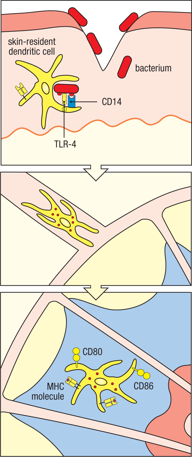
Substances such as LPS that induce co-stimulatory activity have been used for years in mixtures that are co-injected with protein antigens to enhance their immunogenicity. These substances are known as adjuvants (see Appendix I, Section A-1), and it was found empirically that the best adjuvants contain microbial components that induce macrophages and tissue dendritic cells to express co-stimulatory molecules and cytokines. We now know that the effect of these microbial components is due to activation of PRRs. As we shall see in Chapters 9 and 11, the cytokines produced in response to infections influence the functional character of the adaptive immune response that develops. In this way, the ability of the innate immune system to discriminate among different types of pathogens is used by the organism to ensure an appropriate module of the adaptive immune response is induced.
3-13 Toll signaling in Drosophila is downstream of a distinct set of pathogen-recognition molecules.
Before leaving innate sensing, we shall look briefly at how Toll, TLRs, and NODs are used in invertebrate innate immunity. Although Toll is central to defense against both bacterial and fungal pathogens in Drosophila, Toll itself is not a pattern-recognition receptor but is downstream of other proteins that detect pathogens (Fig. 3.23). In Drosophila, there are 13 genes encoding peptidoglycan-recognition proteins (PGRPs) that bind the peptidoglycan components of bacterial cell walls. Another family, the Gram-negative binding proteins (GNBPs), recognizes LPS and β-(1,3)–linked glucans. GNBPs recognize Gram-negative bacteria and fungi rather than Gram-positive bacteria. The family members GNBP1 and PGRP-SA cooperate in the recognition of peptidoglycan from Gram-positive bacteria. They interact with a serine protease called Grass, which initiates a proteolytic cascade that terminates in the cleavage of the protein Spätzle. One of the cleaved fragments forms a homodimer that binds to Toll and induces its dimerization, which in turn stimulates the antimicrobial response. A fungus-specific recognition protein, GNBP3, also activates the proteolytic cascade, causing cleavage of Spätzle and activation of Toll.
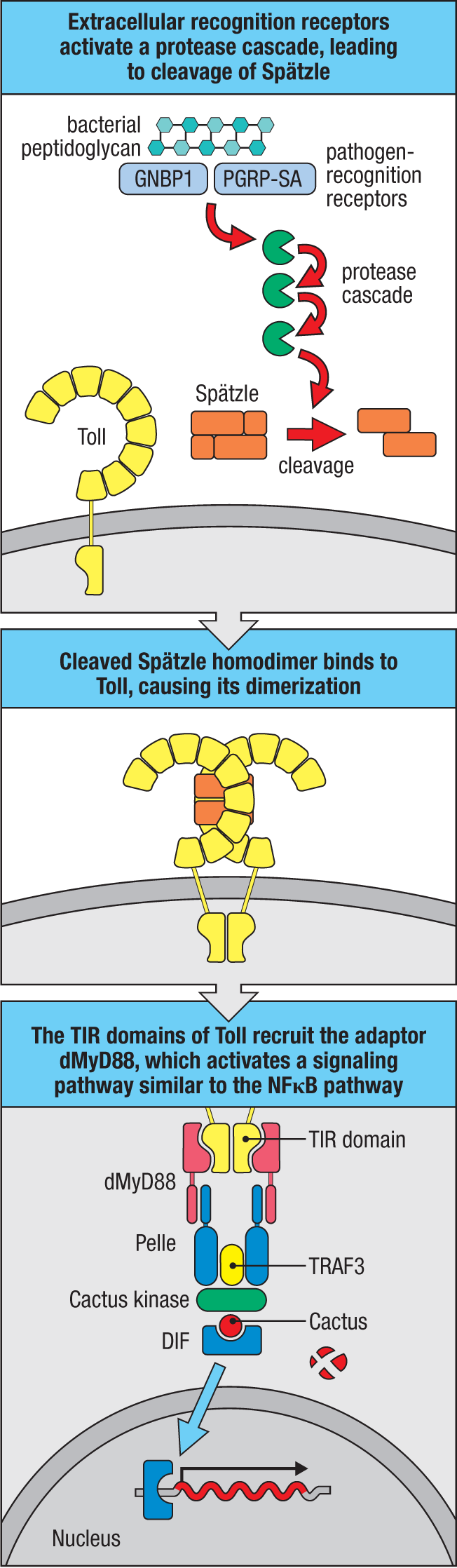
In Drosophila, fat-body cells and hemocytes are phagocytic cells that act as part of the fly’s immune system. When the Spätzle dimer binds to Toll, hemocytes synthesize and secrete antimicrobial peptides. The Toll signaling pathway in Drosophila activates a transcription factor called DIF, which is related to mammalian NFΚB. DIF enters the nucleus and induces the transcription of genes for antimicrobial peptides such as drosomycin. Another Drosophila factor in the NFΚB family, Relish, induces the production of antimicrobial peptides in response to the Imd (immunodeficiency) signaling pathway, which is triggered in Drosophila by particular PGRPs that recognize Gram-negative bacteria. Relish induces expression of the antimicrobial peptides diptericin, attacin, and cecropin, which are distinct from the peptides induced by Toll signaling. Thus, the Toll and Imd pathways activate effector mechanisms to eliminate infection by different kinds of pathogens. Four mammalian PGRP homologs have been identified but act differently than in Drosophila. One, PGLYRP-2, is secreted and functions as an amidase to hydrolyze bacterial peptidoglycans. The others are present in neutrophil granules and exert a bacteriostatic action through interactions with bacterial cell-wall peptidoglycan.
3-14 TLR and NOD genes have undergone extensive diversification in both invertebrates and some primitive chordates.
There are only about a dozen mammalian TLR genes, but some organisms have diversified their repertoire of innate recognition receptors, especially those containing LRR domains, to a much greater degree. The sea urchin Strongylocentrotus purpuratus has an unprecedented 222 different TLR genes, more than 200 NOD-like receptor genes, and more than 200 scavenger receptor genes in its genome. The sea urchin also has an increased number of proteins that are likely to be involved in signaling from these receptors, there being, for example, four genes that are similar to the single mammalian MyD88 gene. However, there is no apparent increase in the number of downstream targets, such as the family of NFΚB transcription factors, suggesting that the ultimate outcome of TLR signaling in the sea urchin may be very similar to that in other organisms.
Sea urchin TLR genes fall into two broad categories. One is a small set of 11 divergent genes. The other is a large family of 211 genes, which show a high degree of sequence variation within particular LRR regions; this, together with the large number of pseudogenes in this family, indicates rapid evolutionary turnover, suggesting rapidly changing receptor specificities, in contrast with the few stable mammalian TLRs. Although the pathogen specificity of sea urchin TLRs is unknown, the hypervariability in the LRR domains could be used to generate a highly diversified pathogen-recognition system based on Toll-like receptors. A similar expansion of innate receptors has occurred in some chordates, the phylum to which vertebrates belong. Amphioxus (the lancelet) is a nonvertebrate chordate lacking an adaptive immune system. The amphioxus genome contains 71 TLRs, more than 100 NOD-like receptors, and more than 200 scavenger receptors. As we will see in Chapter 5, a primitive vertebrate lineage—the jawless fishes, which lack immunoglobulin-based and T cell–based adaptive immunity—uses somatic gene rearrangement of LRR-containing proteins to provide a version of adaptive immunity (see Section 5-18).
Summary.
Innate immune cells express several receptor systems that recognize microbes and induce rapid defenses as well as delayed cellular responses. Several scavenger and lectin-like receptors on neutrophils, macrophages, and dendritic cells help rapidly eliminate microbes through phagocytosis. G protein–coupled receptors for C5a (which can be produced by activation of the complement system’s innate pathogen-recognition ability) and for the bacterial peptide fMLF synergize with phagocytic receptors in activating the NADPH oxidase in phagosomes to generate antimicrobial reactive oxygen intermediates. Toll-like receptors (TLRs) on the cell surface and in the membranes of endosomes detect microbes outside the cell and activate several host-defense signaling pathways. The NFΚB and IRF pathways downstream of these receptors induce pro-inflammatory cytokines, including TNF-α, IL-1β, and IL-6, and antiviral cytokines including type I interferons. Other receptor families detect microbial infection in the cytosol. RIG-I and MDA-5 detect viral infection by sensing the presence of viral RNAs and activate the MAVS pathway, while cGAS senses cytosolic DNA and activates the STING pathway; both of these pathways induce type I interferons. NLR proteins detect pathogen products within the cytosol or sense changes to the cell due to pathogen infection. Some NLRs activate NFΚB and induce the production of pro-inflammatory cytokines, while others form inflammasomes, leading to secretion of pro-inflammatory cytokines and pyroptotic cell death. The signaling pathways activated by all of these primary sensors of pathogens induce a variety of genes, including those for cytokines, chemokines, and co-stimulatory molecules that have essential roles in immediate defense and in directing the course of the adaptive immune response later in infection.
Glossary
- pathogen-associated molecular patterns (PAMPs)
- Molecules specifically associated with groups of pathogens that are recognized by cells of the innate immune system.
- pattern-recognition receptors (PRRs)
- Receptors of the innate immune system that recognize common molecular patterns on pathogen surfaces.
- damage-associated molecular patterns (DAMPs)
- Molecules specifically associated with groups of pathogens that are recognized by cells of the innate immune system.
- macrophages
- Large mononuclear phagocytic cells present in most tissues that have many functions; for example, scavenger cells, pathogen-recognition cells, and production of pro-inflammatory cytokines. Macrophages arise both embryonically and from bone marrow precursors throughout life.
- monocyte
- Type of white blood cell with a bean-shaped nucleus; it is a precursor of tissue macrophages.
- aorta–gonad–mesonephros (AGM)
- An embryonic region in which hematopoietic cells arise during development.
- microglial cell
- An embryonically derived form of tissue macrophage in the central nervous system that is dependent on IL-34 for local self-renewal throughout life.
- Kupffer cells
- Phagocytes lining the hepatic sinusoids; they remove debris and dying cells from the blood but are not known to elicit immune responses.
- classical monocyte
- The major form of monocyte in circulation capable of recruitment to sites of inflammation and differentiation into macrophages.
- patrolling monocyte
- A form of circulating monocyte that adheres to and surveys the vascular endothelium, distinguished from classical monocytes by its low expression of Ly6C.
- neutrophil
- The most numerous type of white blood cell in human peripheral blood. Neutrophils are phagocytic cells with a multilobed nucleus and granules that stain with neutral stains. They enter infected tissues and engulf and kill extracellular pathogens.
- eosinophil
- A type of white blood cell containing granules that stain with eosin. It is thought to be important chiefly in defense against parasitic infections but is also medically important as an effector cell in allergic reactions.
- basophil
- Type of white blood cell containing granules that stain with basic dyes. It is thought to have a function similar to that of mast cells.
- dendritic cells
-
Bone marrow–derived cells found in most tissues, including lymphoid tissues. There are two main functional subsets. Conventional dendritic cells take up antigen in peripheral tissues, are activated by contact with pathogens, and travel to the peripheral lymphoid organs, where they are the most potent stimulators of T-cell responses. Plasmacytoid dendritic cells can also take up and present antigen, but their main function in an infection is to produce large amounts of the antiviral interferons as a result of pathogen recognition through receptors such as TLRs. Both these types of dendritic cells are distinct from the follicular dendritic cell that presents antigen to B cells in lymphoid follicles.
- conventional (or classical) dendritic cells (cDCs)
- The lineage of dendritic cells that mainly participates in antigen presentation to, and activation of, naive T cells. Cf. plasmacytoid dendritic cells.
- plasmacytoid dendritic cells (pDCs)
- A distinct lineage of dendritic cells that secrete large amounts of interferon on activation by pathogens and their products via receptors such as Toll-like receptors. Cf. conventional dendritic cells.
- interferons (IFNs)
- Several related families of cytokines originally named for their interference with viral replication. IFN-α and IFN-β are antiviral in their effects; IFN-γ has other roles in the immune system.
- phagocytosis
- The internalization of particulate matter by cells by a process of engulfment, in which the cell membrane surrounds the material, eventually forming an intracellular vesicle (phagosome) containing the ingested material.
- phagosome
- Intracellular vesicle formed when particulate material is ingested by a phagocyte.
- phagolysosome
- Intracellular vesicle formed by the fusion of a phagosome (containing ingested material) and a lysosome, and in which the ingested material is broken down.
- superoxide radical
- A highly reactive form of oxygen having an unpaired electron (making it a radical) and being negatively charged, making it an ion, formed in the phagolysosome by the action of the NADPH oxidase.
- nitric oxide radical
- The uncharged compound NO, having an unpaired electron conferring its high reactivity, formed in activated phagocytes by the action of inducible nitric oxide synthetase.
- primary granules
- Granules in neutrophils that correspond to lysosomes and contain antimicrobial peptides such as defensins and other antimicrobial agents.
- secondary granules
- Type of granule in neutrophils that stores certain antimicrobial peptides.
- receptor-mediated endocytosis
- The internalization into endosomes of molecules bound to cell-surface receptors.
- macropinocytosis
- A process in which large amounts of extracellular fluid are taken up into an intracellular vesicle. This is one way in which dendritic cells can take up a wide variety of antigens from their surroundings.
- Dectin-1
- A phagocytic receptor on neutrophils and macrophages that recognizes β-(1,3)–linked glucans, which are common components of fungal cell walls.
- mannose receptor
- A receptor on macrophages that is specific for mannose-containing carbohydrates that occur on the surfaces of pathogens but not on host cells.
- scavenger receptors
- Receptors on macrophages and other cells that bind to numerous ligands, such as bacterial cell-wall components, and remove them from the blood. The Kupffer cells in the liver are particularly rich in scavenger receptors. Includes SR-A I, SR-A II, and MARCO.
- SR-A I, SR-A II
- Receptors on macrophages and other cells that bind to numerous ligands, such as bacterial cell-wall components, and remove them from the blood. The Kupffer cells in the liver are particularly rich in scavenger receptors. Includes SR-A I, SR-A II, and MARCO.
- SR-A I, SR-A II
- Receptors on macrophages and other cells that bind to numerous ligands, such as bacterial cell-wall components, and remove them from the blood. The Kupffer cells in the liver are particularly rich in scavenger receptors. Includes SR-A I, SR-A II, and MARCO.
- MARCO (macrophage receptor with a collagenous structure)
- Receptors on macrophages and other cells that bind to numerous ligands, such as bacterial cell-wall components, and remove them from the blood. The Kupffer cells in the liver are particularly rich in scavenger receptors. Includes SR-A I, SR-A II, and MARCO.
- G protein–coupled receptors (GPCRs)
- A large class of seven-span transmembrane cell-surface receptors that associate with intracellular heterotrimeric G proteins after ligand binding and signal by activation of the G protein. Important examples are the chemokine receptors.
- fMet-Leu-Phe (fMLF) receptor
- A pattern-recognition receptor for the peptide fMet-Leu-Phe, which is specific to bacteria, on neutrophils and macrophages. fMet-Leu-Phe acts as a chemoattractant.
- reactive oxygen species (ROS)
- Superoxide anion (O2–) and hydrogen peroxide (H2O2), produced by phagocytic cells such as neutrophils and macrophages after ingestion of microbes, and which help kill the ingested microbes.
- G proteins
- Intracellular GTPases that act as molecular switches in signaling pathways. They bind GTP to induce their active conformation, which is lost when GTP is hydrolyzed to GDP. There are two kinds of G proteins: the heterotrimeric (α, β, γ subunits) receptor-associated G proteins, and the small G proteins, such as Ras and Raf, which act downstream of many transmembrane signaling events.
- heterotrimeric G proteins
- Intracellular GTPases that act as molecular switches in signaling pathways. They bind GTP to induce their active conformation, which is lost when GTP is hydrolyzed to GDP. There are two kinds of G proteins: the heterotrimeric (α, β, γ subunits) receptor-associated G proteins, and the small G proteins, such as Ras and Raf, which act downstream of many transmembrane signaling events.
- Rho family small GTPase proteins
- Several distinct small GTPase family members that regulate the actin cytoskeleton in response to signaling through various receptors. Examples: Rac, Rho, and Cdc42.
- Rac
- Several distinct small GTPase family members that regulate the actin cytoskeleton in response to signaling through various receptors. Examples: Rac, Rho, and Cdc42.
- Rho
- Several distinct small GTPase family members that regulate the actin cytoskeleton in response to signaling through various receptors. Examples: Rac, Rho, and Cdc42.
- guanine nucleotide exchange factors (GEFs)
- Proteins that can remove the bound GDP from G proteins, thus allowing GTP to bind and activate the G protein.
- PREX1
- A guanine nucleotide exchange factor (GEF) activated downstream of small G proteins in response to activation of GPCRs such as the fMLP or C5a receptor.
- NADPH oxidase
- Multicomponent enzyme complex that is assembled and activated in the phagolysosome membrane in stimulated phagocytes. It generates superoxide in an oxygen-requiring reaction called the respiratory burst.
- phagocyte oxidase
- Multicomponent enzyme complex that is assembled and activated in the phagolysosome membrane in stimulated phagocytes. It generates superoxide in an oxygen-requiring reaction called the respiratory burst.
- immunoreceptor tyrosine-based activation motif (ITAM)
- Sequence motifs in the signaling chains of receptors, such as antigen receptors on lymphocytes, that are the site of tyrosine phosphorylation after receptor activation, leading to recruitment of other signaling proteins.
- respiratory burst
- An oxygen-requiring metabolic change in neutrophils and macrophages that have taken up opsonized particles, such as complement- or antibody-coated bacteria, by phagocytosis. It leads to the production of toxic metabolites that are involved in killing the engulfed microorganisms.
- superoxide dismutase (SOD)
- An enzyme that converts the superoxide ion produced in the phagolysosome into hydrogen peroxide, a substrate for further reactive antimicrobial metabolites.
- pus
- Thick yellowish-white liquid typically found at sites of infection with some types of extracellular bacteria, which is composed of the remains of dead neutrophils and other cells.
- pyogenic bacteria
- Capsulated bacteria that result in pus formation at the site of infection. Also called pyogenic (pus-forming) bacteria.
- chronic granulomatous disease (CGD)
- An immunodeficiency in which multiple granulomas form as a result of defective elimination of bacteria by phagocytic cells. It is caused by defects in the NADPH oxidase system of enzymes that generate the superoxide radical involved in bacterial killing.
- neutrophil extracellular traps (NETs)
- A meshwork of nuclear chromatin that is released into the extracellular space by neutrophils undergoing apoptosis at sites of infection, serving as a scaffold that traps extracellular bacteria to enhance their phagocytosis by other phagocytes.
- cytokines
- Proteins made by a cell that affect the behavior of other cells, particularly immune cells. Cytokines made by lymphocytes are often called interleukins (abbreviated ILs). Cytokines and their receptors are listed in eAppendix III. Cf. chemokines.
- chemokines
- Small chemoattractant proteins that stimulate the migration and activation of cells, especially phagocytic cells and lymphocytes. Chemokines have a central role in inflammatory responses. Properties of individual chemokines are listed in eAppendix IV.
- pro-inflammatory
- Tending to induce inflammation.
- cell-adhesion molecules
- Cell-surface proteins of several different types that mediate the binding of one cell to other cells or to extracellular matrix proteins. Integrins, selectins, and members of the immunoglobulin gene superfamily (such as ICAM-1) are among the cell-adhesion molecules important in the operation of the immune system.
- extravasation
- The movement of cells or fluid from within blood vessels into the surrounding tissues.
- inflammatory monocytes
- An activated form of monocytes producing a variety of pro-inflammatory cytokines.
- edema
- Swelling caused by the entry of fluid and cells from the blood into the tissues; it is one of the cardinal features of inflammation.
- endothelial activation
- The changes that occur in the endothelial walls of small blood vessels as a result of inflammation, such as increased permeability and the increased production of cell-adhesion molecules and cytokines.
- prostaglandins
- Lipid products of the metabolism of arachidonic acid that have a variety of effects on tissues, including activities as inflammatory mediators.
- leukotrienes
- Lipid mediators of inflammation that are derived from arachidonic acid. They are produced by macrophages and other cells.
- platelet-activating factor (PAF)
- A lipid mediator that activates the blood clotting cascade and several other components of the innate immune system.
- tumor necrosis factor-α (TNF-α, or TNF)
- Cytokine family, the prototype of which is tumor necrosis factor-α (TNF-α, or TNF). It contains both secreted (e.g., TNF-α and lymphotoxin) and membrane-bound (e.g., CD40 ligand) members.
- mast cell
- A large granule-rich cell found in connective tissues throughout the body, most abundantly in the submucosal tissues and the dermis. The granules store bioactive molecules including the vasoactive amine histamine, which are released on mast-cell activation. Mast cells are thought to be involved in defenses against parasites, and they have a crucial role in allergic reactions.
- kinin system
- An enzymatic cascade of plasma proteins that is triggered by tissue damage to produce several inflammatory mediators, including the vasoactive peptide bradykinin.
- bradykinin
- A vasoactive peptide that is produced as a result of tissue damage and acts as an inflammatory mediator.
- coagulation system
- A collection of proteases and other proteins in the blood that trigger blood clotting when blood vessels are damaged.
- sterile injury
- Damage to tissues due to trauma, ischemia, metabolic stress, or autoimmunity, bearing many immune features similar to infection.
- Toll
- Receptor protein in Drosophila that activates the transcription factor NFκB, leading to the production of antimicrobial peptides.
- Toll-like receptors (TLRs)
- Innate receptors on macrophages, dendritic cells, and some other cells that recognize pathogens and their products, such as bacterial lipopolysaccharide. Recognition stimulates the receptor-bearing cells to produce cytokines that help initiate immune responses.
- lipoteichoic acids
- Components of bacterial cell walls that are recognized by Toll-like receptors.
- lipopolysaccharide (LPS)
- The surface lipopolysaccharide of Gram-negative bacteria, which stimulates TLR-4 on macrophages and dendritic cells.
- leucine-rich repeat (LRR)
- Protein motif that is repeated in series to form, for example, the extracellular portions of Toll-like receptors.
- TIR (for Toll–IL-1 receptor) domain
- Domain in the cytoplasmic tails of the TLRs and in the IL-1 receptor, which interacts with similar domains in intracellular signaling proteins.
- IL-1α, IL-1β
- Interleukin-1α and interleukin-1β are cytokines produced by activated macrophages that have many effects in the immune response, including the activation of vascular endothelium, activation of lymphocytes, and the induction of fever. IL-1β must be cleaved by caspase 1 before having biological activity, while IL-1α does not require cleavage.
- IL-1α, IL-1β
- Interleukin-1α and interleukin-1β are cytokines produced by activated macrophages that have many effects in the immune response, including the activation of vascular endothelium, activation of lymphocytes, and the induction of fever. IL-1β must be cleaved by caspase 1 before having biological activity, while IL-1α does not require cleavage.
- TLR-1
- Cell-surface Toll-like receptor that acts in a heterodimer with TLR-2 to recognize lipoteichoic acid and bacterial lipoproteins.
- TLR-2
- Cell-surface Toll-like receptor that acts in a heterodimer with either TLR-1 or TLR-6 to recognize lipoteichoic acid and bacterial lipoproteins.
- TLR-6
- Cell-surface Toll-like receptor that acts in a heterodimer with TLR-2 to recognize lipoteichoic acid and bacterial lipoproteins.
- diacyl and triacyl lipoproteins
- Ligands for the Toll-like receptors TLR-1–TLR-2 and TLR-2–TLR-6.
- diacyl and triacyl lipoproteins
- Ligands for the Toll-like receptors TLR-1–TLR-2 and TLR-2–TLR-6.
- TLR-5
- Cell-surface Toll-like receptor that recognizes the flagellin protein of bacterial flagella.
- TLR-11, TLR-12
- Mouse Toll-like receptor that recognizes profilin and profilin-like proteins.
- TLR-11, TLR-12
- Mouse Toll-like receptor that recognizes profilin and profilin-like proteins.
- profilin
- An actin-binding protein that sequesters monomeric actin. Protozoan profilins contain sequences recognized by TLR-11 and TLR-12.
- TLR-7
- Endosomal Toll-like receptor that recognizes single-stranded viral RNA.
- TLR-8
- Endosomal Toll-like receptor that recognizes single-stranded viral RNA.
- single-stranded RNA (ssRNA)
- Usually confined to the nucleus and cytoplasm, this normal molecular form serves as a ligand for TLR-7 and TLR-8 when it is present in endosomes, as during parts of a viral life cycle.
- TLR-13
- A member of the Toll-like receptor (TLR) family expressed in mouse and other rodents, but not in human, that has a specificity for certain bacterial ribosomal RNAs.
- TLR-9
- Endosomal Toll-like receptor that recognizes DNA containing unmethylated CpG.
- unmethylated CpG dinucleotides
- While mammalian genomes have heavily methylated the cytosine within CpG sequences, unmethylated CpG is more typically characteristic of bacterial genomes and is recognized by TLR-9 when encountered in the endosomal compartment.
- TLR-3
- Endosomal Toll-like receptor that recognizes double-strand viral RNA.
- double-stranded RNA (dsRNA)
- A chemical structure that is a replicative intermediate of many viruses and is recognized by TLR-3.
- UNC93B1
- A multipass transmembrane protein that is necessary for the normal transport of TLR-3, TLR-7, TLR-8, TLR-9, TLR-11, TLR-12, and TLR-13 from the endoplasmic reticulum, where they are assembled, to the endosome, where they function.
- TLR-4
- Cell-surface Toll-like receptor that, in conjunction with the accessory proteins MD-2 and CD14, recognizes bacterial lipopolysaccharide.
- MD-2
- Accessory protein for TLR-4 activity.
- LPS-binding protein
- Protein in blood and extracellular fluid that binds bacterial lipopolysaccharide (LPS) shed from bacteria.
- shock
- The potentially fatal circulatory collapse caused by the systemic actions of cytokines such as TNF-α.
- septic shock
- Systemic shock reaction that can follow infection of the bloodstream with endotoxin-producing Gram-negative bacteria. It is caused by the systemic release of TNF-α and other cytokines. Also called endotoxic shock.
- sepsis
- Bacterial infection of the bloodstream. This is a very serious and frequently fatal condition.
- IFN-α, IFN-β
- Antiviral cytokines produced by a wide variety of cells in response to infection by a virus, and which also help healthy cells resist viral infection. They act through the same receptor, which signals through a Janus-family tyrosine kinase. Also known as the type I interferons.
- IFN-α, IFN-β
- Antiviral cytokines produced by a wide variety of cells in response to infection by a virus, and which also help healthy cells resist viral infection. They act through the same receptor, which signals through a Janus-family tyrosine kinase. Also known as the type I interferons.
- type I interferons
- The antiviral interferons IFN-α and IFN-β.
- MyD88
- An adaptor protein that functions in signaling by all TLR proteins except TLR-3.
- TRIF
- An adaptor protein that alone is involved in signaling by TLR-3 and which when paired with TRAM functions in signaling by TLR-4.
- MAL
- An adaptor protein that associates with MyD88 in signaling by TLR-2–TLR-1, TLR-2–TLR-6, and TLR-4.
- TRAM
- An adaptor protein that pairs with TRIF in signaling by TLR-4.
- BCAP
- B-cell adaptor for PI 3-kinase (BCAP) is an adaptor that was recently found to link signaling by TLRs to activation of the PI 3-kinase and AKT pathway.
- NFκB
- A heterodimeric transcription factor activated by the stimulation of Toll-like receptors and also by antigen receptor signaling composed of p50 and p65 subunits.
- interferon regulatory factor (IRF)
- A family of nine transcription factors that regulate a variety of immune responses. For example, IRF3 and IRF7 are activated as a result of signaling from some TLRs. Several IRFs promote expression of the genes for type I interferons.
- activator protein 1 (AP-1)
- A transcription factor formed as one of the outcomes of intracellular signaling by antigen receptors of lymphocytes.
- mitogen-activated protein kinases (MAPKs)
- A series of protein kinases that become phosphorylated and activated on cellular stimulation by a variety of ligands and lead to new gene expression by phosphorylating key transcription factors. The MAPKs are part of many signaling pathways, especially those leading to cell proliferation, and have different names in different organisms.
- death domain
- A protein domain involved in protein–protein interactions with death domains in other proteins. The death domain was first identified in proteins involved in apoptosis.
- IRAK1, IRAK4
- Protein kinases that are part of the intracellular signaling pathways leading from TLRs.
- IRAK1, IRAK4
- Protein kinases that are part of the intracellular signaling pathways leading from TLRs.
- signaling scaffold
- A configuration of proteins and modifications, such as phosphorylation or ubiquitination, that facilitates signaling by binding various enzymes and their substrates.
- TRAF6 (tumor necrosis factor receptor–associated factor 6)
- An E3 ligase that produces a K63 polyubiquitin signaling scaffold in TLR-4 signaling to activate the NFκB pathway.
- ubiquitin ligase
- Enzyme that attaches ubiquitin covalently to exposed lysine residues on the surfaces of other proteins.
- UBC13
- A complex of the E2 ubiquitin ligase UBC13 and cofactor Uve1A that interacts with TRAF6 in forming the K63 polyubiquitin signaling scaffold in TLR signaling downstream of MyD88.
- Uve1A
- A complex of the E2 ubiquitin ligase UBC13 and cofactor Uve1A that interacts with TRAF6 in forming the K63 polyubiquitin signaling scaffold in TLR signaling downstream of MyD88.
- TRIKA1
- A complex of the E2 ubiquitin ligase UBC13 and cofactor Uve1A that interacts with TRAF6 in forming the K63 polyubiquitin signaling scaffold in TLR signaling downstream of MyD88.
- K63 linkages
- In polyubiquitin chains, the covalent ligation of the lysine 63 amino group of one ubiquitin protein with the carboxy terminus of a second ubiquitin. This type of linkage is most associated with activation of signaling by formation of a scaffold recognized by signaling adaptors such as TAB1/2.
- polyubiquitin chains
- Polymers of ubiquitin covalently linked from lysine residues within one ubiquitin monomer to the carboxy terminus of a second ubiquitin.
- TAB1, TAB2
- An adaptor complex that binds K63-linked polyubiquitin chains. TAB1 and TAB2 complex with TAK1, targeting TAK1 to signaling scaffolds where it phosphorylates substrates such as IKKα.
- TAB1, TAB2
- An adaptor complex that binds K63-linked polyubiquitin chains. TAB1 and TAB2 complex with TAK1, targeting TAK1 to signaling scaffolds where it phosphorylates substrates such as IKKα.
- TAK1
- A serine/threonine kinase that is activated by phosphorylation by the IRAK complex and which activates downstream targets such as IKKβ and MAPKs.
- IκB kinase (IKK)
- The IκB kinase, IKK, is a multi-subunit protein complex composed of IKKα, IKKβ, and IKKγ (the latter also known as NEMO).
- NEMO
- The IκB kinase, IKK, is a multi-subunit protein complex composed of IKKα, IKKβ, and IKKγ (the latter also known as NEMO).
- IκB
- A cytoplasmic protein that constitutively associates with the NFκB homodimer, which is composed of p50 and p65 subunits. When IκB is phosphorylated by activated IKK (IκB kinase), IκB becomes degraded and allows the NFκB dimer to be released as an active transcription factor.
- p50
- See NFκB.
- p65
- See NFκB.
- IL-6
- Interleukin-6, a cytokine produced by activated macrophages and which has many effects, including lymphocyte activation, the stimulation of antibody production, and the induction of fever.
- IRAK4 deficiency
- An immunodeficiency characterized by recurrent bacterial infections, caused by inactivating mutations in the IRAK4 gene that result in a block in TLR signaling.
- X-linked hypohidrotic ectodermal dysplasia and immunodeficiency
- A syndrome with some features resembling hyper IgM syndrome. It is caused by mutations in the protein NEMO, a component of the NFκB signaling pathway. Also called NEMO deficiency.
- NEMO deficiency
- A syndrome with some features resembling hyper IgM syndrome. It is caused by mutations in the protein NEMO, a component of the NFκB signaling pathway. Also called NEMO deficiency.
- TRAF3
- An E3 ligase that produces a K63 polyubiquitin signaling scaffold in TLR-3 signaling to induce type I interferon gene expression.
- IKKϵ
- A kinase that interacts with TBK1 (TANK-binding kinase 1) in the phosphorylation of IRF3 downstream of TLR-3 signaling.
- TBK1 (TANK-binding kinase 1)
- A serine/threonine kinase activated during signaling by TLR-3 and MAVS and serving to phosphorylate and activate IRF3 for induction of type I interferon gene expression.
- RIG-I–like receptors (RLRs)
- A small family of intracellular viral sensors that use a carboxy-terminal RNA helicase–like domain in detection of various forms of viral RNA. These signal through MAVS to activate antiviral immunity. Examples include RIG-I, MDA-5, and LGP2.
- RIG-I (retinoic acid–inducible gene I)
- A small family of intracellular viral sensors that use a carboxy-terminal RNA helicase–like domain in detection of various forms of viral RNA. These signal through MAVS to activate antiviral immunity. Examples include RIG-I, MDA-5, and LGP2.
- caspase recruitment domain (CARD)
- A protein domain present in some receptor tails that can dimerize with other CARD-containing proteins, including caspases, thus recruiting them into signaling pathways.
- caspases
- A family of cysteine proteases that cleave proteins at aspartic acid residues. They have important roles in apoptosis and in the processing of cytokine pro-polypeptides.
- capping
- A process occurring in the nucleus in which the modified purine 7-methylguanosine is added to the 5′-phosphate of the first nucleotide of the RNA transcript.
- MDA-5 (melanoma differentiation–associated 5, also helicard)
- This protein contains an RNA helicase–like domain similar to RIG-I and senses double-stranded RNA for detection of intracellular viral infections.
- LGP2
- A member of the RLR family, it cooperates with RIG-I and MDA-5 in the recognition of viral RNA.
- Riplet
- An E3 ubiquitin ligase involved in signaling by RIG-I and MDA-5 for the activation of MAVS.
- TRIM25
- An E3 ubiquitin ligase involved in signaling by RIG-I and MDA-5 for the activation of MAVS.
- cGAS (cyclic GAMP synthase)
- A cytosolic enzyme that is activated by double-stranded DNA to form cyclic guanosine monophosphate–adenosine monophosphate. See cyclic dinucleotides (CDNs).
- cyclic guanosine monophosphate–adenosine monophosphate (cyclic GMP–AMP, or cGAMP)
- Cyclic dimers of guanylate and/or adenylate monophosphate that are produced by various bacteria as second messengers and detected by STING.
- STING (stimulator of interferon genes)
- A dimeric protein complex in the cytoplasm anchored to the endoplasmic reticulum membrane that functions in intracellular sensing for infection. It is activated by specific cyclic dinucleotides to activate TBK1, which phosphorylates IRF3 to induce transcription of type I interferon genes.
- cyclic dinucleotides (CDNs)
- Cyclic dimers of guanylate and/or adenylate monophosphate that are produced by various bacteria as second messengers and detected by STING.
- Trex1
- Three-prime repair exonuclease 1 (Trex1) is a DNA exonuclease with 3′-to-5′ activity, which, while playing a proofreading role for DNA polymerase, also has immune activity in degrading cytosolic DNA. The absence of Trex1 can lead to activation of cGAS, causing autoinflammatory disorders resembling lupus erythematosus.
- NOD-like receptors (NLRs)
- Large family of proteins containing a nucleotide-binding and oligomerization domain (NOD) associated with various other domains, and whose general function is the detection of microbes and of cellular stress.
- nucleotide-binding and oligomerization domain (NOD)
- A type of conserved domain originally recognized in ATP-binding cassette (ABC) transporters present in a large number of proteins, but which also mediates protein homo-oligomerization.
- NOD1, NOD2
- Intracellular proteins of the NOD subfamily that contain a leucine-rich repeat (LRR) domain that binds components of bacterial cell walls to activate the NFκB pathway and initiate inflammatory responses.
- γ-glutamyl diaminopimelic acid (iE-DAP)
- A product of degradation of the peptidoglycan of Gram-negative bacteria. It is sensed by NOD1.
- NOD1, NOD2
- Intracellular proteins of the NOD subfamily that contain a leucine-rich repeat (LRR) domain that binds components of bacterial cell walls to activate the NFκB pathway and initiate inflammatory responses.
- muramyl dipeptide (MDP)
- A component of the peptidoglycan of most bacteria that is recognized by the intracellular sensor NOD2.
- RIP2
- A CARD containing serine/threonine kinase that functions in signaling by NOD proteins to activate the NFκB transcription factor.
- nitric oxide (NO)
- A reactive molecular gas species produced by cells—particularly macrophages—during infection and which is toxic to bacteria and intracellular microbes.
- Crohn’s disease
- Chronic inflammatory bowel disease thought to result from an abnormal overresponsiveness to the commensal gut microbiota.
- early-onset sarcoidosis
- Disease associated with activating NOD2 mutations characterized by inflammation in tissues such as liver.
- Blau syndrome
- An inherited granulomatous disease caused by gain-of-function mutations in the NOD2 gene.
- inflammasome
- A pro-inflammatory protein complex that is formed after stimulation of the intracellular NOD-like receptors. Production of an active caspase in the complex processes cytokine proproteins into active cytokines.
- caspase 1
- Also known as interleukin-1 converting enzyme (ICE), caspase 1 is a protease that becomes activated in the inflammasome response, and which then functions to process a variety of targets, such as the pro-forms of IL-1β and IL-18, and gasdermin D.
- gasdermin D
- A cytosolic protein that undergoes proteolytic cleavage by caspases (caspase 1) and inserts into the plasma membrane, forming a pore. The pore is required for release of the active forms of IL-1β and IL-18 and also leads to a form of cell death called pyroptosis.
- pyroptosis
- A form of programmed cell death that is associated with abundant pro-inflammatory cytokines such as IL-1β and IL-18 produced through inflammasome activation.
- NAIPs (NLR family, apoptosis inhibitory proteins)
- A family of NLR-containing proteins that serve as intracellular pattern-recognition receptors (PRRs).
- NLRC4
- An NLR family member that cooperates with NAIP2 and NAIP5.
- NAIP5
- An NLR protein that, together with NLRC4, recognizes intracellular flagellin to activate an inflammasome pathway in response to infection.
- NAIP2
- An NLR protein that, together with NLRC4, recognizes the PrgJ protein of the Salmonella enterica ssp. typhimurium type III secretion system to activate an inflammasome pathway in response to infection.
- type III secretion system (T3SS)
- Specialized appendage of Gram-negative bacteria used to aid infection of eukaryotic cells by direct secretion of effector proteins into their cytoplasm.
- NLRP3
- A member of the family of intracellular NOD-like receptor proteins that have pyrin domains. It acts as a sensor of cellular damage and is part of the inflammasome. Sometimes called NALP3.
- pyrin
- One of several protein-interaction domains, structurally related to but distinct from CARD, TIR, DD, and DED domains.
- Pyrin
- An inflammasome sensor protein that becomes dephosphorylated when Rho GTPase is inactivated by toxins produced by certain bacteria such as Clostridium difficile, hereby acquiring an active conformation that recruits ASC and leads to caspase 1 activation.
- NLRP
- A group of 14 NOD-like receptor (NLR) proteins that contain a pyrin domain and function in the formation of a signaling complex called the inflammasome.
- purinergic receptor P2X7
- An ATP-activated ion channel that allows potassium efflux from cells when activated, which can trigger inflammasome activation in response to excessive extracellular ATP.
- ASC
- An adaptor protein containing pyrin and CARD domains involved in activating caspase 1 in the inflammasome. Also called PYCARD.
- NLRP1
- An NLR family member that assembles into an inflammasome that recruits and activates caspase 1.
- lethal factor protease
- An endopeptidase produced by Bacillus anthracis that cleaves NLRP1, inducing cell death within the infected cell, typically a macrophage.
- AIM2 (absent in melanoma 2)
- A member of the PYHIN subfamily of the NLR (NOD-like receptor) family containing an N-terminal HIN domain. It activates caspase 1 in response to viral double-stranded DNA.
- PYHIN
- A family of four intracellular sensor proteins containing an HIN (hematopoietic expression, interferon-inducible nature, and nuclear localization) domain in place of the LRR domain found in most other NLR proteins. The HIN domain functions in recognition of cytoplasmic dsDNA. Examples are AIM2 and IFI16.
- caspase 11
- This caspase is homologous to human caspase 4 and caspase 5. Its expression is induced by TLR signaling. Intracellular LPS can directly activate it, leading to pyroptosis.
- caspase 4, caspase 5
- These human proteases are cytosolic sensors, counterparts to mouse caspase 11, that are activated by LPS and directly cleave and activate gasdermin D, leading to pyroptosis.
- caspase 4, caspase 5
- These human proteases are cytosolic sensors, counterparts to mouse caspase 11, that are activated by LPS and directly cleave and activate gasdermin D, leading to pyroptosis.
- gout
- Disease caused by monosodium urate crystals deposited in the cartilaginous tissues of joints, causing inflammation. Urate crystals activate the NLRP3 inflammasome, which induces inflammatory cytokines.
- autoinflammatory diseases
- Diseases due to unregulated inflammation in the absence of infection; they can have a variety of causes, including inherited genetic defects.
- familial cold autoinflammatory syndrome (FCAS)
- An episodic autoinflammatory disease caused by gain-of-function mutations in the genes NLRP3 or NLRP12, encoding NLRP3 or NLRP12, respectively, members of the NOD-like receptor family and components of inflammasomes. The symptoms are caused by unregulated overproduction of inflammatory cytokines and are induced by exposure to cold.
- Muckle–Wells syndrome
- An inherited episodic autoinflammatory disease caused by mutations in the gene encoding NLRP3, a component of the inflammasome.
- co-stimulatory molecules
- Cell-surface proteins on antigen-presenting cells that deliver co-stimulatory signals to naive T cells. Examples are the B7 molecules on dendritic cells, which are ligands for CD28 on naive T cells.
- co-stimulatory receptors
- Cell-surface receptors on naive lymphocytes through which the cells receive signals additional to those received through the antigen receptor, and which are necessary for the full activation of the lymphocyte. Examples are CD30 and CD40 on B cells and CD27 and CD28 on T cells.
- adjuvant
- Any substance that enhances the immune response to an antigen with which it is mixed.
- peptidoglycan-recognition proteins (PGRPs)
- A family of Drosophila proteins that bind peptidoglycans from bacterial cell walls and serve to initiate the proteolytic cascade of the Toll pathway.
- Gram-negative binding proteins (GNBPs)
- Proteins that act as the pathogen-recognition proteins in the Toll pathway of immune defense in Drosophila.
- Grass
- A serine protease of Drosophila that functions downstream of peptidoglycan-recognition proteins (PGRPs) and Gram-negative binding proteins (GNBPs) to initiate the proteolytic cascade leading to Toll activation.
- Relish
- A distinct member of the Drosophila NFκB transcription factor family that induces the expression of several antimicrobial peptides in response to Gram-negative bacteria.
- Imd (immunodeficiency) signaling pathway
- A defense against Gram-negative bacteria in insects that results in the production of antimicrobial peptides such as diptericin, attacin, and cecropin.