 2.1 Complement System
The complement system and innate immunity.
When a pathogen breaches the host’s epithelial barriers and initial antimicrobial defenses, it next encounters a major component of innate immunity known as the complement system, or complement. Complement is a collection of soluble proteins present in blood and other body fluids. It was discovered in the 1890s by Jules Bordet as a heat-labile substance in normal plasma whose activity could ‘complement’ the bactericidal activity of immune sera. Part of the process is opsonization, which refers to coating a pathogen with antibodies and/or complement proteins so that it can be more readily taken up and destroyed by phagocytic cells. Although complement was first discovered as an effector arm of the antibody response, we now understand that it originally evolved as part of the innate immune system and that it still provides protection early in infection, in the absence of antibodies, through more ancient pathways of complement activation.
The complement system is composed of more than 30 different plasma proteins, which are produced mainly by the liver. In the absence of infection, these proteins circulate in an inactive form. In the presence of pathogens or of antibody bound to pathogens, the complement system becomes ‘activated.’ Particular complement proteins interact with each other to form several different pathways of complement activation, all of which have the final outcome of killing the pathogen, either directly or by facilitating its phagocytosis, and inducing inflammatory responses that help to fight infection. There are three pathways of complement activation. As the antibody-triggered pathway of complement activation was discovered first, this became known as the classical pathway of complement activation. The next to be discovered, the so-called alternative pathway, can be activated by the presence of the pathogen alone; and the most recently discovered is the lectin pathway, which is activated by lectin-type proteins that recognize and bind to carbohydrates on pathogen surfaces.
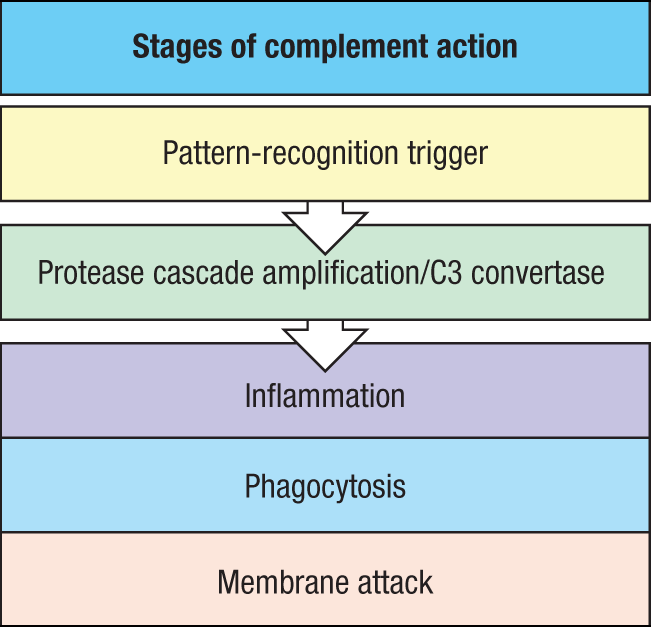
We learned in Section 2-4 that proteolysis can be used as a means of activating antimicrobial proteins. In the complement system, activation by proteolysis is inherent, with many of the complement proteins being proteases that successively cleave and activate one another. The proteases of the complement system are synthesized as inactive pro-enzymes, or zymogens, which become enzymatically active only after proteolytic cleavage, usually by another complement protein. The complement pathways are triggered by proteins that act as pattern-recognition receptors to detect the presence of pathogens. This detection activates an initial zymogen, triggering a cascade of proteolysis in which complement zymogens are activated sequentially, each becoming an active protease that cleaves and activates many molecules of the next zymogen in the pathway, amplifying the signal as the cascade proceeds. This results in activation of three distinct effector pathways—inflammation, phagocytosis, and membrane attack—that help eliminate the pathogen. In this way, the detection of even a small number of pathogens produces a rapid response that is greatly amplified at each step. This overall scheme for complement is shown in Fig. 2.13.
Nomenclature for complement proteins can seem confusing, so we will start by explaining their names. The first proteins discovered belong to the classical pathway, and they are designated by the letter C followed by a number. The native complement proteins—such as the inactive zymogens—have a simple number designation; for example, C1 and C2. Unfortunately, they were named in the order of their discovery rather than the sequence of reactions. The reaction sequence in the classical pathway, for example, is C1, C4, C2, C3, C5, C6, C7, C8, and C9 (note that not all of these are proteases). Products of cleavage reactions are designated by adding a lowercase letter as a suffix. For example, cleavage of C3 produces a small protein fragment called C3a and the remaining larger fragment, C3b. By convention, the larger fragment for other factors is designated by the suffix b. There is an exception to this convention. For C2, the larger fragment was originally named C2a in the literature by its discoverers. Although there have been discussions regarding changes to this nomenclature, there has not been uniform agreement and so we will preserve the original usage of C2a as the large active fragment of C2. Another exception is the naming of C1q, C1r, and C1s: these are not cleavage products of C1 but are distinct proteins that together compose C1. The proteins of the alternative pathway were discovered later and are designated by different capital letters; for example, factor B and factor D. The factor B cleavage products are also designated by the addition of lowercase a and b; thus, the large fragment of B is called Bb and the small fragment is called Ba. Activated complement components were often designated in the past by a horizontal line, for example, C2a, but we will not use this convention. All the components of the complement system are listed in Fig. 2.14.
|
Functional protein classes in the complement system |
|
|
Binding to antigen:antibody complexes and pathogen surfaces |
C1q |
|
Binding to carbohydrate structures such as mannose or GlcNAc on microbial surfaces |
MBL Ficolins Properdin (P) |
|
Activating enzymes* |
C1r C1s C2a Bb Factor D MASP-1 MASP-2 MASP-3 |
|
Surface-binding proteins and opsonins |
C4b C3b |
|
Peptide mediators of inflammation |
C5a C3a C4a |
|
Membrane-attack proteins |
C5b C6 C7 C8 C9 |
|
Complement receptors |
CR1 CR2 CR3 CR4 CRIg |
|
Complement-regulatory proteins |
C1INH C4BP CR1/CD35 MCP/CD46 DAF/CD55 Factor H Factor I Properdin (P) CD59 |
Fig. 2.14 Functional protein classes in the complement system. *In this book, C2a is used to denote the larger, active fragment of C2.
Besides acting in innate immunity, complement also influences adaptive immunity. Opsonization of pathogens by complement facilitates their uptake by phagocytic antigen-presenting cells that express complement receptors; this enhances the presentation of pathogen antigens to T cells, which we discuss in more detail in Chapter 6. B cells express receptors for complement proteins that enhance their responses to complement-coated antigens, as we describe later in Chapter 10. In addition, several of the complement fragments can act to influence cytokine production by antigen-presenting cells, thereby influencing the direction and extent of the subsequent adaptive immune response, as we describe in Chapter 11.
2-5 The complement system recognizes features of microbial surfaces and marks them for destruction by coating them with C3b.
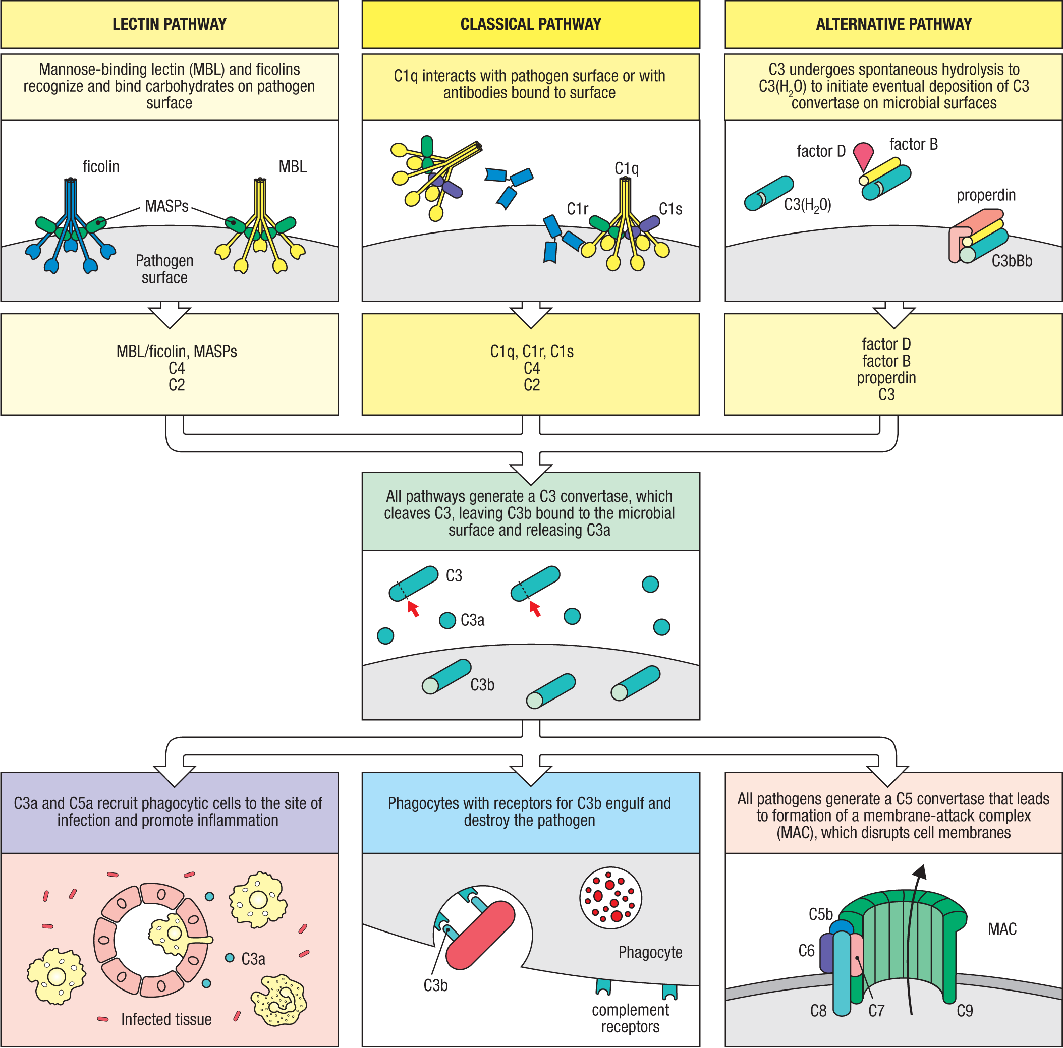
Figure 2.15 gives a highly simplified preview of the initiation mechanisms and outcomes of complement activation. The three pathways of complement activation are initiated in different ways. The lectin pathway is initiated by soluble carbohydrate-binding proteins—mannose-binding lectin (MBL) and ficolins—that bind to particular carbohydrate structures on microbial surfaces. Specific proteases, called MBL-associated serine proteases (MASPs), that associate with these recognition proteins trigger the cleavage of complement proteins and activation of the pathway. The classical pathway is initiated when complement component 1 (C1), which comprises a recognition protein (C1q) associated with proteases (C1r and C1s), either recognizes a microbial surface directly or binds to antibodies already bound to a pathogen. Finally, the alternative pathway can be initiated by spontaneous hydrolysis and activation of complement component 3 (C3), which can then bind directly to microbial surfaces.
These three pathways converge at the central and most important step in complement activation. When any of the pathways interacts with a pathogen surface, the enzymatic activity of a C3 convertase is generated. There are various types of C3 convertase, depending on the complement pathway activated, but each is a multi-subunit protein with protease activity that cleaves C3. The C3 convertase is bound covalently to the pathogen surface, where it cleaves C3 to generate large amounts of C3b, the main effector molecule of the complement system, and C3a, a small peptide that binds to specific receptors and helps induce inflammation. Cleavage of C3 is the critical step in complement activation and leads directly or indirectly to all the effector activities of the complement system (see Fig. 2.15). C3b binds covalently to the microbial surface and acts as an opsonin, enabling phagocytes that carry receptors for complement to take up and destroy the C3b-coated microbe. Later in the chapter, we will describe the different complement receptors that bind C3b and are involved in this function of complement and explain how C3b is degraded by a serum protease into inactive smaller fragments called C3f and C3dg and C3d. C3b can also bind to the C3 convertases produced by the classical and lectin pathways and form another multi-subunit enzyme, the C5 convertase, which cleaves C5, liberating the highly inflammatory peptide C5a and generating C5b. C5b initiates the ‘late’ events of complement activation, also called the terminal pathway, in which additional complement proteins interact with C5b to form a membrane-attack complex (MAC) on the pathogen surface, creating a pore in the cell membrane that leads to cell lysis (see Fig. 2.15, bottom right).
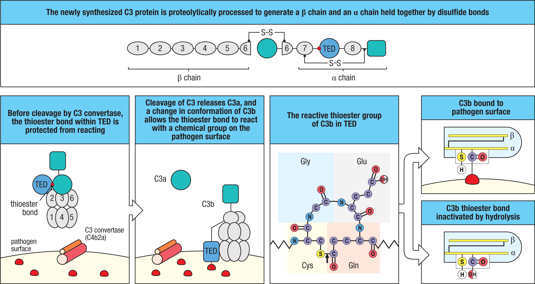
The key feature of C3b is its ability to form a covalent bond with microbial surfaces, which allows the innate recognition of microbes to be translated into effector responses. Covalent bond formation is due to a highly reactive thioester bond that is hidden inside the folded C3 protein and cannot react until C3 is cleaved. When C3 convertase cleaves C3 and releases the C3a fragment, large conformational changes occur in C3b that allow the thioester bond to react with a hydroxyl or amino group on the nearby microbial surface (Fig. 2.16). If no bond is made, the thioester is rapidly hydrolyzed, inactivating C3b, which is one way that alternative pathway activity is confined to a target area in healthy individuals. As we will see below, some of the individual components of C3 and C5 convertases differ between the various complement pathways; the components that are different are listed in Fig. 2.17.
|
C3 convertase |
|
|
Lectin pathway |
C4b2a |
|
Classical pathway |
C4b2a |
|
Alternative pathway |
C3bBb |
|
Fluid phase |
C3(H2O)Bb |
|
C5 convertase |
|
|
Lectin pathway |
C4b2a3b |
|
Classical pathway |
C4b2a3b |
|
Alternative pathway |
C3b2Bb |
Fig. 2.17 C3 and C5 convertases of the complement pathways. Note the C5 convertase of the alternative pathway consists of two C3b subunits and one Bb subunit.
Pathways leading to such potent inflammatory and destructive effects—and which have a series of built-in amplification steps—are potentially dangerous and must be tightly regulated. One important safeguard is that the key activated complement components are rapidly inactivated unless they bind to the pathogen surface on which their activation was initiated. There are also several points in the pathway at which regulatory proteins act to prevent the activation of complement on the surfaces of healthy host cells, thereby protecting them from accidental damage, as we shall see later in the chapter. Complement can, however, be activated by dying cells, such as those at sites of ischemic injury, and by cells undergoing apoptosis, or programmed cell death. In these cases, the complement coating helps phagocytes dispose of the dead and dying cells neatly, thus limiting the risk of cell contents being released and triggering an autoimmune response (discussed in Chapter 15).
Having introduced some of the main complement components, we are ready for a more detailed account of the three pathways. To help indicate the types of functions carried out by each of the complement components in the tables throughout the rest of the chapter, we will use the color code introduced in Fig. 2.13 and Fig. 2.14: yellow for recognition and activation, green for amplification, purple for inflammation, blue for phagocytosis, and pink for membrane attack.
2-6 The lectin pathway uses soluble receptors that recognize microbial surfaces to activate the complement cascade.
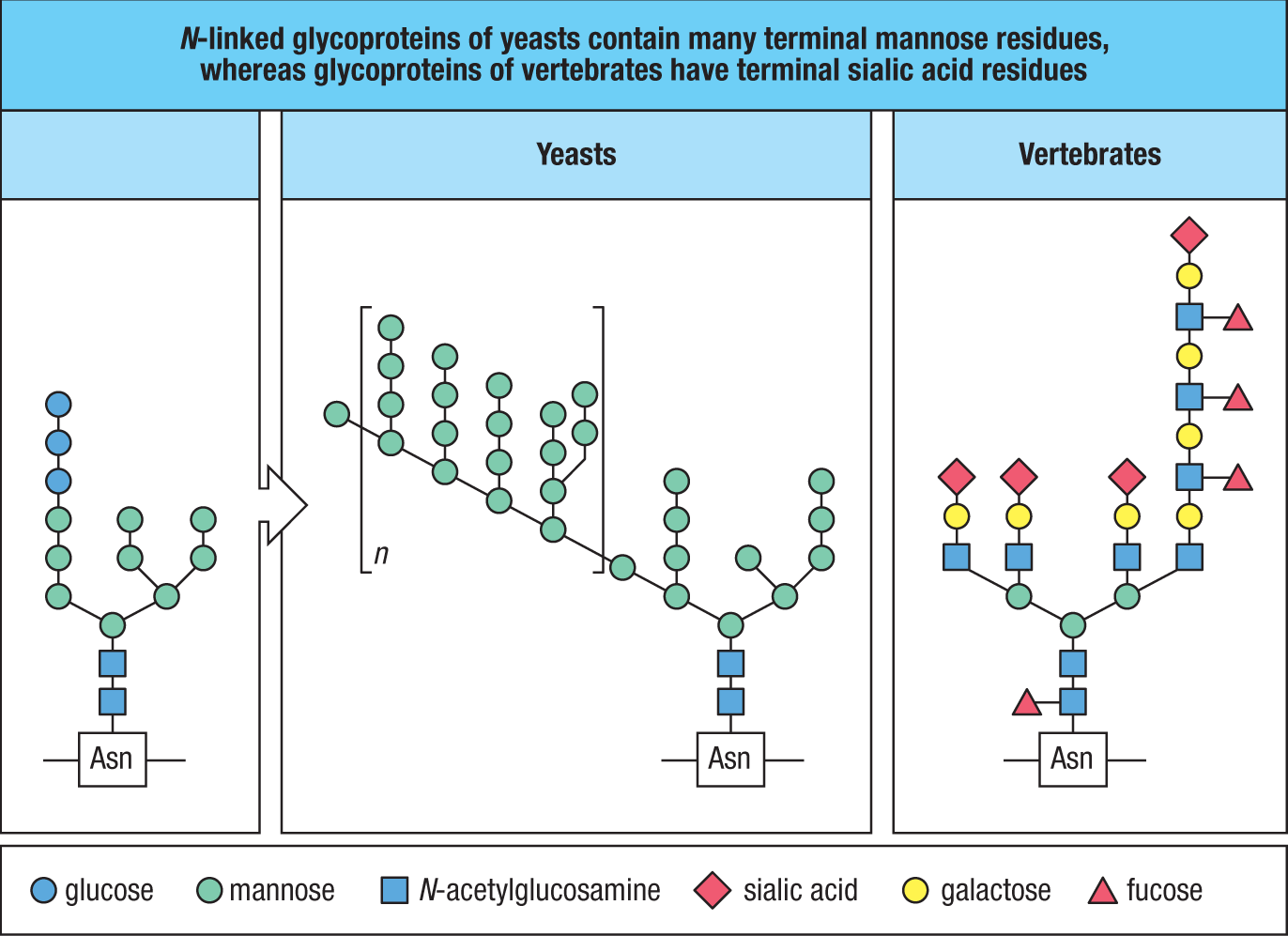
Microorganisms typically bear on their surface repeating patterns of molecular structures, known generally as pathogen-associated molecular patterns (PAMPs). The cell walls of Gram-positive and Gram-negative bacteria, for example, are composed of a matrix of proteins, carbohydrates, and lipids in a repetitive array (see Fig. 2.9). The lipoteichoic acids of Gram-positive bacterial cell walls and the lipopolysaccharide of the outer membrane of Gram-negative bacteria are not present on animal cells and are important in the recognition of bacteria by the innate immune system. Similarly, the glycans of yeast surface proteins commonly terminate in mannose residues rather than the sialic acid residues (N-acetylneuraminic acid) that terminate the glycans of vertebrate cells (Fig. 2.18). The lectin pathway uses these features of microbial surfaces to detect and respond to pathogens.
The lectin pathway can be triggered by any of four different pattern-recognition receptors that circulate in blood and extracellular fluids and recognize carbohydrates on microbial surfaces. The first such receptor to be discovered was mannose-binding lectin (MBL), which is shown in Fig. 2.19, and which is synthesized in the liver. MBL is an oligomeric protein built up from a monomer that contains an amino-terminal collagen-like domain and a carboxyl-terminal C-type lectin domain (see Section 2-4). Proteins of this type are called collectins. MBL monomers assemble into trimers through the formation of a triple helix by their collagen-like domains. Trimers then assemble into oligomers by disulfide bonding between the cysteine-rich collagen domains. The MBL present in the blood is composed of two to six trimers, with the major forms of human MBL being trimers and tetramers. A single carbohydrate-recognition domain of MBL has a low affinity for mannose, fucose, and N-acetylglucosamine (GlcNAc) residues, which are common on microbial glycans, but does not bind sialic acid residues, which terminate vertebrate glycans. Thus, multimeric MBL has high total binding strength, or avidity, for repetitive carbohydrate structures on a wide variety of microbial surfaces, including Gram-positive and Gram-negative bacteria, mycobacteria, yeasts, and some viruses and parasites, while not interacting with host cells. MBL is present at low concentrations in the plasma of most individuals, but in the presence of infection, its production is increased during the acute-phase response. This is part of the induced phase of the innate immune response and is discussed in Chapter 3.
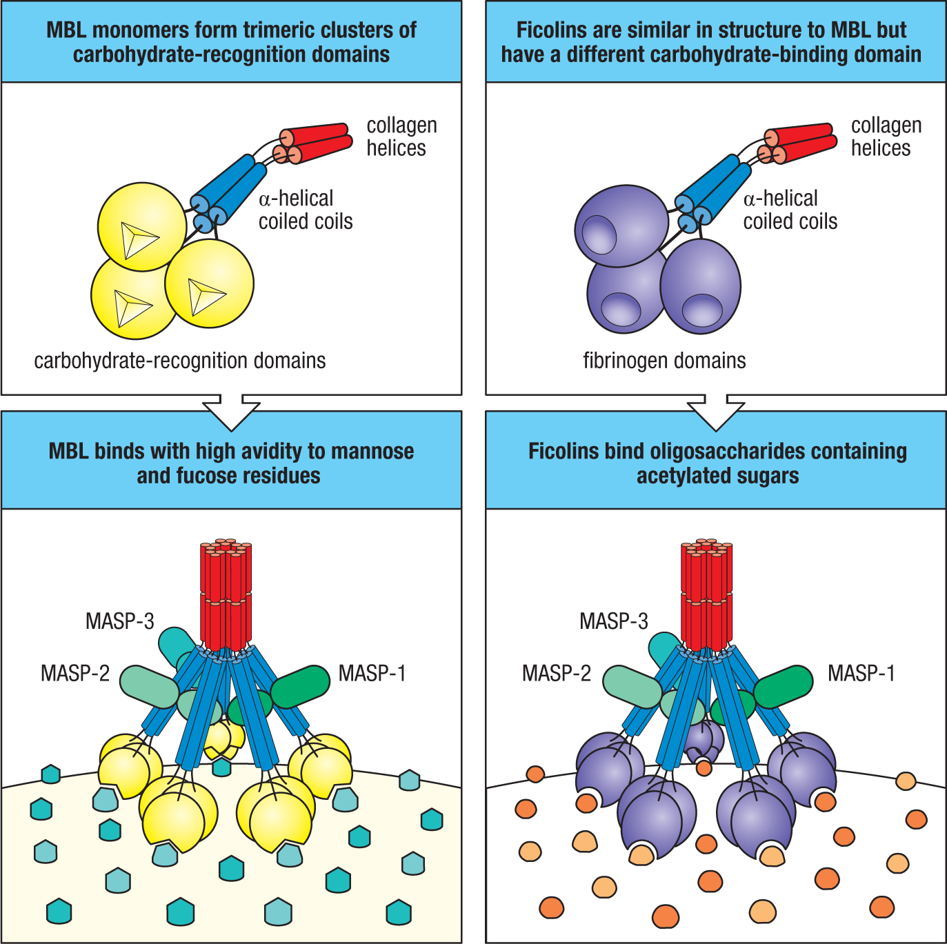
The other pathogen-recognition molecules used by the lectin pathway are known as ficolins. Although related in overall shape and function to MBL, they have a fibrinogen-like domain, rather than a lectin domain, attached to the collagen-like stalk (see Fig. 2.19). The fibrinogen-like domain gives ficolins a general specificity for oligosaccharides containing acetylated sugars, but it does not bind mannose-containing carbohydrates. Humans have three ficolins: ficolin-1 (M-ficolin), ficolin-2 (L-ficolin), and ficolin-3 (H-ficolin). Each binds to a spectrum of bacteria. Ficolins are secreted into the blood and tissues by various cells, including cells of the liver and lung, and by neutrophils.
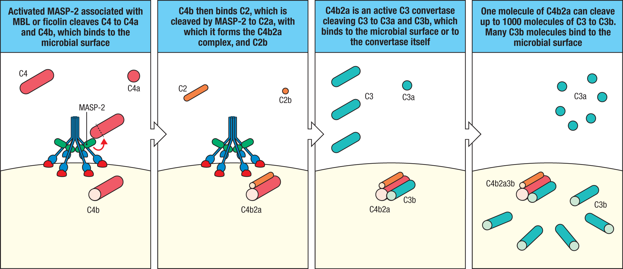
MBL in plasma can bind to five proteins: three MBL-associated serine proteases, MASP-1, MASP-2, and MASP-3, and two nonenzymatic proteins, MAp19 and MAp44. These five proteins are encoded by alternative splice variants of two genes: MASP1, which encodes MASP-1, MASP-3, and MAp44, and MASP2, which encodes MASP-2 and MAp19. MASP-1 and MASP-3 use different serine protease domains and have different substrate specificity. When MBL bound to MASP-1 and MASP-2 interacts with a pathogen surface, a conformational change occurs in MASP-1 that enables it to cleave and activate a MASP-2 molecule in the same MBL complex. Activated MASP-2 can then cleave complement components 4 and 2 (C4 and C2) to initiate the lectin and classical complement pathways (Fig. 2.20). When MBL bound to MASP-3 and MAp44 binds a pathogen surface, MASP-3 cleaves a pro-form of factor D, producing active factor D, which is a key event in the initiation of the alternative complement pathway. Like MBL, ficolins form oligomers that make a complex with MASP-1 and MASP-2, which similarly activate complement upon recognition of a microbial surface by the ficolin. C4, like C3, contains a buried thioester bond. When MASP-2 cleaves C4, it releases C4a, allowing a conformational change in C4b that exposes the reactive thioester as described for C3b (see Fig. 2.16). C4b bonds covalently via this thioester to the microbial surface nearby, where it then binds one molecule of C2 (see Fig. 2.20). C2 is cleaved by MASP-2, producing C2a, an active serine protease that remains bound to C4b to form C4b2a, which is the C3 convertase of the lectin and classical pathways. (Remember, we use the original convention by which C2a is the active large C2 fragment.) C4b2a now cleaves many molecules of C3 into C3a and C3b. The C3b fragments bond covalently to the nearby pathogen surface, and the released C3a initiates a local inflammatory response. The complement-activation pathway initiated by ficolins proceeds like the MBL lectin pathway (see Fig. 2.20).
Individuals deficient in MBL or MASP-2 experience substantially more respiratory infections by common extracellular bacteria during early childhood, indicating the importance of the lectin pathway for host defense. This susceptibility illustrates the particular importance of innate defense mechanisms in early childhood, when adaptive immune responses are not yet fully developed but the maternal antibodies transferred across the placenta and present in the mother’s milk are gone. Other members of the collectin family are the surfactant proteins A and D (SP-A and SP-D), which are present in the fluid that bathes the epithelial surfaces of the lung. There they coat the surfaces of pathogens, making them more susceptible to phagocytosis by macrophages that have left the subepithelial tissues to enter the alveoli. Because SP-A and SP-D do not associate with MASPs, they do not activate complement.
We have used MBL here as our prototype activator of the lectin pathway, but the ficolins are more abundant than MBL in plasma and so may actually be more important. Ficolin-2 recognizes acetylated sugars, such as GlcNAc and N-acetylgalactosamine (GalNAc), and particularly recognizes lipoteichoic acid, a component of the cell walls of Gram-positive bacteria that contains GalNAc. It can also activate complement after binding to a variety of capsulated bacteria. Ficolin-1 also recognizes acetylated sugar residues; ficolin-3 shows a more restricted binding specificity, for d-fucose and d-galactose, and has been linked to activity against the Gram-positive bacterium Aerococcus viridans, a cause of bacterial endocarditis.
2-7 The classical pathway is initiated by activation of the C1 complex and is homologous to the lectin pathway.
In its overall scheme, the classical pathway is similar to the lectin pathway, except that it uses a pathogen sensor known as the C1 complex, or C1. Because C1 interacts directly with some pathogens but can also interact with antibodies, C1 allows the classical pathway to function both in innate immunity, which we describe now, and in adaptive immunity, which we examine in more detail in Chapter 10.
Like the MBL:MASP complex, the C1 complex is composed of a large subunit (C1q), which acts as the pathogen sensor, and two serine proteases (C1r and C1s), which are initially in their inactive form (Fig. 2.21). C1q is a hexamer of trimers, composed of monomers that contain an amino-terminal globular domain and a carboxyl-terminal collagen-like domain. The trimers assemble through interactions of the collagen-like domains, bringing the globular domains together to form a globular head. Six of these trimers assemble to form a complete C1q molecule, which has six globular heads held together by their collagen-like tails. C1r and C1s are closely related to MASP-2 and somewhat more distantly related to MASP-1 and MASP-3; all five enzymes are likely to have evolved from the duplication of a gene for a common precursor. C1r and C1s interact noncovalently and form tetramers that fold into the arms of C1q, with at least part of the C1r:C1s complex being external to C1q, as illustrated in Fig. 2.21.
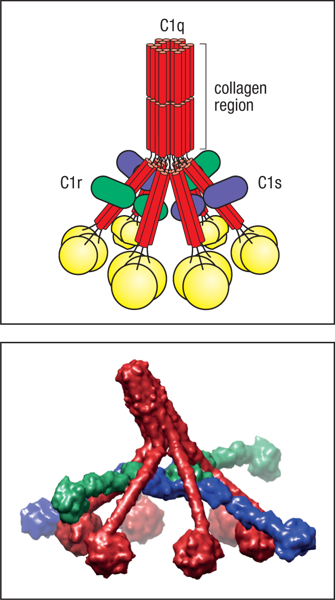
The recognition function of C1 resides in the six globular heads of C1q. When two or more of these heads interact with a ligand, this causes a conformational change in the C1r:C1s complex, which leads to the activation of an autocatalytic enzymatic activity in C1r; the active form of C1r then cleaves its associated C1s to generate an active serine protease. The activated C1s acts on the next two components of the classical pathway, C4 and C2. C1s cleaves C4 to produce C4b, which binds covalently to the pathogen surface as described earlier for the lectin pathway (see Fig. 2.20). C4b then also binds one molecule of C2, which is cleaved by C1s to produce the serine protease C2a. This produces the active C3 convertase C4b2a, which is the C3 convertase of both the lectin and the classical pathways. However, because it was first discovered as part of the classical pathway, C4b2a is often known as the classical C3 convertase (see Fig. 2.17). The proteins involved in the classical pathway, along with their active forms, are listed in Fig. 2.22.
|
Proteins of the classical pathway of complement activation |
|||
|
Native component |
Active form |
Function of the active form |
|
|
C1 (C1q:C1r2:C1s2) |
C1q |
Binds directly to pathogen surfaces or indirectly to antibody bound to pathogens, thus allowing autoactivation of C1r |
|
|
C1r |
Cleaves C1s to active protease |
||
|
C1s |
Cleaves C4 and C2 |
||
|
C4 |
C4b |
Covalently binds to pathogen and opsonizes it. Binds C2 for cleavage by C1s |
|
|
C4a |
Peptide mediator of inflammation (weak activity) |
||
|
C2 |
C2a |
Active enzyme of classical pathway C3/C5 convertase: cleaves C3 and C5 |
|
|
C2b |
Inactive small fragment |
||
|
C3 |
C3b |
Binds to pathogen surface and acts as opsonin. Initiates amplification via the alternative pathway. Binds C5 for cleavage by C2a |
|
|
C3a |
Peptide mediator of inflammation (intermediate activity) |
||
Fig. 2.22 The proteins of the classical pathway of complement activation.
C1q can attach itself to the surface of pathogens in several different ways. One is by binding directly to surface components on some bacteria, including certain proteins of bacterial cell walls and polyanionic structures, such as the lipoteichoic acid on Gram-positive bacteria. A second is through binding to C-reactive protein, an acute-phase protein in human plasma that binds to phosphocholine residues in bacterial surface molecules such as pneumococcal C polysaccharide—hence the name C-reactive protein. We discuss the acute-phase proteins in detail in Chapter 3. However, a main function of C1q in an immune response is to bind to the constant, or Fc, regions of antibodies (see Section 1-9) that have bound pathogens via their antigen-binding sites. C1q thus links the effector functions of complement to recognition provided by adaptive immunity. This might seem to limit the usefulness of C1q in fighting the first stages of an infection, before the adaptive immune response has generated pathogen-specific antibodies. However, some antibodies, called natural antibodies, are produced by the immune system in the apparent absence of infection. These antibodies have a low affinity for many microbial pathogens and are highly cross-reactive, recognizing common membrane constituents such as phosphocholine and even recognizing some antigens of the body’s own cells (that is, self antigens). Natural antibodies may be produced in response to commensal microbiota or to self antigens, but do not seem to be the consequence of an adaptive immune response to infection by pathogens. Most natural antibody is of the isotype, or class, known as IgM (see Chapter 5) and represents a considerable amount of the total IgM circulating in humans. IgM is the class of antibody most efficient at binding C1q, making natural antibodies an effective means of activating complement on microbial surfaces immediately after infection and leading to the clearance of bacteria such as Streptococcus pneumoniae (the pneumococcus) before they become dangerous.
2-8 Complement activation is largely confined to the surface on which it is initiated.
We have seen that both the lectin and the classical pathways of complement activation are initiated by proteins that bind to pathogen surfaces. During the triggered enzyme cascade that follows, it is important that activating events are confined to this same site, so that C3 activation also occurs on the surface of the pathogen and not in the plasma or on host-cell surfaces. This is achieved principally by the covalent binding of C4b to the pathogen surface. In innate immunity, C4 cleavage is catalyzed by a ficolin or MBL complex that is bound to the pathogen surface, and so the C4b cleavage product can bind adjacent proteins or carbohydrates on the pathogen surface. If C4b does not rapidly form this bond, the thioester bond is cleaved by reaction with water, and C4b is irreversibly inactivated. This helps to prevent C4b from diffusing from its site of activation on the microbial surface and becoming attached to healthy host cells.
C2 becomes susceptible to cleavage by C1s only when it is bound by C4b, and the active C2a serine protease is thereby also confined to the pathogen surface, where it remains associated with C4b, forming the C3 convertase C4b2a. Cleavage of C3 to C3a and C3b is thus also confined to the surface of the pathogen. Like C4b, C3b is inactivated by hydrolysis unless its exposed thioester rapidly makes a covalent bond (see Fig. 2.16), and it therefore opsonizes only the surface on which complement activation has taken place. Opsonization by C3b is more effective when antibodies are also bound to the pathogen surface, as phagocytes have receptors for both complement and Fc receptors that bind the Fc region of antibody (see Sections 1-20 and 10-20). Because the reactive forms of C3b and C4b are able to form a covalent bond with any adjacent protein or carbohydrate, when complement is activated by bound antibody a proportion of the reactive C3b or C4b will become linked to the antibody molecules themselves. Antibody that is chemically cross-linked to complement is likely the most efficient trigger for phagocytosis.
2-9 The alternative pathway is an amplification loop for C3b formation that is accelerated by properdin in the presence of pathogens.
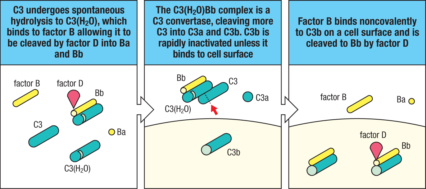
Although probably the most ancient of the complement pathways, the alternative pathway is so named because it was discovered as a second, or ‘alternative,’ pathway for complement activation after the classical pathway had been defined. Its key feature is its ability to be spontaneously activated. It has a unique C3 convertase, the alternative pathway C3 convertase, which differs from the C4b2a convertase of the lectin or classical pathways (see Fig. 2.17). The alternative pathway C3 convertase is composed of C3b itself bound to Bb, which is a cleavage fragment of the plasma protein factor B. This C3 convertase, designated C3bBb, has a special place in complement activation because, by producing C3b, it can generate more of itself. This means that once some C3b has been formed, by whichever pathway, the alternative pathway can act as an amplification loop to increase C3b production rapidly.
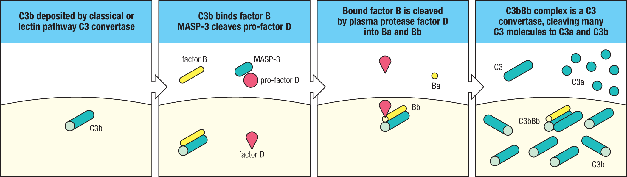
The alternative pathway can be activated in two different ways. The first is by the action of the lectin or classical pathway. C3b generated by either of these pathways and covalently linked to a microbial surface can bind factor B (Fig. 2.23). This alters the conformation of factor B, enabling a plasma protease called factor D to cleave it into Ba and Bb. Bb remains stably associated with C3b, forming the C3bBb C3 convertase. The second way of activating the alternative pathway involves the spontaneous hydrolysis (known as ‘tickover’) of the thioester bond in C3 to form C3(H2O), as shown in Fig. 2.24. C3 is abundant in plasma, and tickover causes a steady, low-level production of C3(H2O). This C3(H2O) can bind factor B, which is then cleaved by factor D, producing a short-lived fluid-phase C3 convertase, C3(H2O)Bb. Although formed in only small amounts by C3 tickover, fluid-phase C3(H2O)Bb can cleave many molecules of C3 to C3a and C3b. Much of this C3b is inactivated by hydrolysis, but some attaches covalently via its thioester bond to the surfaces of any microbes present. C3b formed in this way is no different from C3b produced by the lectin or classical pathways and binds factor B, leading to the formation of C3 convertase and a stepping up of C3b production (see Fig. 2.23).
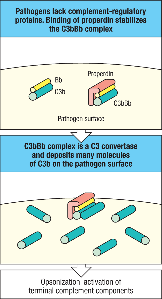
On their own, the alternative pathway C3 convertases C3bBb and C3(H2O)Bb are very short-lived (~90 seconds). They are stabilized, however, by around 5- to 10-fold by binding the plasma protein properdin (Fig. 2.25). Properdin is made by neutrophils and stored in secondary granules. It is released when neutrophils are activated by the presence of pathogens. Properdin may have some properties of a pattern-recognition receptor, as it can bind to some microbial surfaces. Properdin-deficient individuals are particularly susceptible to infections with Neisseria meningitidis, the main agent of bacterial meningitis. Properdin’s ability to bind to bacterial surfaces may direct the activity of the alternative complement pathway to these pathogens, thus aiding their removal by phagocytosis. Properdin can also bind to mammalian cells that are undergoing apoptosis or have been damaged or modified by ischemia, viral infection, or antibody binding, leading to the deposition of C3b on these cells and facilitating their removal by phagocytosis. The distinctive components of the alternative pathway are listed in Fig. 2.26.
|
Proteins of the alternative pathway of complement activation |
|||
|
Native component |
Active fragments |
Function |
|
|
C3 |
C3b |
Binds to pathogen surface; binds B for cleavage by D; C3bBb is a C3 convertase and C3b2Bb is a C5 convertase |
|
|
Factor B (B) |
Ba |
Small fragment of B, unknown function |
|
|
Bb |
Bb is the active enzyme of the C3 convertase C3bBb and the C5 convertase C3b2Bb |
||
|
Factor D (D) |
D |
Plasma serine protease, cleaves B when it is bound to C3b to Ba and Bb |
|
|
Properdin (P) |
P |
Plasma protein that binds to bacterial surfaces and stabilizes the C3bBb convertase |
|
Fig. 2.26 The proteins of the alternative pathway of complement activation.
2-10 Membrane and plasma proteins that regulate the formation and stability of C3 convertases determine the extent of complement activation.
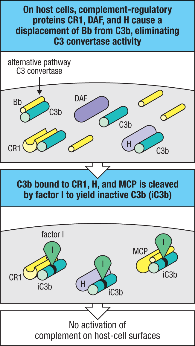
Several mechanisms ensure that complement activation will proceed only on the surface of a pathogen or on damaged host cells, and not on normal host cells and tissues. After initial complement activation by any pathway, the extent of amplification via the alternative pathway is critically dependent on the stability of the C3 convertase C3bBb. This stability is controlled by both positive and negative regulatory proteins. We have already described how properdin acts as a positive regulatory protein on foreign surfaces, such as those of bacteria or damaged host cells, by stabilizing C3bBb.
Several negative regulatory proteins, present in plasma and in host-cell membranes, protect healthy host cells from the injurious effects of inappropriate complement activation on their surfaces. These complement-regulatory proteins interact with C3b and either prevent the convertase from forming or promote its rapid dissociation (Fig. 2.27). For example, a membrane-attached protein known as decay-accelerating factor (DAF or CD55) competes with factor B for binding to C3b on the cell surface and can displace Bb from a convertase that has already formed. Convertase formation can also be prevented by cleaving C3b to an inactive derivative, iC3b. This is achieved by a plasma protease, factor I, in conjunction with C3b-binding proteins that act as cofactors, such as membrane cofactor of proteolysis (MCP or CD46), another host-cell membrane protein (see Fig. 2.27). Cell-surface complement receptor type 1 (CR1, also known as CD35) behaves similarly to DAF and MCP in that it inhibits C3 convertase formation and promotes the catabolism of C3b to inactive products, respectively, but it has a more limited tissue distribution. Factor H is another complement-regulatory protein in plasma that binds C3b, and like CR1, it is able to compete with factor B to displace Bb from the convertase; in addition, it acts as a cofactor for factor I. Factor H binds preferentially to C3b bound to vertebrate cells because it has an affinity for the sialic acid residues present on their cell surfaces (see Fig. 2.18). Thus, the amplification loop of the alternative pathway is allowed to proceed on the surface of a pathogen or on damaged host cells, but not on normal host cells or on tissues that express these negative regulatory proteins.
The C3 convertase of the classical and lectin pathways (C4b2a) is molecularly distinct from that of the alternative pathway (C3bBb). However, understanding of the complement system is simplified somewhat by recognition of the close evolutionary relationships between the different complement proteins (Fig. 2.28). Thus the complement zymogens factor B and C2 are closely related proteins encoded by homologous genes located in tandem within the major histocompatibility complex (MHC) on human chromosome 6. Furthermore, their respective binding partners, C3 and C4, both contain thioester bonds that provide the means of covalently attaching the C3 convertases to a pathogen surface.
|
Step in pathway |
Protein serving function in pathway |
Relationship |
||
|
Alternative |
Lectin |
Classical |
||
|
Initiating serine protease |
D |
MASP |
C1s |
Homologous (C1s and MASP) |
|
Covalent binding to cell surface |
C3b |
C4b |
Homologous |
|
|
C3/C5 convertase |
Bb |
C2a |
Homologous |
|
|
Control of activation |
CR1 H |
CR1 C4BP |
Identical Homologous |
|
|
Opsonization |
C3b |
Identical |
||
|
Initiation of effector pathway |
C5b |
Identical |
||
|
Local inflammation |
C5a, C3a |
Identical |
||
|
Stabilization |
P |
None |
Unique |
|
Fig. 2.28 There is a close evolutionary relationship among the factors of the alternative, lectin, and classical pathways of complement activation. Most of the factors are either identical or the homologous products of genes that have duplicated and then diverged in sequence. The proteins C4 and C3 are homologous and contain the unstable thioester bond by which their large fragments, C4b and C3b, bind covalently to membranes. The genes encoding proteins C2 and factor B are adjacent in the MHC region of the genome and arose by gene duplication. The regulatory proteins factor H, CR1, and C4BP share a repeat sequence common to many complement-regulatory proteins. The greatest divergence between the pathways is in their initiation: in the classical pathway the C1 complex binds either to certain pathogens or to bound antibody, and in the latter circumstance it serves to convert antibody binding into enzyme activity on a specific surface; in the lectin pathway, mannose-binding lectin (MBL) associates with a serine protease, activating MBL-associated serine protease (MASP), to serve the same function as C1r:C1s; in the alternative pathway this enzyme activity is provided by factor D.
Only one component of the alternative pathway seems entirely unrelated to its functional equivalents in the classical and lectin pathways: the initiating serine protease, factor D. Factor D can also be singled out as the only activating protease of the complement system to circulate as an active enzyme rather than a zymogen. This is both necessary for the initiation of the alternative pathway (through the cleavage of factor B bound to spontaneously activated C3) and safe for the host, because factor D has no other substrate than factor B bound to C3b. This means factor D finds its substrate only at pathogen surfaces and at a very low level in plasma, where the alternative pathway of complement activation can be allowed to proceed.
2-11 Complement developed early in the evolution of multicellular organisms.
The complement system was originally known only from vertebrates, but homologs of C3 and factor B and a prototypical ‘alternative pathway’ have been discovered in nonchordate invertebrates. This is not altogether surprising as C3, which is cleaved and activated by serine proteases, is evolutionarily related to the serine protease inhibitor α2-macroglobulin, whose first appearance likely was in an ancestor to all modern vertebrates. The amplification loop of the alternative pathway also has an ancestral origin, as it is present in echinoderms (which include sea urchins and sea stars) and is based on a C3 convertase formed by the echinoderm homologs of C3 and factor B. These factors are expressed by phagocytic cells called amoeboid coelomocytes present in the coelomic fluid. Expression of C3 by these cells increases when bacteria are present. This simple system seems to function to opsonize bacterial cells and other foreign particles and facilitate their uptake by coelomocytes. C3 homologs in invertebrates are clearly related to each other. They all contain the distinctive thioester linkage and form a family of proteins, the thioester-containing proteins, or TEPs. In the mosquito Anopheles, the production of protein TEP1 is induced in response to infection, and the protein may directly bind to bacterial surfaces to mediate phagocytosis of Gram-negative bacteria. Some form of C3 activity may even predate the evolution of the Bilateria—animals with bilateral symmetry, flatworms being the most primitive modern representatives—because genomic evidence of C3, factor B, and some later-acting complement components has been found in the Anthozoa (corals and sea anemones).
After its initial appearance, the complement system seems to have evolved by the acquisition of new activation pathways that allow specific targeting of microbial surfaces. The first to evolve was likely the ficolin pathway, which is present both in vertebrates and in some closely related invertebrates, such as the urochordates. Evolutionarily, the ficolins may predate the collectins, which are also first seen in the urochordates. Homologs of MBL and of the classical pathway complement component 1q (C1q), both collectins, have been identified in the genome of the ascidian urochordate Ciona (sea squirt). Two invertebrate homologs of mammalian MASPs also have been identified in Ciona, and it seems likely that they may be able to cleave and activate C3. Thus, the minimal complement system of the echinoderms appears to have been expanded in the urochordates by the recruitment of a specific activation system that may target C3 deposition onto microbial surfaces. This also suggests that when adaptive immunity evolved, much later, the ancestral antibody molecule used an already diversified C1q-like collectin member to activate the complement pathway, and that the complement activation system evolved further by use of this collectin and its associated MASPs to become the initiating components of the classical complement pathway; namely, C1q, C1r, and C1s.
2-12 Surface-bound C3 convertase deposits large numbers of C3b fragments on pathogen surfaces and generates C5 convertase activity.
We now return to the present-day complement system. The formation of C3 convertases is the point at which the three pathways of complement activation converge. The convertase of the lectin and classical pathways, C4b2a, and the convertase of the alternative pathway, C3bBb, initiate the same subsequent events—they cleave C3 to C3b and C3a. C3b binds covalently through its thioester bond to adjacent molecules on the pathogen surface; otherwise it is inactivated by hydrolysis. C3 is the most abundant complement protein in plasma, occurring at a concentration of 1.2 mg/ml, and up to 1000 molecules of C3b can bind in the vicinity of a single active C3 convertase (see Fig. 2.23). Thus the main effect of complement activation is to deposit large quantities of C3b on the surface of the infecting pathogen, where the C3b forms a covalently bonded coat that can signal the ultimate destruction of the pathogen by phagocytes.
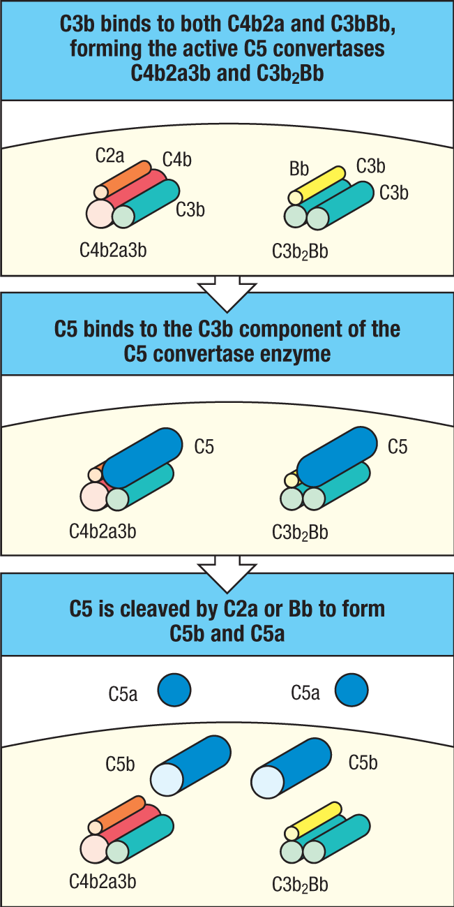
The next step in the complement cascade is the generation of the C5 convertases. C5 is a member of the same family of proteins as C3, C4, α2-macroglobulin, and the thioester-containing proteins of invertebrates. C5 does not form an active thioester bond during its synthesis, but, like C3 and C4, it is cleaved by a specific protease into C5a and C5b fragments, each of which exerts specific downstream actions that are important in propagating the complement cascade. In the classical and the lectin pathways, a C5 convertase is formed by the binding of C3b to C4b2a to yield C4b2a3b. The C5 convertase of the alternative pathway is formed by the binding of C3b to the C3bBb convertase to form C3b2Bb. A C5 is captured by these C5 convertase complexes through binding to an acceptor site on C3b and is thus rendered susceptible to cleavage by the serine protease activity of C2a or Bb. This reaction, which generates C5b and C5a, is much more limited than cleavage of C3, because C5 can be cleaved only when it binds to C3b that is in turn bound to C4b2a or C3bBb to form the active C5 convertase complex. Thus, complement activated by all three pathways leads to the binding of large numbers of C3b molecules on the surface of the pathogen, the generation of a more limited number of C5b molecules, and the release of C3a and a smaller amount of C5a (Fig. 2.29).
2-13 Ingestion of complement-tagged pathogens by phagocytes is mediated by receptors for the bound complement proteins.
The most important action of complement is to facilitate the uptake and destruction of pathogens by phagocytic cells. This occurs by the specific recognition of bound complement components by complement receptors (CRs) on phagocytes. These complement receptors bind pathogens opsonized with complement components: opsonization of pathogens is a major function of C3b and its proteolytic derivatives. C4b also acts as an opsonin but has a relatively minor role, largely because so much more C3b than C4b is generated.
The known receptors for bound complement components and for C5a and C3a are listed, with their functions and distributions, in Fig. 2.30. The C3b receptor CR1, described in Section 2-10, is a negative regulator of complement activation (see Fig. 2.27). CR1 is expressed on many types of immune cells, including macrophages and neutrophils. Binding of C3b to CR1 cannot by itself stimulate phagocytosis, but it can lead to phagocytosis in the presence of other immune mediators that activate macrophages. For example, the small complement fragment C5a can activate macrophages to ingest bacteria that are bound to their CR1 receptors (Fig. 2.31). C5a accomplishes this by binding to another receptor expressed by macrophages, the C5a receptor 1 (C5aR1, CD88), which has seven membrane-spanning domains. Receptors of this type transduce their signals via intracellular guanine nucleotide–binding proteins called G proteins and are known generally as G protein–coupled receptors (GPCRs); they are discussed in Section 3-2. C5a receptor 2 (C5L2, GPR77) is expressed by neutrophils and macrophages, but its function is not firmly established. It is a nonsignaling receptor that may act as a decoy receptor for C5a and regulate activity of the C5a receptor. Proteins associated with the extracellular matrix, such as fibronectin, can also contribute to phagocyte activation; these are encountered when phagocytes are recruited to connective tissue and activated there.
|
Receptor |
Specificity |
Functions |
Cell types |
|
CR1 (CD35) |
C3b, C4b |
Promotes C3b and C4b decay |
Erythrocytes, macrophages, monocytes, neutrophils, B cells, FDCs |
|
CR2 (CD21) |
C3d, iC3b, C3dg |
Enhances B-cell response to antigens bearing C3d, iC3b, or C3dg, part of B-cell co-receptor |
B cells, FDCs |
|
CR3 (Mac-1) (CD11b: CD18) |
iC3b |
Stimulates phagocytosis |
Macrophages, monocytes, polymorphonuclear leukocytes, FDCs |
|
CR4 (gp150, 95) (CD11c: CD18) |
iC3b |
Stimulates phagocytosis |
Macrophages, monocytes, polymorphonuclear leukocytes, dendritic cells |
|
CRIg |
C3b, iC3b |
Phagocytosis of circulating pathogens |
Tissue-resident macrophages |
|
C5aR1 (CD88) |
C5a |
Binding of C5a, activates G protein |
Neutrophils, macrophages, endothelial cells, mast cells |
|
C5aR2 (C5L2, GPR77) |
C5a |
Decoy receptor, regulates C5a receptor |
Neutrophils, macrophages |
|
C3a receptor |
C3a |
Binding of C3a, activates G protein |
Macrophages, endothelial cells, mast cells |
|
PAR1 PAR4 |
C4a |
Increases vascular permeability |
Epithelium, platelets |
Fig. 2.30 Distribution and function of cell-surface receptors for complement proteins. A variety of complement receptors are specific for bound C3b and its cleavage products (iC3b and C3dg). CR1 and CR3 are important in inducing the phagocytosis of bacteria with complement components bound to their surface. CR2 is found mainly on B cells, where it is part of the B-cell co-receptor complex. CR1 and CR2 share structural features with the complement-regulatory proteins that bind C3b and C4b. CR3 and CR4 are integrins composed of integrin β2 (also known as CD18, and sometimes gp95) paired with either integrin αM (CD11b) or integrin αX (CD11c), respectively (see eAppendix II, found in the ebook and on the Digital Landing Page for this book); CR3, also called Mac-1, is also important for leukocyte adhesion and migration, as we shall see in Chapter 3, whereas CR4 is only known to function in phagocytosis. The receptors for C5a and C3a are seven-span G protein–coupled receptors. FDCs (follicular dendritic cells) are not involved in innate immunity and are discussed in later chapters.
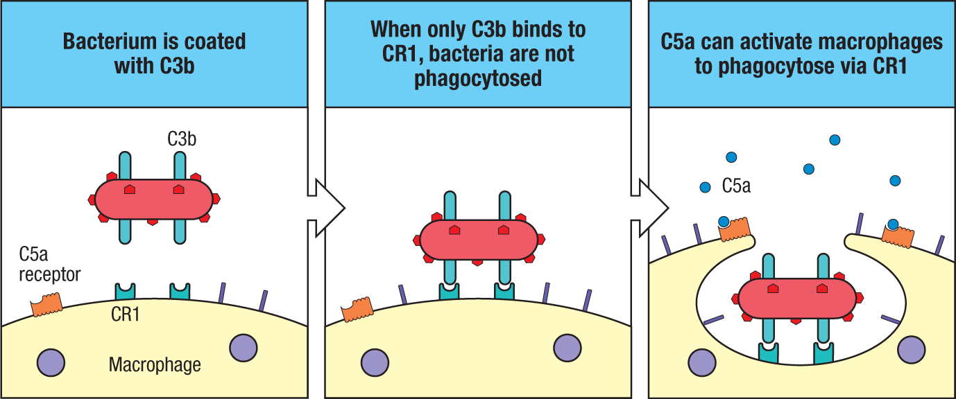
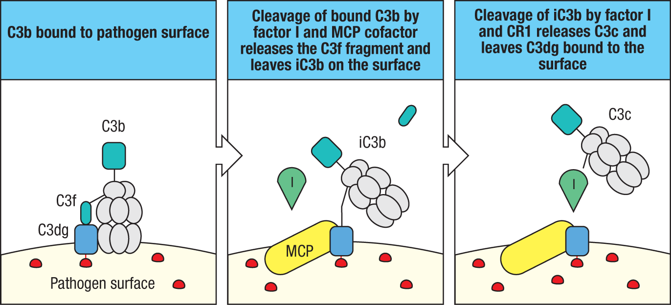
Four other complement receptors—CR2 (also known as CD21), CR3 (CD11b:CD18), CR4 (CD11c:CD18), and CRIg (complement receptor of the immunoglobulin superfamily)—bind to forms of C3b that have been cleaved by factor I but that remain attached to the pathogen surface. Like several other key components of complement, C3b is subject to regulatory mechanisms that cleave it into derivatives, such as iC3b, that cannot form an active convertase. C3b bound to the microbial surface can be cleaved by factor I and MCP to remove the small fragment C3f, leaving the inactive iC3b form bound to the surface (Fig. 2.32). iC3b is recognized by several complement receptors—CR2, CR3, CR4, and CRIg. Unlike the binding of C3b to CR1, the binding of iC3b to the receptor CR3 is sufficient on its own to stimulate phagocytosis. Factor I and CR1 cleave iC3b to release C3c, leaving C3dg bound to the pathogen. C3dg is recognized only by CR2. CR2 is found on B cells as part of a co-receptor complex that can augment the signal received through the antigen-specific immunoglobulin receptor. Thus, a B cell whose antigen receptor is specific for an antigen of a pathogen will receive a strong signal on binding this antigen if it or the pathogen is also coated with C3dg. The activation of complement can therefore contribute to producing a strong antibody response.
The importance of opsonization by C3b and its inactive fragments in destroying extracellular pathogens can be seen in the effects of various complement deficiencies. For example, individuals deficient in C3 or in molecules that catalyze C3b deposition show an increased susceptibility to infection by a wide range of extracellular bacteria, including Streptococcus pneumoniae. We describe the effects of various defects in complement and the diseases they cause in Chapter 13.
2-14 The small fragments of some complement proteins initiate a local inflammatory response.
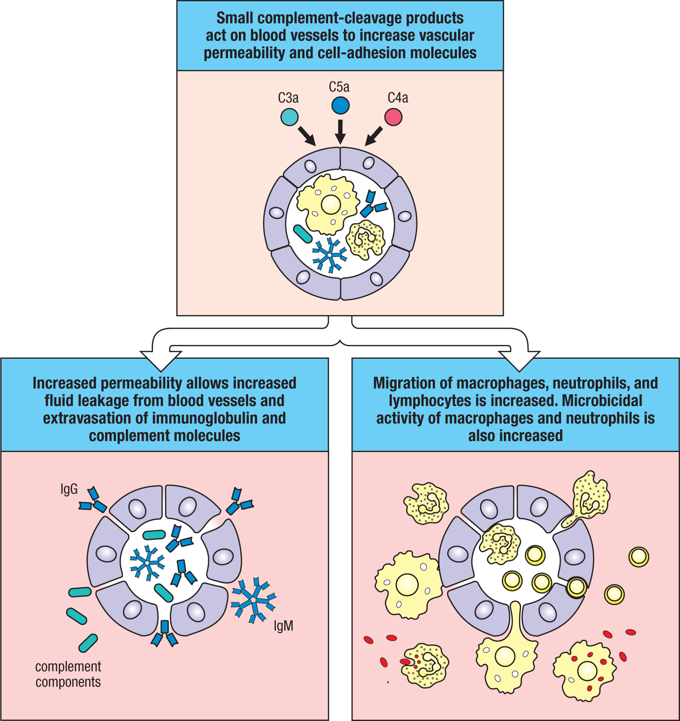
The small complement fragments C3a and C5a act on specific receptors on endothelial cells and mast cells (see Fig. 2.30) to produce local inflammatory responses. Like C5a, C3a also signals through a GPCR family member. When produced in large amounts or injected systemically, C3a and C5a induce a generalized circulatory collapse, producing a shocklike syndrome similar to that seen in a systemic allergic reaction involving antibodies of the IgE class, discussed in Chapter 14. Such a reaction is termed anaphylactic shock, and these small fragments of complement are therefore often referred to as anaphylatoxins. C5a has the highest specific biological activity, but both C3a and C5a induce the contraction of smooth muscle, increase vascular permeability, and act on the endothelial cells lining blood vessels to induce the synthesis of adhesion molecules. In addition, C3a and C5a can activate the mast cells that populate submucosal tissues to release inflammatory molecules such as histamine and the cytokine tumor necrosis factor-α (TNF-α), which cause similar effects. C4a is also generated during C4 cleavage and is inactive at C3a and C5a receptors. Instead, C4a acts at two other GPCRs, protease-activated receptor 1 (PAR1) and protease-activated receptor 4 (PAR4), which are expressed on endothelial cells and platelets and are also activated by thrombin, a protease activated in the clotting cascade. C4a activates signaling in these cells to increase endothelial-cell permeability and by actions on platelets may link complement activation with the clotting cascade.
The changes induced by C5a, C3a, and C4a combine to recruit antibody, complement, and phagocytic cells to the site of an infection (Fig. 2.33). The increased fluid in the tissues hastens the movement of pathogen-bearing antigen-presenting cells to the local lymph nodes, contributing to the prompt initiation of the adaptive immune response. C5a also acts directly on neutrophils and monocytes to increase their adherence to vessel walls, their migration toward sites of antigen deposition, and their ability to ingest particles; it also increases the expression of CR1 and CR3 on the surfaces of these cells. In this way, C5a, and to a lesser extent C3a and C4a, act in concert with other complement components to hasten the destruction of pathogens by phagocytes.
2-15 The terminal complement proteins polymerize to form pores in membranes that can kill certain pathogens.
 Case Study: Deficiency of the C8 Complement Component
One of the important effects of complement activation is the assembly of the terminal components of complement (Fig. 2.34) to form a membrane-attack complex. The reactions leading to the formation of this complex are shown schematically in Fig. 2.35. The end result is a pore in the lipid bilayer membrane that destroys membrane integrity. This is thought to kill the pathogen by destroying the proton gradient across the pathogen’s cell membrane.
|
The terminal complement components that form the membrane-attack complex |
|||
|
Native protein |
Active component |
Function |
|
|
C5 |
C5a |
Small peptide mediator of inflammation (high activity) |
|
|
C5b |
Initiates assembly of the membrane-attack system |
||
|
C6 |
C6 |
Binds C5b; forms acceptor for C7 |
|
|
C7 |
C7 |
Binds C5b6; amphiphilic complex inserts into lipid bilayer |
|
|
C8 |
C8 |
Binds C5b67; initiates C9 polymerization |
|
|
C9 |
C9n |
Polymerizes to C5b678 to form a membrane-spanning channel, lysing the cell |
|
Fig. 2.34 The terminal complement components.
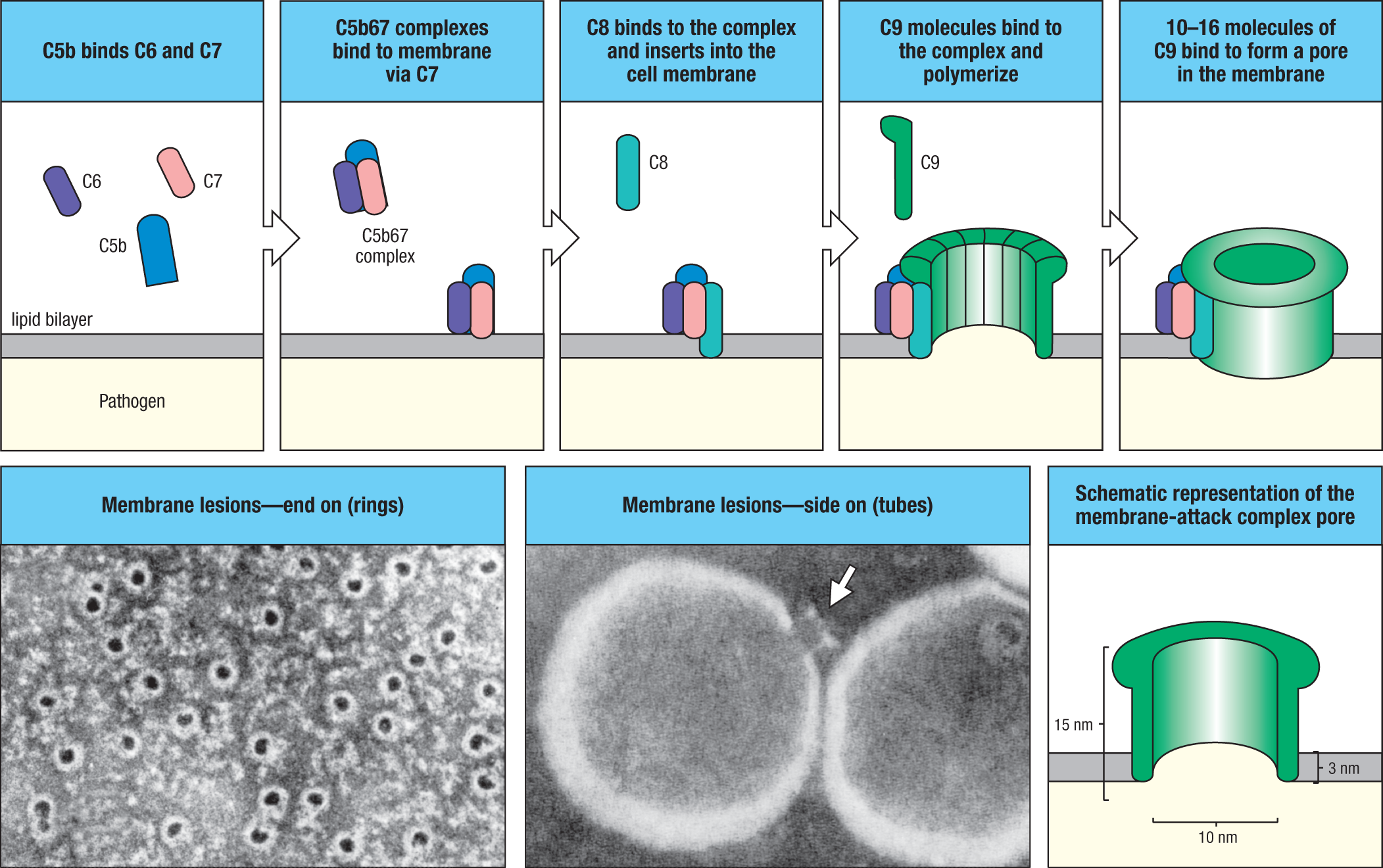
The first step in the formation of the membrane-attack complex is the cleavage of C5 by a C5 convertase to release C5b (see Fig. 2.29). In the next stages (see Fig. 2.35), C5b initiates the assembly of the terminal complement components and their insertion into the cell membrane. The process begins when one molecule of C5b binds one molecule of C6, and the C5b6 complex then binds one molecule of C7. This reaction leads to a conformational change that exposes a hydrophobic site on C7, which inserts into the lipid bilayer. Similar hydrophobic sites are exposed on the later components C8 and C9 when they are bound to the complex, allowing these proteins also to insert into the lipid bilayer. C8 is a complex of two proteins, C8β and C8α-γ. The C8β protein binds to C5b, and this binding of C8β to the membrane-associated C5b67 complex allows the hydrophobic domain of C8α-γ to insert into the lipid bilayer. Finally, C8α-γ induces the polymerization of 10–16 molecules of C9 into a pore-forming structure called the membrane-attack complex. The membrane-attack complex has a hydrophobic external face, allowing it to associate with the lipid bilayer, but a hydrophilic internal channel. The diameter of this channel is about 10 nm, allowing the free passage of solutes and water across the lipid bilayer. The pore damage to the lipid bilayer leads to the loss of cellular homeostasis, the disruption of the proton gradient across the membrane, the penetration of enzymes such as lysozyme into the cell, and the eventual destruction of the pathogen.
Although the effect of the membrane-attack complex is very dramatic, particularly in experimental demonstrations in which antibodies against red blood cell membranes are used to trigger the complement cascade, the significance of these components in host defense seems to be quite limited. So far, deficiencies in C5–C9 have been associated with susceptibility only to Neisseria species, the bacteria that cause the sexually transmitted disease gonorrhea and a common form of bacterial meningitis. Thus, the opsonizing and inflammatory actions of the earlier components of the complement cascade are clearly more important for host defense against infection. Formation of the membrane-attack complex seems to be important only for the killing of a few pathogens, although, as we will see in Chapter 15, this complex might well have a major role in immunopathology.
2-16 Complement-control proteins regulate all three pathways of complement activation and protect the host from their destructive effects.
Complement activation usually is initiated on a pathogen surface, and the activated complement fragments that are produced usually bind nearby on the pathogen surface or are rapidly inactivated by hydrolysis. Even so, all complement components are activated spontaneously at a low rate in plasma, and these activated complement components will sometimes bind proteins on host cells. Section 2-10 introduced the soluble host proteins factor I and factor H and the membrane-bound proteins MCP and DAF that regulate the alternative pathway of complement activation. In addition, several other soluble and membrane-bound complement-control proteins can regulate the complement cascade at various steps to protect normal host cells while allowing complement activation to proceed on pathogen surfaces (Fig. 2.36).
|
Regulatory proteins of the classical and alternative pathways |
|||
|
Soluble factors regulating complement |
|||
|
Name |
Ligand/Binding factor |
Action |
Pathology if defective |
|
C1 inhibitor (C1INH) |
C1r, C1s (C1); MASP-2 (MBL) |
Binds and deactivates C1r, C1s, and MASP-2 |
Hereditary angiodema |
|
C4-binding protein (C4BP) |
C4b |
Displaces C2a from C4b cofactor for C4b cleavage by factor I |
|
|
CPN1 (Carboxypeptidase N) |
C3a, C5a |
Inactivates C3a and C5a |
Recurrent angioedema |
|
Factor H |
C3b |
Displaces Bb from C3b, cofactor for C3b cleavage by factor I |
Age-related macular degeneration, atypical hemolytic uremic syndrome |
|
Factor I |
C3b, C4b |
Serine protease, cleaves C3b and C4b |
Low C3 levels, atypical hemolytic uremic syndrome |
|
Membrane-bound factors regulating complement |
|||
|
Name |
Ligand/Binding factor |
Action |
Pathology if defective |
|
CRIg |
C3b, iC3b |
Inhibits activation of alternative pathway |
Increased susceptibiity to blood-borne infections |
|
Complement receptor 1 (CR1, CD35) |
C3b, C4b |
Cofactor for factor I; displaces Bb from C3b, and C2a from C4b |
|
|
Decay-accelerating factor (DAF, CD55) |
C3 convertase |
Displaces Bb and C2a from C3b and C4b respectively |
Paroxysmal nocturnal hemoglobinuria |
|
Membrane cofactor of proteolysis (MCP, CD46) |
C3b, C4b |
Cofactor for factor I |
Atypical hemolytic uremic syndrome |
|
Protectin (CD59) |
C8 |
Inhibits MAC formation |
Paroxysmal nocturnal hemoglobinuria |
Fig. 2.36 The soluble and membranebound proteins that regulate the activity of complement.
The activation of C1 is controlled by the C1 inhibitor (C1INH), which is a plasma serine protease inhibitor, or serpin. C1INH binds to the catalytic sites of C1r and C1s and irreversibly inactivates them (Fig. 2.37). In this way, C1INH limits cleavage and activation of C4 and C2 by C1. By the same means, C1INH limits the spontaneous activation of C1 in plasma. Its importance can be seen in the C1INH deficiency disease hereditary angioedema (HAE), in which chronic spontaneous complement activation leads to the production of excess cleaved fragments of C4 and C2. The large activated fragments from this cleavage (C2a and C4b), which normally combine to form the C3 convertase, do not damage host cells in such individuals because C4b is rapidly inactivated by hydrolysis in plasma, and the convertase does not form. C1INH also regulates the plasma protease kallikrein, a component of the kinin system (discussed in Section 3-3). Kallikrein is normally activated by tissue damage, but in HAE the lack of C1INH leads to increased kallikrein activity and thus to the uncontrolled production of bradykinin. Hereditary angioedema is fully corrected by replacing C1INH. A similar, extremely rare human disease stems from a partial deficiency of carboxypeptidase N (CPN), a metalloproteinase that inactivates the anaphylatoxins C3a and C5a as well as bradykinin and kallikrein. Humans with partial CPN deficiency exhibit recurrent angioedema due to delayed inactivation of serum C3a and bradykinin.
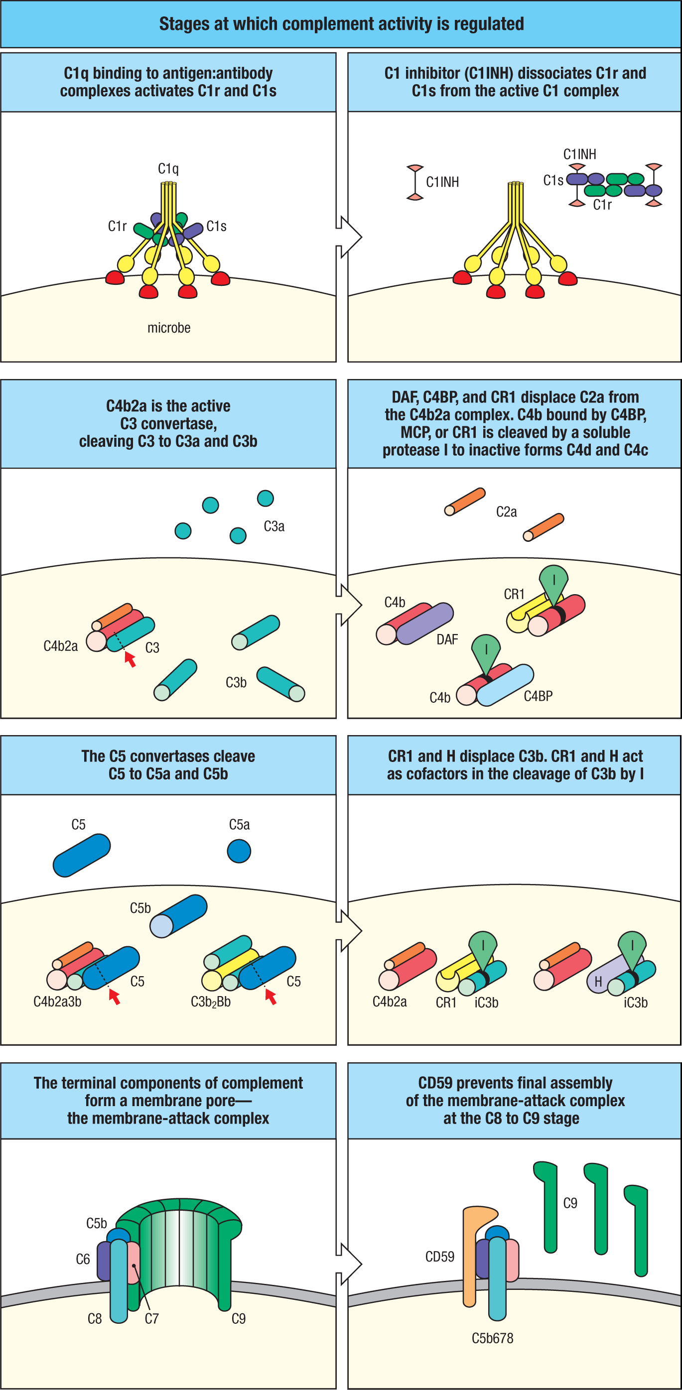
Because the highly reactive thioester bond of activated C3 and C4 cannot distinguish acceptor groups on a host cell from those on the surface of a pathogen, mechanisms have evolved to prevent the small amounts of C3 or C4 molecules deposited on host cells from fully triggering complement activation. We introduced these mechanisms in the context of control of the alternative pathway (see Fig. 2.27), but they are also important regulators of the classical pathway convertase (see Fig. 2.37, second and third rows). Section 2-10 described the proteins that inactivate any C3b or C4b that has bound to host cells. These are the plasma factor I and its cofactors MCP and CR1, which are membrane proteins. Circulating factor I is an active serine protease, but it can cleave C3b and C4b only when they are bound to MCP, factor H, or CR1 (see Fig. 2.27). In these circumstances, factor I cleaves C3b, first into iC3b and then further to C3dg, thus permanently inactivating it. C4b is similarly inactivated by cleavage into C4c and C4d. Microbial cell walls lack MCP and CR1 and thus cannot promote the breakdown of C3b and C4b, which instead act as binding sites for factor B and C2, promoting complement activation. The importance of factor I can be seen in people with genetically determined factor I deficiency. Because of uncontrolled complement activation, complement proteins rapidly become depleted, and such people suffer repeated bacterial infections, especially with ubiquitous pyogenic bacteria.
There are also plasma proteins with cofactor activity for factor I, most notably C4b-binding protein (C4BP) (see Fig. 2.36). It binds C4b and acts mainly as a regulator of the classical pathway in the fluid phase. Factor H binds C3b in the fluid phase, as well as at cell membranes, and helps to distinguish the C3b that is bound to host cells from that bound to microbial surfaces. The higher affinity of factor H for sialic acid residues on host-membrane glycoproteins allows it to displace factor B in binding to C3b on host cells. Also, C3b at host-cell membranes is bound by cofactor protein MCP. Factor H competes with factor B for binding to C3b bound to host cells, so that the bound C3b is catabolized by factor I into iC3b and C3dg, and complement activation is inhibited. In contrast, microbial membranes do not express MCP and lack the sialic acid modifications that attract factor H, and so factor B is favored for binding C3b on these surfaces. The greater amount of factor B on a microbial surface stimulates formation of more C3bBb, the alternative pathway C3 convertase, and thus amplifies complement activation.
2-17 Genetic and acquired disorders in complement regulation can produce various inflammatory conditions.
The critical balance between the inhibition and the activation of complement on cell surfaces is illustrated in individuals heterozygous for mutations in various complement-regulatory proteins. Several renal diseases are caused by complement dysregulation. A condition known as atypical hemolytic uremic syndrome (aHUS) can be caused by relatively rare mutations in factor H, factor I, and MCP. The reduced action of these regulatory factors can increase the alternative pathway activation on the surface of platelets and red blood cells. The damage that this produces can lead to low levels of platelets (thrombocytopenia) and red blood cells (anemia) and can damage the kidneys. aHUS can also be caused by an acquired autoantibody that binds to factor H and reduces its activity, again leading to increased alternative pathway activity. A related condition, called C3 glomerulopathy, can be caused by similar mutations in these factors. It is characterized by deposition of C3b in the renal glomerulus but is not associated with thrombocytopenia or anemia. Like aHUS, C3 glomerulopathy is more commonly caused by autoantibodies that inhibit factor H. Such autoantibodies are called nephritic factors, as they lead to inflammation in the kidney, and they can be directed at other components of complement. One type of nephritic factor is targeted to C3, and stabilizes the alternative pathway C3 convertase C3bBb, rather than inhibiting a complement regulatory protein.
Another serious health problem related to complement malfunction is a significantly increased risk of age-related macular degeneration (AMD). AMD is the leading cause of blindness in the elderly in developed countries. It has been linked to single-nucleotide polymorphisms in the factor H gene, where tyrosine 402 is changed to histidine (Y402H). This polymorphism in factor H is not the primary cause of AMD, but it influences the response to retinal injury. Cellular debris produced during injury or renewal of photoreceptors, such as oxidized lipids, can activate complement and induce local inflammation. Normal factor H can bind these oxidized lipids and inactivate C3b that would otherwise trigger further complement activation. The Y402H variant of factor H interacts more weakly with oxidized lipids than does normal factor H, and consequently allows a chronic background of complement activation and inflammation to take place, which damages retinal epithelial cells and activates macrophages. Polymorphisms in other complement genes have also been found to be either detrimental or protective for this disease. Thus, even small alterations in the efficiency of either the activation or the regulation of this powerful effector system can contribute to the progression of degenerative or inflammatory disorders.
DAF and MCP also mediate protection against complement through a third mechanism, which augments the dissociation of C4b2a and C3bBb convertases that have already formed. CR1 is among the host-cell membrane molecules that regulate complement through both these mechanisms; that is, by promoting the dissociation of convertase and exhibiting cofactor activity. All the proteins that bind the homologous C4b and C3b molecules contain three or more tandem copies of a structural element called the short consensus repeat (SCR), the complement-control protein (CCP) repeat, or (especially in Japan) the sushi domain.
In addition to the mechanisms for preventing C3 convertase formation and C4 and C3 deposition on cell membranes, there are also inhibitory mechanisms that prevent the inappropriate insertion of the membrane-attack complex (MAC) into membranes. We saw in Section 2-15 that the membrane-attack complex polymerizes onto C5b molecules created by the action of C5 convertase. The membrane-attack complex mainly inserts into cell membranes adjacent to the site of the C5 convertase; that is, close to the site of complement activation on a pathogen. However, some newly formed membrane-attack complexes may diffuse from the site of complement activation and insert into adjacent host-cell membranes. Several plasma proteins, including, notably, vitronectin (also known as S-protein), bind to the C5b67, C5b678, and C5b6789 complexes and thereby inhibit their random insertion into cell membranes. Host-cell membranes also contain the intrinsic protein CD59, or protectin, which inhibits the binding of C9 to the C5b678 complex (see Fig. 2.37, bottom row). CD59 and DAF are both linked to the cell surface by a glycosylphosphatidylinositol (GPI) tail, like many other peripheral membrane proteins. One of the enzymes involved in the synthesis of GPI tails is encoded by a gene, PIGA, on the X chromosome. In people with a somatic mutation in this gene in a clone of hematopoietic cells, both CD59 and DAF fail to function. This causes the disease paroxysmal nocturnal hemoglobinuria, which is characterized by episodes of intravascular red blood cell lysis by complement. Red blood cells that lack only CD59 are also susceptible to destruction as a result of spontaneous activation of the complement cascade.
2-18 Pathogens produce several types of proteins that can inhibit complement activation.
Bacterial pathogens have evolved various strategies to avoid activation of complement, and thereby to avoid elimination by this first line of innate defense (Fig. 2.38). One strategy that many pathogens employ is to mimic host surfaces by attracting host complement regulators to their own surfaces. A mechanism to achieve this is for the pathogen to express surface proteins that bind to soluble complement-regulatory proteins such as C4BP and factor H; for example, the Gram-negative pathogen Neisseria meningitidis produces factor H binding protein (fHbp), which recruits factor H (see Section 2-10), and the outer membrane protein PorA, which binds to C4BP. By recruiting factor H and C4BP to the pathogen membrane, the pathogen is able to inactivate C3b that is deposited on its surface and thereby avoid the consequences of complement activation. Complement is important in defense against Neisseria species, and several complement deficiencies are associated with increased susceptibility to this pathogen.
|
Pathogen |
Evasion molecule |
Host target |
Mechanism of action |
|
Membrane proteins |
|||
|
Neisseria meningitidis |
Factor H binding protein (fHbp) |
Factor H |
Inactivates bound C3b |
|
Borrelia burgdorferi |
Outer surface protein E (OspE) |
Factor H |
Inactivates bound C3b |
|
Streptococcus pneumoniae |
Pneumococcal surface protein C (PspC) |
Factor H |
Inactivates bound C3b |
|
Secreted proteins |
|||
|
Neisseria meningitides |
PorA |
C4BP |
Inactivates bound C3b |
|
Staphylococcus aureus |
Clumping factor A (ClfA) |
Factor I |
Inactivates bound C3b |
|
Staphylococcus aureus |
Staphylococcal protein A (Spa) |
Immunoglobulin |
Binds to Fc regions and interferes with C1 activation |
|
Staphylococcus aureus |
Staphylokinase (SAK) |
Immunoglobulin |
Cleaves immunoglobulins |
|
Staphylococcus aureus |
Complement inhibitor (SCIN, CINB, SCINC) |
C3 convertase (C4b2a, C3bBb) |
Inhibition of convertase activity |
Fig. 2.38 Complement evasion proteins produced by various pathogens.
Another strategy employed by pathogens is to secrete proteins that directly inhibit components of complement. The Gram-positive pathogen Staphylococcus aureus provides several examples of this type of strategy. Staphylococcal protein A (Spa) binds to the Fc regions of immunoglobulins and interferes with the recruitment and activation of C1. This binding specificity was used as an early biochemical technique in the purification of antibodies. The staphylococcal protein staphylokinase (SAK) acts by cleaving immunoglobulins bound to the pathogen membrane, preventing complement activation and avoiding phagocytosis. The staphylococcal complement inhibitor (SCIN) protein binds to the classical C3 convertase, C4b2a, and the alternative pathway C3 convertase, C3bBb, and inhibits their activity. Other stages of complement activation, including formation of the C5 convertase, are targets of inhibition by proteins produced by these and other pathogens. We will return to this topic of complement regulation in Chapter 13 when we discuss how the immune system sometimes fails or is evaded by pathogens.
Summary.
The complement system is one of the major mechanisms by which pathogen recognition is converted into an effective host defense against initial infection. Complement is a system of plasma proteins that can be activated directly by pathogens or indirectly by pathogen-bound antibody, leading to a cascade of reactions that occurs on the surface of pathogens and generates active components with various effector functions. There are three pathways of complement activation: the lectin pathway, triggered by the pattern-recognition receptors MBL and the ficolins; the classical pathway, triggered directly by antibody binding to the pathogen surface; and the alternative pathway, which utilizes spontaneous C3 deposition onto microbial surfaces, is augmented by properdin, and provides an amplification loop for the other two pathways. The early events in all pathways consist of a sequence of cleavage reactions in which the larger cleavage product binds covalently to the pathogen surface and contributes to the activation of the next component. The pathways converge with the formation of a C3 convertase enzyme, which cleaves C3 to produce the active cleavage product C3b. The binding of large numbers of C3b molecules to the pathogen is the central event in complement activation. Bound complement components, especially bound C3b and its inactive fragments, are recognized by specific complement receptors on phagocytic cells, which engulf pathogens opsonized by C3b and its inactive fragments. The small cleavage fragments of C3, C4, and C5 act on specific trimeric G protein–coupled receptors to recruit phagocytes, such as neutrophils, to sites of infection or to alter local vascular permeability and coagulation. Together, these activities promote the uptake and destruction of pathogens by phagocytes. The molecules of C3b that bind the C3 convertase itself initiate the late events of complement, binding C5 to make it susceptible to cleavage by C2a or Bb. The larger C5b fragment triggers the assembly of a membrane-attack complex, which can result in the lysis of certain pathogens. A system of soluble and membrane-bound complement-regulatory proteins acts to limit complement activation on host tissues, in order to prevent tissue damage from the inadvertent binding of activated complement components to host cells or from the spontaneous activation of complement components in plasma. Many pathogens produce a variety of soluble and membrane-associated proteins that can counteract complement activation and contribute to infection of the host by the microbe.
Summary to Chapter 2.
This chapter has described the preexisting, constitutive components of innate immunity. The body’s epithelial surfaces are a constant but imperfect barrier to pathogen entry. In addition to their other specialized functions, epithelia have specialized adaptations, such as cilia, various antimicrobial molecules, and mucus that provide the simplest form of innate immunity. The complement system is a more specialized system that combines direct recognition of microbes with a complex effector system. The lectin pathway relies on pattern-recognition receptors that detect microbial membranes, while the alternative pathway relies on spontaneous complement activation that is down-regulated by host molecules expressed on self membranes. The main event in complement activation is accumulation of C3b on microbial membranes, which is recognized by complement receptors on phagocytic cells to promote microbial clearance by cells recruited to sites of infection by C3a and C5a. In addition, C5b initiates the membrane-attack complex that is able to lyse some microbes directly. The complement cascade is under regulation to prevent attack on host tissues, and genetic variation in regulatory pathways can result in autoimmune syndromes and age-related tissue damage.
Glossary
- complement
- A set of plasma proteins that act together as a defense against pathogens in extracellular spaces. The pathogen becomes coated with complement proteins that facilitate its removal by phagocytes and that can also kill certain pathogens directly. Activation of the complement system can be initiated in several different ways. See classical pathway, alternative pathway, lectin pathway.
- opsonization
- The coating of the surface of a pathogen by antibody and/or complement that makes it more easily ingested by phagocytes.
- complement proteins
- See C1, C2, C3, etc.
- complement activation
- The activation of the normally inactive proteins of the complement system that occurs on infection. See classical pathway, alternative pathway, lectin pathway.
- zymogen
- An inactive form of an enzyme, usually a protease, that must be modified in some way, for example by selective cleavage of the protein chain, before it can become active.
- inflammation
- General term for the local accumulation of fluid, plasma proteins, and white blood cells that is initiated by physical injury, infection, or a local immune response.
- phagocytosis
- The internalization of particulate matter by cells by a process of engulfment, in which the cell membrane surrounds the material, eventually forming an intracellular vesicle (phagosome) containing the ingested material.
- membrane attack
- Effector pathway of complement that is based on formation of the membrane-attack complex (MAC).
- C2
- Complement protein of the classical and lectin pathways that is cleaved by the C1 complex to yield C2b and C2a. C2a is an active protease that forms part of the classical C3 convertase C4b2a.
- C2a
- The large fragment of C2 formed when C2 is cleaved by C1s. When bound to C4b, C2a is the serine protease of the C3 convertase (C4b2a) of the classical and the lectin pathways.
- lectin pathway
- Complement-activation pathway that is triggered by mannose-binding lectin (MBL) or ficolins bound to bacteria.
- classical pathway
- The complement-activation pathway that is initiated by C1 binding either directly to bacterial surfaces or to antibody bound to the bacteria, thus flagging the bacteria as foreign. See also alternative pathway, lectin pathway.
- alternative pathway
- A form of complement activation that is initiated by spontaneous hydrolysis of C3 and which uses factor B and factor D to form the unique C3 convertase C3bBb.
- C3 convertase
- Enzyme complex that cleaves C3 to C3b and C3a on the surface of a pathogen. The C3 convertase of the classical and lectin pathways is formed from membrane-bound C4b complexed with the protease C2a. The alternative pathway C3 convertase is formed from membrane-bound C3b complexed with the protease Bb.
- C3b
- Complement protein on which all complement activation pathways converge. C3 cleavage forms C3b, which can bind covalently to microbial surfaces, where it promotes destruction by phagocytes.
- C3b2Bb
- The C5 convertase of the alternative pathway of complement activation.
- C3a
- Pro-inflammatory complement fragments C5a and C3a released by cleavage during complement activation. They are recognized by specific receptors and recruit fluid and inflammatory cells to the site of their release.
- C3f
- A small fragment of C3b that is removed by factor I and MCP to leave iC3b on the microbial surface.
- C3dg
- Breakdown product of iC3b that remains attached to the microbial surface, where it can bind complement receptor 2 (CR2).
- C5 convertase
- Enzyme complex that cleaves C5 to C5a and C5b.
- C5a
- Pro-inflammatory complement fragments C5a and C3a released by cleavage during complement activation. They are recognized by specific receptors and recruit fluid and inflammatory cells to the site of their release.
- C5b
- Fragment of C5 that initiates the formation of the membrane-attack complex (MAC).
- membrane-attack complex (MAC)
- Protein complex composed of C5b to C9 that assembles a membrane-spanning hydrophilic pore on pathogen surfaces, causing cell lysis.
- mannose-binding lectin (MBL)
- Mannose-binding protein present in the blood. It can opsonize pathogens bearing mannose on their surfaces and can activate the complement system via the lectin pathway, an important part of innate immunity.
- collectins
- A family of calcium-dependent sugar-binding proteins (lectins) containing collagen-like sequences. An example is mannose-binding lectin (MBL).
- avidity
- The sum total of the strength of binding of two molecules or cells to one another at multiple sites. It is distinct from affinity, which is the strength of binding of one site on a molecule to its ligand.
- acute-phase response
- A change in the proteins present in the blood that occurs during the early phases of an infection. It includes the production of acute-phase proteins, many of which are produced in the liver.
- ficolins
- Carbohydrate-binding proteins that can initiate the lectin pathway of complement activation. They are members of the collectin family and bind to the N-acetylglucosamine present on the surface of some pathogens.
- MBL-associated serine proteases
- See MASP-1, MASP-2, MASP-3.
- MAp19
- A non-enzymatically active splice variant encoded by the MASP-2 gene that can associate with mannose-binding lectin (MBL) and may regulate lectin pathway complement activation.
- MAp44
- A non-enzymatically active splice variant encoded by the MASP-1 gene that can associate with mannose-binding lectin (MBL) and may regulate lectin pathway complement activation.
- C4
- Complement protein of the classical and lectin pathways. C4 is cleaved by C1s to C4b, which forms part of the classical C3 convertase.
- C4b2a
- C3 convertase of the classical and lectin pathways of complement activation.
- surfactant proteins A and D (SP-A and SP-D)
- Acute-phase proteins that help protect the epithelial surfaces of the lung against infection.
- C1 complex (C1)
- Protein complex activated as the first step in the classical pathway of complement activation, composed of C1q bound to two molecules each of the proteases C1r and C1s. Binding of a pathogen or antibody to C1q activates C1r, which cleaves and activates C1s, which cleaves C4 and C2.
- C1 complex (C1)
- Protein complex activated as the first step in the classical pathway of complement activation, composed of C1q bound to two molecules each of the proteases C1r and C1s. Binding of a pathogen or antibody to C1q activates C1r, which cleaves and activates C1s, which cleaves C4 and C2.
- C1q
- A component of the C1 complex formed by a hexamer of trimers of a polypeptide with a globular domain that can bind bacterial surfaces or interact with Fc regions of antibodies and a carboxy-terminal collagen-like domain that mediates its assembly and association with C1r and C1s.
- C1r
- A serine protease that associates with C1q and C1s to help form the C1 complex. Its autocatalytic activity is triggered by conformational changes resulting from C1q binding to Fc or bacterial surfaces; C1r then cleaves C1s.
- C1s
- A serine protease that associates with C1q and C1r to help form the C1 complex. It is cleaved and activated by C1r and then functions to activate the downstream complement components 4 and 2 (C4 and C2).
- classical C3 convertase
- The complex of activated complement components C4b2a, which cleaves C3 to C3b on pathogen surfaces in the classical pathway of complement activation.
- natural antibodies
- Antibodies produced by the immune system in the apparent absence of any infection. They have a broad specificity for self and microbial antigens, can react with many pathogens, and can activate complement.
- alternative pathway C3 convertase
- The C3 convertase composed of C4b and C2a. This is also the C3 convertase of the classical complement pathway.
- factor B
- Protein in the alternative pathway of complement activation, in which it is cleaved to Ba and an active protease, Bb, the latter binding to C3b to form the alternative pathway C3 convertase, C3bBb.
- C3bBb
- The C3 convertase of the alternative pathway of complement activation.
- factor D
- A serine protease in the alternative pathway of complement activation, which cleaves factor B into Ba and Bb.
- tickover
- The low-level generation of C3b continually occurring in the blood in the absence of infection.
- properdin
- Plasma protein released by activated neutrophils that stabilizes the C3 convertase C3bBb of the alternative pathway. Also known as factor P.
- complement-regulatory proteins
- Proteins that control complement activity and prevent complement from being activated on the surfaces of host cells.
- iC3b
- Inactive complement fragment produced by cleavage of C3b.
- factor I
- Complement-regulatory protease in plasma that cleaves C3b to the inactive derivative iC3b, thus preventing the formation of a C3 convertase.
- CR1 (CD35)
- A receptor expressed by phagocytic cells that binds to C3b. It stimulates phagocytosis and inhibits C3 convertase formation on host-cell surfaces. It also acts in conjunction with factor I to cleave C3b to its inactive derivative iC3b and thus prevent convertase formation.
- CR1 (CD35)
- A receptor expressed by phagocytic cells that binds to C3b. It stimulates phagocytosis and inhibits C3 convertase formation on host-cell surfaces. It also acts in conjunction with factor I to cleave C3b to its inactive derivative iC3b and thus prevent convertase formation.
- factor H
- Complement-regulatory protein in plasma that binds C3b and competes with factor B to displace Bb from the convertase. It also acts in conjunction with factor I to cleave C3b to its inactive derivative iC3b and thus prevent convertase formation.
- amoeboid coelomocytes
- Phagocytic cells found in more primitive organisms (such as echinoderms and others) that carry out basic protective functions; these cells are a source of C3 homologs, thioester-containing proteins (TEPs), during infection by bacteria.
- thioester-containing proteins (TEPs)
- Homologs of complement component 3 (C3) that are found in insects and are thought to have some function in insect innate immunity.
- thioester-containing proteins (TEPs)
- Homologs of complement component 3 (C3) that are found in insects and are thought to have some function in insect innate immunity.
- C4b2a3b
- C5 convertase of the classical and lectin pathways of complement activation.
- complement receptors (CRs)
- Cell-surface proteins of various types that recognize and bind complement proteins that have become bound to an antigen such as a pathogen. Complement receptors on phagocytes enable them to identify and bind pathogens coated with complement proteins and to ingest and destroy them. See CR1, CR2, CR3, CR4, CRIg, and the C1 complex.
- C5a receptor 1
- A G protein–coupled receptor expressed by macrophages and activated by C5a that facilitates ingestion of opsonized bacteria bound to CR1.
- G protein–coupled receptors (GPCRs)
- A large class of seven-span transmembrane cell-surface receptors that associate with intracellular heterotrimeric G proteins after ligand binding and signal by activation of the G protein. Important examples are the chemokine receptors.
- C5a receptor 2
- A nonsignaling surface receptor expressed by neutrophils and macrophages and which may act as a decoy receptor for C5a and thus regulate activity of the C5a receptor.
- CR2 (CD21)
- Complement receptor that is part of the B-cell co-receptor complex. It binds to antigens coated with breakdown products of C3b, especially C3dg, and, by cross-linking the B-cell receptor, enhances sensitivity to antigen at least 100-fold. It is also the receptor used by the Epstein–Barr virus to infect B cells.
- CR2 (CD21)
- Complement receptor that is part of the B-cell co-receptor complex. It binds to antigens coated with breakdown products of C3b, especially C3dg, and, by cross-linking the B-cell receptor, enhances sensitivity to antigen at least 100-fold. It is also the receptor used by the Epstein–Barr virus to infect B cells.
- CR3 (CD11b:CD18)
- Complement receptor 3. A β2 integrin that acts both as an adhesion molecule and as a complement receptor. CR3 on phagocytes binds iC3b, a breakdown product of C3b on pathogen surfaces, and stimulates phagocytosis.
- CR4 (CD11c:CD18)
- A β2 integrin that acts both as an adhesion molecule and as a complement receptor. CR4 on phagocytes binds iC3b, a breakdown product of C3b on pathogen surfaces, and stimulates phagocytosis.
- CRIg (complement receptor of the immunoglobulin superfamily)
- A complement receptor that binds to inactivated forms of C3b.
- anaphylactic shock
- A rapid-onset and systemic allergic reaction to antigen; for example, to insect venom injected directly into the bloodstream or to foods such as peanuts. Severe systemic reactions can potentially be fatal due to circulatory collapse and suffocation from tracheal swelling. Anaphylaxis usually results from antigens binding to IgE bound by Fcε receptors on mast cells, leading to systemic release of inflammatory mediators.
- anaphylatoxins
- Pro-inflammatory complement fragments C5a and C3a released by cleavage during complement activation. They are recognized by specific receptors and recruit fluid and inflammatory cells to the site of their release.
- C1 inhibitor (C1INH)
- An inhibitor protein for C1 that binds and inactivates C1r:C1s enzymatic activity. Deficiency in C1INH causes hereditary angioedema through production of vasoactive peptides that cause subcutaneous and laryngeal swelling.
- serine protease inhibitor (serpin)
- Class of proteins that inhibit various proteases, originally referring to those specific to serine proteases.
- serine protease inhibitor (serpin)
- Class of proteins that inhibit various proteases, originally referring to those specific to serine proteases.
- hereditary angioedema (HAE)
- A genetic deficiency of the C1 inhibitor of the complement system. In the absence of C1 inhibitor, spontaneous activation of the complement system can cause diffuse fluid leakage from blood vessels, the most serious consequence of which is swelling of the larynx, leading to suffocation.
- carboxypeptidase N (CPN)
- A metalloproteinase that inactivates C3a and C5a. CPN deficiency causes a condition of recurrent angioedema.
- factor I deficiency
- A genetically determined lack of the complement-regulatory protein factor I. This results in uncontrolled complement activation, so that complement proteins rapidly become depleted. Those with the deficiency suffer repeated bacterial infections, especially with ubiquitous pyogenic bacteria.
- C4b-binding protein (C4BP)
- A complement-regulatory protein that inactivates the classical pathway C3 convertase formed on host cells by displacing C2a from the C4b2a complex. C4BP binds C4b attached to host cells, but cannot bind C4b attached to pathogens.
- atypical hemolytic uremic syndrome (aHUS)
- A condition characterized by damage to platelets and red blood cells and inflammation of the kidneys that is caused by uncontrolled complement activation in individuals with inherited deficiencies in complement-regulatory proteins.
- C3 glomerulopathy
- An inflammatory condition in the kidney in which C3b is abnormally deposited in the glomerulus leading to proteinuria and hematuria. It can be associated with autoantibodies, called nephritic factors, that can inhibit factor H, other complement-regulatory components, or that stabilize C3 convertase, leading to increased C3 deposition.
- nephritic factor
- An autoantibody that leads to inflammation in the kidney by increasing C3 deposition. Nephritic factors may inhibit the activity of complement-regulatory proteins, such as factor H, or may stabilize the alternative pathway C3 convertase C3bBb.
- vitronectin
- Plasma protein that binds incompletely formed MAC complexes, such as C5b67, preventing bystander complement damage to host membranes.
- S-protein (vitronectin)
- Plasma protein that binds incompletely formed MAC complexes, such as C5b67, preventing bystander complement damage to host membranes.
- glycosylphosphatidylinositol (GPI) tail
- A glycolipid modification of proteins that can allow attachment to host membranes without the requirement of a transmembrane protein domain.
- paroxysmal nocturnal hemoglobinuria
- A disease in which complement-regulatory proteins are defective, so that activation of complement binding to red blood cells leads to episodes of spontaneous hemolysis.
- factor H binding protein (fHbp)
- A protein produced by the pathogen Neisseria meningitidis that recruits factor H to its membrane, thereby inactivating C3b deposited on its surface, and evading destruction by complement.
- PorA
- Outer membrane protein of Neisseria meningitidis that binds C4BP, thereby inactivating C3b deposited on its surface.
- staphylococcal protein A (Spa)
- Staphylococcal protein that blocks the binding of the antibody Fc region with C1, thereby preventing complement activation.
- staphylokinase (SAK)
- Staphylococcal protease that cleaves immunoglobulins bound to its surface, thereby preventing complement activation.
- staphylococcal complement inhibitor (SCIN)
- Staphylococcal protein that inhibits the activity of the classical and alternative C3 convertases, promoting the evasion of destruction by complement.
- MASP-1, MASP-2, MASP-3
- Serine proteases of the classical and lectin pathways of complement activation that bind to C1q, ficolins, and mannose-binding lectin, and function in their activation to cleave C4.
- MASP-1, MASP-2, MASP-3
- Serine proteases of the classical and lectin pathways of complement activation that bind to C1q, ficolins, and mannose-binding lectin, and function in their activation to cleave C4.
- fluid-phase C3 convertase
- Short-lived alternative pathway C3 convertase, C3(H20)Bb, that is continually produced at a low level in the plasma that can initiate activation of the alternative pathway of complement activation.
- decay-accelerating factor (DAF or CD55)
- A cell-surface protein that protects cells from lysis by complement. Its absence causes the disease paroxysmal nocturnal hemoglobinuria.
- membrane cofactor of proteolysis (MCP or CD46)
- A complement-regulatory protein; a host-cell membrane protein that acts in conjunction with factor I to cleave C3b to its inactive derivative iC3b and thus prevent convertase formation.
- C6, C7, C8, C9
- Complement proteins that act with C5b to form the membrane-attack complex, producing a pore that leads to lysis of the target cell.
- C6, C7, C8, C9
- Complement proteins that act with C5b to form the membrane-attack complex, producing a pore that leads to lysis of the target cell.
- C6, C7, C8, C9
- Complement proteins that act with C5b to form the membrane-attack complex, producing a pore that leads to lysis of the target cell.
- C6, C7, C8, C9
- Complement proteins that act with C5b to form the membrane-attack complex, producing a pore that leads to lysis of the target cell.
- CD59, protectin
- Cell-surface protein that protects host cells from complement damage by blocking binding of C9 to the C5b678 complex, thus preventing MAC formation.
- CD59, protectin
- Cell-surface protein that protects host cells from complement damage by blocking binding of C9 to the C5b678 complex, thus preventing MAC formation.