Anatomic barriers and initial chemical defenses.
Microorganisms that cause disease in humans and animals enter the body at different sites and produce disease symptoms by a variety of mechanisms. Microorganisms that cause disease and produce damage, or pathology, to tissues are referred to as pathogenic microorganisms, or simply pathogens. As innate immunity eliminates most microorganisms that may occasionally cross an anatomic barrier, pathogens are microorganisms that have evolved ways of overcoming the body’s innate defenses more effectively than have other microorganisms. Once infection is established, both innate and adaptive immune responses are typically required to eliminate pathogens from the body. Even in these cases, the innate immune system performs a valuable function by reducing pathogen numbers during the time needed for the adaptive immune system to gear up for action. In the first part of this chapter, we briefly describe the different types of pathogens and their invasive strategies and then examine the immediate innate defenses that, in most cases, prevent microorganisms from establishing an infection.
2-1 Infectious diseases are caused by diverse living agents that replicate in their hosts.
The agents that cause disease fall into five groups: viruses, bacteria, fungi, protozoa, and helminths (worms). Protozoa and worms are usually grouped together as parasites and are the subject of the discipline of parasitology, whereas viruses, bacteria, and fungi are the subject of microbiology. Figure 2.2 lists some examples of the different classes of microorganisms and parasites, and the diseases they cause. The characteristic features of each pathogen are its mode of transmission, its mechanism of replication, its mechanism of pathogenesis—the means by which it causes disease—and the response it elicits from the host. The distinct pathogen habitats and life cycles mean that a range of different innate and adaptive immune mechanisms have to be deployed for pathogen destruction.
|
Routes of infection for pathogens |
||||
|
Route of entry |
Mode of transmission |
Pathogen |
Disease |
Type of pathogen |
|
Mucosal surfaces |
||||
|
Mouth and respiratory tract |
Inhalation or ingestion of infective material (e.g., saliva droplets) |
Measles virus Influenza virus Varicella-zoster Epstein–Barr virus Streptococcus pyogenes Haemophilus influenzae Neisseria meningitidis |
Measles Influenza Chickenpox Mononucleosis Tonsilitis Pneumonia, meningitis Meningococcal meningitis |
Paramyxovirus Orthomyxovirus Herpesvirus Herpesvirus Gram-positive bacterium Gram-negative bacterium Gram-negative bacterium |
|
Spores |
Bacillus anthracis |
Inhalation anthrax |
Gram-positive bacterium |
|
|
Gastrointestinal tract |
Contaminated water or food |
Rotavirus Hepatitis A Salmonella enterica ssp. enteritidis, Salmonella enterica ssp. typhimurium Vibrio cholerae Salmonella enterica ssp. typhi Trichuris trichiura |
Diarrhea Jaundice Food poisoning Cholera Typhoid fever Trichuriasis |
Rotavirus Picornavirus Gram-negative bacterium Gram-negative bacterium Gram-negative bacterium Helminth |
|
Reproductive tract and other routes |
Sexual transmission/ infected blood |
Hepatitis B virus Human immunodeficiency virus (HIV) |
Hepatitis B Acquired immune deficiency syndrome (AIDS) |
Hepadnavirus Retrovirus |
|
Sexual transmission |
Neisseria gonorrhoeae Treponema pallidum |
Gonorrhea Syphilis |
Gram-negative bacterium Bacterium (spirochete) |
|
|
Opportunistic infections |
Resident microbiota |
Candida albicans |
Candidiasis, thrush |
Fungus |
|
Resident lung microbiota |
Pneumocystis jirovecii |
Pneumonia |
Fungus |
|
|
External epithelia |
||||
|
External surface |
Physical contact |
Trichophyton |
Athlete’s foot |
Fungus |
|
Wounds and abrasions |
Minor skin abrasions Puncture wounds Handling infected animals |
Bacillus anthracis Clostridium tetani Francisella tularensis |
Cutaneous anthrax Tetanus Tularemia |
Gram-positive bacterium Gram-positive bacterium Gram-negative bacterium |
|
Insect bites |
Mosquito bites (Aedes aegypti) Deer tick bites Mosquito bites (Anopheles) |
Flavivirus Borrelia burgdorferi Plasmodium spp. |
Yellow fever Lyme disease Malaria |
Virus Bacterium (spirochete) Protozoan |
Fig. 2.2 A variety of microorganisms can cause disease. Pathogenic organisms are of five main types: viruses, bacteria, fungi, protozoa, and helminths (worms). Some well-known pathogens are listed.
Infectious agents can grow in all body compartments, as shown schematically in Fig. 2.3. We saw in Chapter 1 that two major compartments can be defined: extracellular and intracellular. Both innate and adaptive immune responses have different ways of dealing with pathogens found in these two compartments. Many bacterial pathogens live and replicate in extracellular spaces, either within tissues or on the surface of the epithelia that line body cavities. Extracellular bacteria are usually susceptible to killing by phagocytes, an important arm of the innate immune system, but some pathogens, such as Staphylococcus and Streptococcus species, achieve a very basic form of immune evasion by having a protective polysaccharide capsule that resists engulfment. This can be overcome to some extent by the help of another component of innate immunity (complement), which deposits specific molecules on the surface of the bacteria (opsonization), rendering them more susceptible to phagocytosis. In the adaptive immune response, bacteria are rendered more susceptible to phagocytosis by a combination of antibodies and complement.
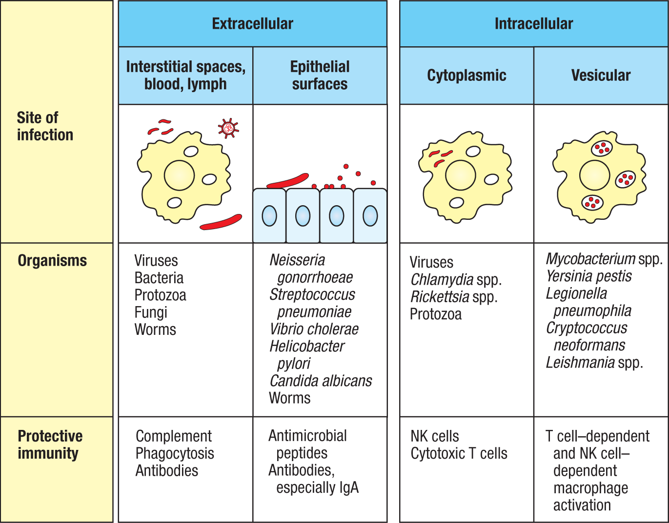
Infectious diseases differ in their symptoms and outcome depending on where the causal pathogen replicates within the body—the intracellular or the extracellular compartment—and what damage it does to the tissues (Fig. 2.4). Pathogens that live intracellularly frequently cause disease by damaging or killing the cells they infect. Obligate intracellular pathogens, such as viruses, must invade host cells to replicate. Facultative intracellular pathogens, such as mycobacteria, can replicate either intracellularly or outside the cell. Two strategies of innate immunity defend against intracellular pathogens. One strategy aims to destroy pathogens before they infect cells and includes soluble defenses such as antimicrobial peptides, as well as phagocytic cells that can engulf and destroy pathogens before they become intracellular. Alternatively, the innate immune system can recognize and kill cells infected by some pathogens. This is the role of the natural killer cells (NK cells), which act to limit certain viral infections before cytotoxic T cells of the adaptive immune system become functional. Intracellular pathogens can be subdivided further into those that replicate freely in the cell, such as viruses and certain bacteria (for example, Chlamydia, Rickettsia, and Listeria), and those that replicate inside intracellular vesicles, such as mycobacteria. Pathogens that live inside macrophage vesicles may become more susceptible to being killed after activation of the macrophage as a result of NK-cell or T-cell actions (see Fig. 2.3).
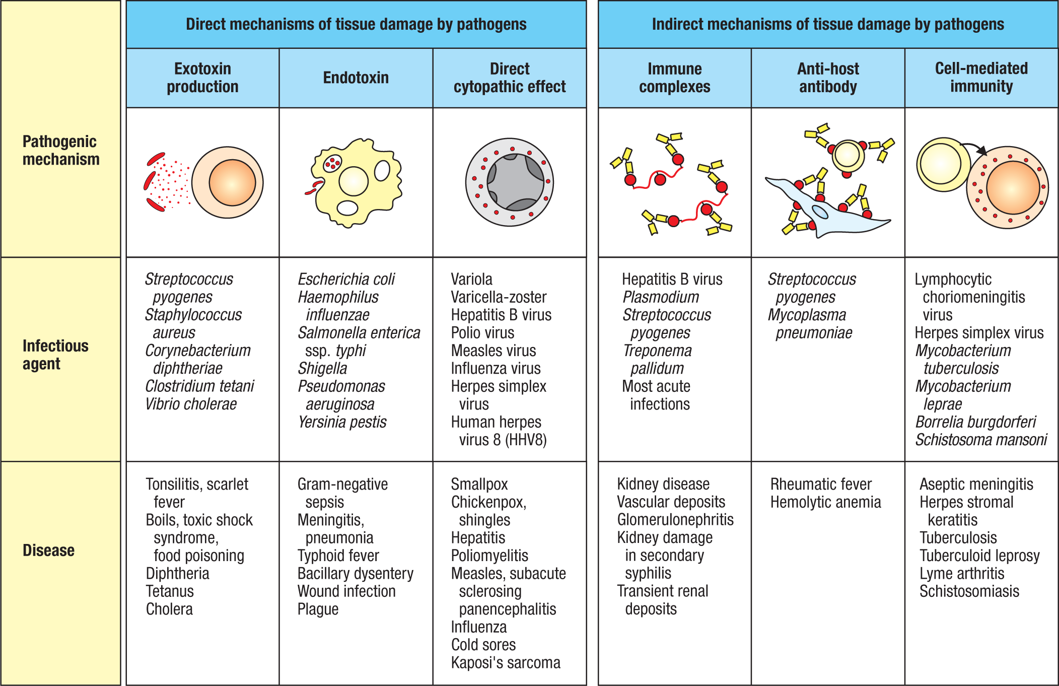
Many of the most dangerous extracellular bacterial pathogens cause disease by releasing protein toxins; these secreted toxins are called exotoxins (see Fig. 2.4). The innate immune system has little defense against such toxins, and highly specific antibodies produced by the adaptive immune system are required to neutralize their action (see Fig. 1.28). The damage caused by a particular infectious agent also depends on where it grows; Streptococcus pneumoniae in the lung causes pneumonia, for example, whereas in the blood it causes a potentially fatal systemic illness, pneumococcal sepsis. In contrast, nonsecreted constituents of bacterial structure that trigger phagocytes to release cytokines with local and systemic effects are called endotoxins. An endotoxin of major medical importance is the lipopolysaccharide (LPS) of the outer cell membrane of Gram-negative bacteria, such as Salmonella. Many of the clinical symptoms of infection by such bacteria—including fever, pain, rash, hemorrhage, septic shock—are due largely to LPS.
Most pathogenic microorganisms can overcome innate immune responses and continue to grow, making us ill. An adaptive immune response is required to eliminate them and to prevent subsequent reinfections. Certain pathogens are never entirely eliminated by the immune system and persist in the body for years. But most pathogens are not universally lethal. Those that have lived for thousands of years in the human population are highly evolved to exploit their human hosts; they cannot alter their pathogenicity without upsetting the compromise they have achieved with the human immune system. Rapidly killing every host it infects is no better for the long-term survival of a pathogen than being wiped out by the immune response before the microbe has had time to infect someone else. In short, we have adapted to live with many microbes, and they with us. Nevertheless, highly pathogenic strains of avian influenza, the episode in 2002–2003 of SARS (severe acute respiratory syndrome), a severe pneumonia in humans that is caused by the coronavirus SARS-CoV from bats, and the SARS-CoV-2 pandemic of 2020 all remind us that new and deadly infections can transfer from animal reservoirs to humans. Even though infection with SARS-CoV-2 has a case fatality rate substantially lower than that seen in SARS, the 2020 pandemic has had an enormous negative impact on the global economy. Such transmission appears responsible for the Ebola virus epidemic in West Africa in 2014–2015. These are known as zoonotic infections—and we must be on the alert at all times for the emergence of new pathogens and new threats to health. The human immunodeficiency virus that causes AIDS (discussed in Chapter 13) serves as a warning that we remain constantly vulnerable.
 Case Study: Acquired Immune Deficiency Syndrome (AIDS)
2-2 Epithelial surfaces of the body provide the first barrier against infection.
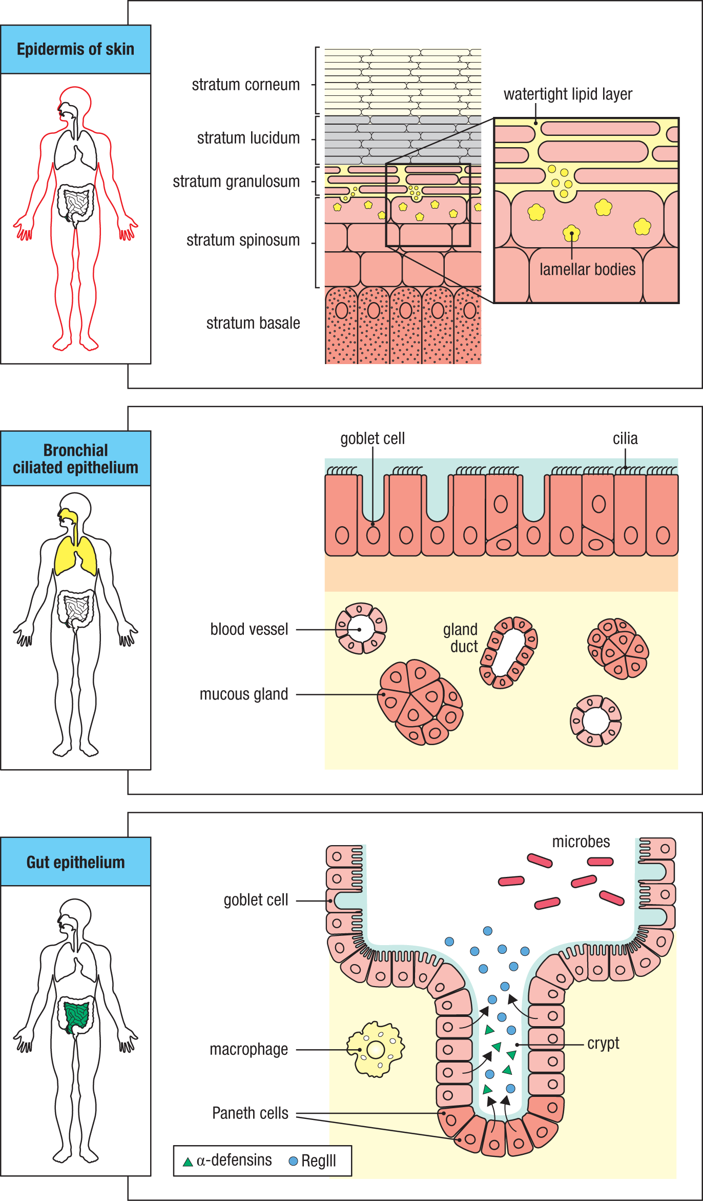
Our body surfaces are composed of epithelia with diverse functions. They can provide a protective physical barrier between the internal milieu and the external world that contains pathogens. Epithelia compose the skin and the linings of the body’s tubular structures—the respiratory, urogenital, and gastrointestinal tracts. Epithelia in these locations are specialized for their particular functions and possess unique innate defense strategies against the microbes they typically encounter (Fig. 2.5 and Fig. 2.6). The necessity to carry out various functions in these locations, such as absorption of nutrients or oxygen for example, means that these epithelia are not perfect barriers to infection.
|
Skin |
Gut |
Lungs |
Eyes/nose/oral cavity |
|
|
Stratified epithelium |
Single cell layer of columnar epithelium |
Upper airway: pseudostratified columnar epithelium Lower airway: single cell layer of columnar epithelium |
Pseudostratified columnar epithelium |
|
|
Mechanical |
Epithelial cells joined by tight junctions |
|||
|
Longitudinal flow of air or fluid |
Longitudinal flow of air or fluid |
Movement of mucus by cilia |
Tears Nasal cilia |
|
|
Chemical |
Fatty acids |
Low pH |
Pulmonary surfactant |
Enzymes in tears and saliva (lysozyme) |
|
Enzymes (pepsin) |
||||
|
β-defensins Lamellar bodies Cathelicidin |
α-defensins (cryptdins) RegIII (lecticidins) Cathelicidin |
α-defensins Cathelicidin |
Histatins β-defensins |
|
|
Microbiological |
Normal microbiota |
|||
Fig. 2.5 Many barriers prevent pathogens from crossing epithelia and colonizing tissues. Surface epithelia provide mechanical, chemical, and microbiological barriers to infection. The epithelium of the skin is stratified, having multiple layers. Epithelia in the various locations in the intestine have a single cell layer. The epithelia of the upper airway have a single cell layer, but the cell nuclei are staggered in a way that suggests multiple layers.
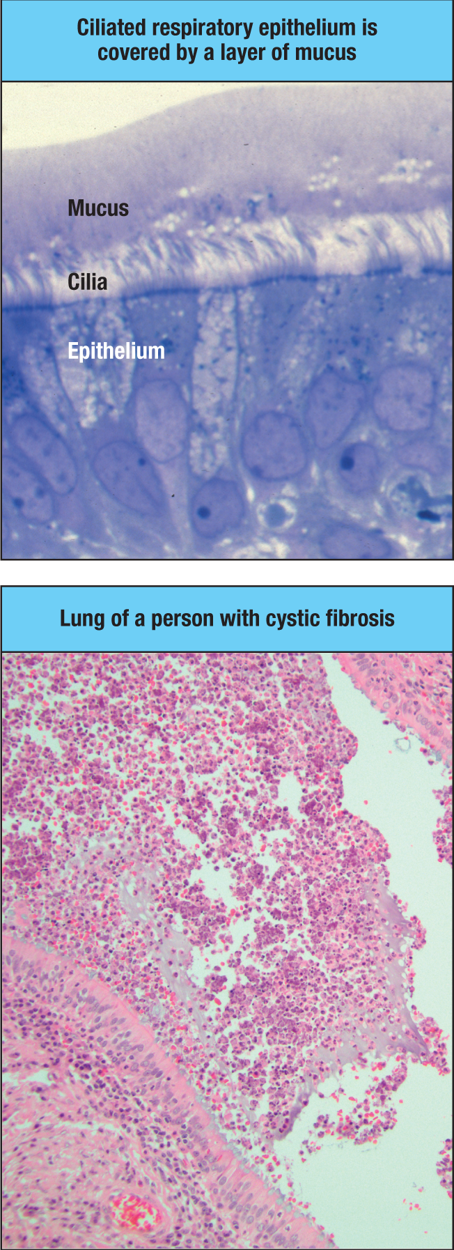
Epithelia can be a layer of single cells or multiple layers of cells with varying geometry. Epithelial cells are held together by tight junctions, which effectively form a seal against the external environment. The body’s internal epithelia are known as mucosal epithelia because they secrete a viscous fluid called mucus, which contains many glycoproteins called mucins. Mucus has a number of protective functions. The mucus layer establishes a viscous physical barrier that impedes microbial access to various epithelial surfaces. In the respiratory tract, microorganisms can be expelled in the outward flow of mucus driven by the beating of cilia on the mucosal epithelium (Fig. 2.7). The importance of mucus flow in clearing infection is illustrated by people with the inherited disease cystic fibrosis, in which the mucus becomes abnormally thick and dehydrated because of defects in a gene, CFTR, encoding a chloride channel in the epithelium. Such individuals frequently develop lung infections caused by bacteria that colonize the epithelial surface but do not cross it (see Fig. 2.7). Mucus also plays an important role in the gut. Peristalsis is an important mechanism for keeping both food and infectious agents moving through the body. Failure of peristalsis is typically accompanied by the overgrowth of pathogenic bacteria within the lumen of the gut. The colon of the large intestine has two layers of mucus. The inner layer, nearest the epithelium, has a denser, less porous composition that excludes the passage of bacteria from the colonic lumen to the epithelial surface. An outer mucus layer, farther from the epithelium, is more porous, forming the habitat for colonic bacteria.
Most healthy epithelial surfaces are associated with populations of normally nonpathogenic bacteria, known as commensal bacteria or the microbiota, which help keep pathogens at bay. The microbiota can also make antimicrobial substances, such as the lactic acid produced by vaginal lactobacilli, some strains of which also produce antimicrobial peptides (bacteriocins). Commensal microorganisms in the outer mucus layer generate short-chain fatty acids (SCFAs) and other metabolites that can strengthen the barrier functions of epithelia by stimulating production of antimicrobial peptides (see Section 2-4). When commensal microorganisms are killed by antibiotic treatment, pathogens frequently replace them and cause disease (see Fig. 12.13). Under some circumstances commensal microbes themselves can cause disease if their growth is not kept in check or if the immune system is compromised. In Chapter 12, we will discuss how commensal microorganisms play an important role in the setting of normal immunity, particularly in the intestine; and in Chapter 15, we will see how these normally nonpathogenic organisms can cause disease in the context of inherited immunodeficiencies.
2-3 Infectious agents must overcome innate host defenses to establish a focus of infection.
Our bodies are constantly exposed to microorganisms present in our environment, including infectious agents that have been shed by other individuals. Contact with these microorganisms may occur through external or internal epithelial surfaces. To establish an infection, a microorganism must first invade the body by binding to or crossing an epithelium (Fig. 2.8). Infection is a major cause of mortality and morbidity in cases of epithelial damage caused by wounds, burns, or loss of the integrity of the body’s internal epithelia. The body rapidly repairs damaged epithelial surfaces, but even without epithelial damage, pathogens may establish infection by specifically adhering to and colonizing epithelial surfaces, using the attachment to avoid being dislodged by the flow of air or fluid across the surface.
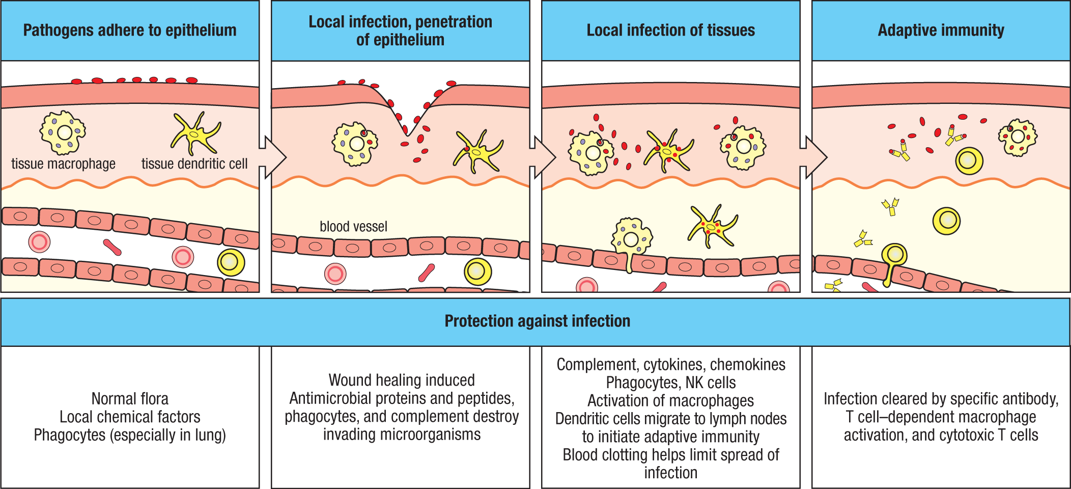
Disease occurs when a microorganism succeeds in evading or overwhelming innate host defenses to establish a local site of infection, and then replicates there to allow its further transmission within our bodies. The epithelium lining the respiratory tract provides a route of entry into tissues for airborne microorganisms, and the lining of the gastrointestinal tract does the same for microorganisms ingested in food and water. The intestinal pathogens Salmonella enterica ssp. typhi, which causes typhoid fever, and Vibrio cholerae, which causes cholera, are spread through fecally contaminated food and water, respectively. Insect bites and wounds allow microorganisms to penetrate the skin, and direct contact between individuals offers opportunities for infection through the skin, the gut, and the reproductive tract (see Fig. 2.2).
In spite of this exposure, infectious disease is fortunately quite infrequent. Most of the microorganisms that succeed in crossing an epithelial surface are efficiently removed by innate immune mechanisms that function in the underlying tissues, preventing infection from becoming established. It is difficult to know how many infections are repelled in this way, because they cause no symptoms and pass undetected.
In general, pathogenic microorganisms are distinguished from the mass of microorganisms in the environment by having special adaptations that evade the immune system. In some cases, such as the fungal disease athlete’s foot, the initial infection remains local and does not cause significant pathology. In other cases, such as tetanus, the bacterium (Clostridium tetani in this case) secretes a powerful neurotoxin, and the infection causes serious illness as it spreads through the lymphatics or the bloodstream, invades and destroys tissues, and disrupts the body’s workings.
The spread of a pathogen is often initially countered by an inflammatory response that recruits more effector cells and molecules of the innate immune system out of the blood and into the tissues, while inducing clotting in small blood vessels further downstream so that the microbe cannot spread through the circulation (see Fig. 2.8). The cellular responses of innate immunity act over several days. During this time, the adaptive immune response may also begin if antigens derived from the pathogen are delivered to local lymphoid tissues by dendritic cells (see Section 1-15). While an innate immune response may eliminate some infections, an adaptive immune response can target particular strains and variants of pathogens and protect the host against reinfection by using either effector T cells or antibodies to generate immunological memory.
2-4 Epithelial cells and phagocytes produce several kinds of antimicrobial molecules.
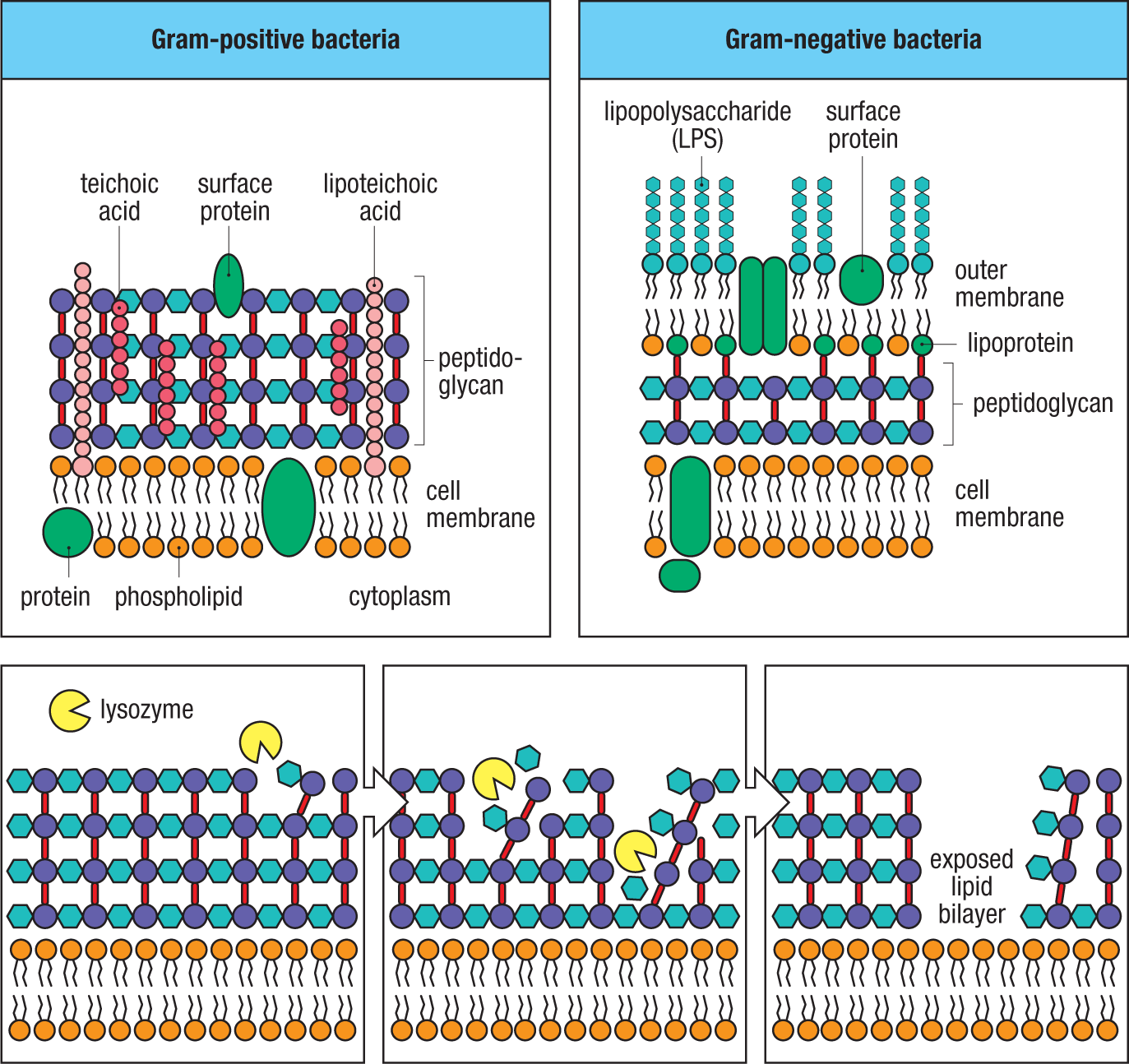
Our surface epithelia are more than mere physical barriers to infection; they also produce a wide variety of chemical substances that are microbicidal or that inhibit microbial growth. For example, the acid pH of the stomach and the digestive enzymes, bile salts, fatty acids, and lysolipids present in the upper gastrointestinal tract create a substantial chemical barrier to infection (see Fig. 2.5). One important group of antimicrobial proteins comprises enzymes that attack chemical features specific to bacterial cell walls. Such antimicrobial enzymes include lysozyme and secretory phospholipase A2, which are secreted in tears and saliva and by phagocytes. Lysozyme is a glycosidase that breaks a specific chemical bond in the peptidoglycan layer of the bacterial cell wall. Peptidoglycan is composed of a network of polymers that consist of alternating residues of N-acetylglucosamine (GlcNAc) and N-acetylmuramic acid (MurNAc); the peptidoglycan layer is formed by peptide bridges that cross-link these polysaccharide polymers (Fig. 2.9). Lysozyme selectively cleaves the β-(1,4) linkage between the two sugars of the polymer and is more effective in acting against Gram-positive bacteria, in which the peptidoglycan cell wall is exposed, than against Gram-negative bacteria, which have an outer layer of LPS covering the peptidoglycan layer. Lysozyme is also produced by Paneth cells, specialized epithelial cells in the base of the crypts in the small intestine that secrete many antimicrobial proteins into the gut (see Fig. 2.6). Paneth cells also produce secretory phospholipase A2, a highly basic enzyme that can enter the bacterial cell wall to access and hydrolyze phospholipids in the cell membrane, killing the bacteria.
A second group of antimicrobial agents secreted by epithelial cells and phagocytes is the antimicrobial peptides. These represent one of the most ancient forms of defense against infection. These peptides can be produced by phagocytic immune cells, such as neutrophils, where they may be retained within phagocytic granules. They are also produced by epithelial cells, such as in the lung, where they can be secreted onto mucosal surfaces. Three important classes of antimicrobial peptides in mammals are defensins, cathelicidins, and histatins.
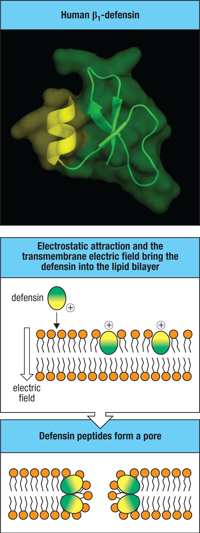
Defensins are an ancient, evolutionarily conserved class of antimicrobial peptides made by many eukaryotic organisms, including mammals, insects, and plants (Fig. 2.10). They are short cationic peptides of around 30–40 amino acids that usually have three disulfide bonds stabilizing a common amphipathic structure—a positively charged region separated from a hydrophobic region. Defensins act within minutes to disrupt the cell membranes of bacteria and fungi, as well as the membrane envelopes of some viruses. The mechanism is thought to involve insertion of the hydrophobic region into the membrane bilayer and the formation of a pore that makes the membrane leaky (see Fig. 2.10). Most multicellular organisms make many different defensins—the plant Arabidopsis thaliana produces 13 and the fruit fly Drosophila melanogaster at least 15. Human Paneth cells make as many as 21 different defensins, many of which are encoded by a cluster of genes on chromosome 8.
Three subfamilies of defensins—α-, β-, and θ-defensins—are distinguished on the basis of amino acid sequence. Each family has members with distinct activities, some being active against Gram-positive bacteria and some against Gram-negative bacteria, while others are specific for fungal pathogens. All the antimicrobial peptides, including the defensins, are generated by proteolytic processing from inactive propeptides (Fig. 2.11). In humans, developing neutrophils produce α-defensins through the processing of an initial propeptide of about 90 amino acids by cellular proteases to remove an anionic propiece, generating a mature cationic defensin that is stored in so-called primary granules. The primary granules of neutrophils are specialized membrane-enclosed vesicles, rather similar to lysosomes, that contain a number of other antimicrobial agents as well as defensins. We will explain in Chapter 3 how these primary granules are induced to fuse with phagocytic vesicles (phagosomes) after the cell has engulfed a pathogen, helping to kill the microbe. The Paneth cells of the gut constitutively produce α-defensins, called cryptdins, which are processed by proteases such as the metalloprotease matrilysin in mice or trypsin in humans, before being secreted into the gut lumen. The β-defensins lack the long propiece of α-defensins and are generally produced specifically in response to the presence of microbial products. β-Defensins (and some α-defensins) are made by epithelia outside the gut, primarily in the respiratory and urogenital tracts, skin, and tongue. β-Defensins made by keratinocytes in the epidermis and by type II pneumocytes in the lungs are packaged into lamellar bodies (see Fig. 2.6), lipid-rich secretory organelles that release their contents into the extracellular space to form a watertight lipid sheet in the epidermis and the pulmonary surfactant layer in the lung. The θ-defensins arose in the primates, but the single human θ-defensin gene has been inactivated by a mutation.
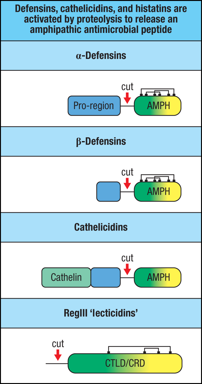
The antimicrobial peptides belonging to the cathelicidin family lack the disulfide bonds that stabilize the defensins. Humans and mice have one cathelicidin gene, but some other mammals, including cattle and sheep, have several. Cathelicidins are made constitutively by neutrophils and macrophages, and in response to infection by keratinocytes in the skin and epithelial cells in the lungs and intestine. They are made as inactive propeptides composed of two linked domains and are processed before secretion (see Fig. 2.11). In neutrophils, the inactive cathelicidin propeptides are stored in another type of specialized cytoplasmic granules called secondary granules. Cathelicidin is activated by proteolytic cleavage only when primary and secondary granules are induced to fuse with phagosomes, where it is cleaved by neutrophil elastase that has been stored in primary granules. Cleavage separates the two domains, and the cleavage products either remain in the phagosome or are released from the neutrophil by exocytosis. The carboxyl-terminal peptide is a cationic amphipathic peptide that disrupts membranes and is toxic to a wide range of microorganisms. The amino-terminal peptide is similar in structure to a protein called cathelin, an inhibitor of cathepsin L (a lysosomal enzyme involved in antigen processing and protein degradation), but its role in immune defense is unclear. In keratinocytes, cathelicidins, like β-defensins, are stored and processed in the lamellar bodies.
A class of antimicrobial peptides called histatins are constitutively produced in the oral cavity by the parotid, sublingual, and submandibular glands. These short, histidine-rich, cationic peptides are active against pathogenic fungi such as Cryptococcus neoformans and Candida albicans. More recently, histatins were found to promote the rapid wound healing that is typical in the oral cavity, but the mechanism of this effect is unclear.
Another type of bactericidal proteins made by epithelia is carbohydrate-binding proteins, or lectins. Most C-type lectins require calcium for the binding activity of their carbohydrate-recognition domain (CRD), which provides a variable interface for binding carbohydrate structures. C-type lectins of the RegIII family include several bactericidal proteins expressed by intestinal epithelium in humans and mice. However, carbohydrate recognition by RegIII proteins is not calcium dependent, but instead is inhibited by calcium. In mice, RegIIIγ is produced by Paneth cells and secreted into the gut, where it binds to peptidoglycans in bacterial cell walls and exerts direct bactericidal activity. Like other bactericidal peptides, RegIIIγ is produced in inactive form but is cleaved by the protease trypsin, which removes a short amino-terminal fragment to activate the bactericidal potential of RegIIIγ within the intestinal lumen (see Fig. 2.11). Human RegIIIα (also called HIP/PAP for hepatocarcinoma intestine pancreas/pancreatitis–associated protein) kills bacteria directly by forming a hexameric pore in the bacterial membrane (Fig. 2.12). RegIII family proteins preferentially kill Gram-positive bacteria, in which the peptidoglycan is exposed on the outer surface (see Fig. 2.9). In fact, the LPS of Gram-negative bacteria inhibits the pore-forming ability of RegIIIα, further enforcing the selectivity of RegIII proteins for Gram-positive bacteria.
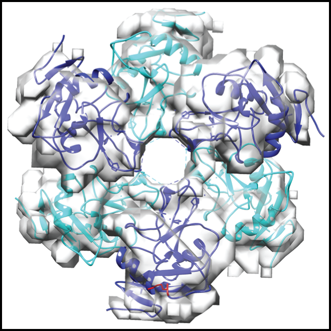
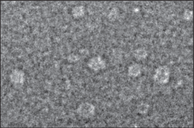
Fig. 2.12 Pore formation by human RegIIIα. Panel a: A model of the RegIIIα pore was generated by docking the human pro-RegIIIα structure (PDB ID: 1UV0), shown as individual purple and turquoise ribbon diagrams, into the cryo-electron microscopic map of the RegIIIα filament. LPS blocks the pore-forming activity of RegIIIα, explaining its selective bactericidal activity against Gram-positive but not Gram-negative bacteria. Panel b: Electron microscopic images of RegIIIα pores assembled in the presence of lipid bilayers.
Finally, we briefly mention the peptides S100A8 and S100A9, which heterodimerize to form calprotectin, which we will discuss in Section 11-11 in the context of production by T cells. Calprotectin acts to sequester magnesium and iron required by microorganisms and exerts an antimicrobial effect at sites of its production. This activity requires binding of calcium to one of calprotectin’s two metal-binding sites, which allows its second site to bind other metal ions. Calprotectin is produced in high amounts by neutrophils, and by T cells, but it is also made by intestinal epithelia, and so is part of the innate barrier defense system.
Summary.
The mammalian immune response to invading organisms proceeds in three phases, beginning with immediate innate defenses, then the induced innate defenses, and finally adaptive immunity. The first phase of host defense consists of those mechanisms that are present and ready to resist an invader at any time. Epithelial surfaces provide a physical barrier against pathogen entry, but they also have other, more specialized strategies. Mucosal surfaces have a protective barrier of mucus. Through particular cell-surface interactions, highly differentiated epithelia protect against both microbial colonization and invasion. Defense mechanisms of epithelia include the prevention of pathogen adherence, secretion of antimicrobial enzymes and bactericidal peptides, and the flow caused by the actions of cilia. Antimicrobial peptides and the bactericidal lectins of the RegIII family are made as inactive proproteins that require a proteolytic step to complete their activation, whereupon they become capable of killing microbes by forming pores in the microbial cell membranes. The actions of antimicrobial enzymes and peptides described in this part of the chapter often involve binding to unique glycan/carbohydrate structures on the microbe. Thus, these soluble molecular defenses are both pattern-recognition receptors and effector molecules at the same time, representing the simplest form of innate immunity.
Glossary
- pathogenic microorganism
- Microorganism that typically causes disease when it infects a host.
- pathogen
- Microorganism that typically causes disease when it infects a host.
- pathogenesis
- The origin or cause of the pathology of a disease.
- immune evasion
- Mechanisms used by pathogens to avoid detection and/or elimination by host immune defenses.
- exotoxin
- A protein toxin produced and secreted by a bacterium.
- endotoxins
- Toxins derived from bacterial cell walls released by damaged cells. They can potently induce cytokine synthesis and in large amounts can cause a systemic reaction called septic shock or endotoxic shock.
- lipopolysaccharide (LPS)
- The surface lipopolysaccharide of Gram-negative bacteria, which stimulates TLR-4 on macrophages and dendritic cells.
- Gram-negative bacteria
- Bacteria that fail to retain crystal violet stain after alcohol wash because of a thin peptidoglycan layer.
- zoonotic
- Describes a disease of animals that can be transmitted to humans.
- mucosal epithelia
- Mucus-coated epithelia lining the body’s internal cavities that connect with the outside (such as the gut, airways, and vaginal tract).
- mucus
- Sticky solution of proteins (mucins) secreted by goblet cells of internal epithelia, forming a protective layer on the epithelial surface.
- mucins
- Highly glycosylated cell-surface proteins. Mucin-like molecules are bound by L-selectin in lymphocyte homing.
- cystic fibrosis
- Disease caused by defect in the CFTR gene, leading to abnormally thick mucus and causing serious recurrent infections of the lung.
- commensal bacteria
- Microorganisms (predominantly bacteria) that normally live harmlessly in symbiosis with their host (e.g., the gut bacteria in humans and other animals). Many commensals confer a positive benefit on their host in some way.
- microbiota
- Microorganisms (predominantly bacteria) that normally live harmlessly in symbiosis with their host (e.g., the gut bacteria in humans and other animals). Many commensals confer a positive benefit on their host in some way.
- inflammatory response
- General term for the local accumulation of fluid, plasma proteins, and white blood cells that is initiated by physical injury, infection, or a local immune response.
- lysozyme
- Antimicrobial enzyme that degrades bacterial cell walls.
- peptidoglycan
- A component of bacterial cell walls that is recognized by certain receptors of the innate immune system.
- Paneth cells
- Specialized epithelial cells at the base of the crypts in the small intestine that secrete antimicrobial peptides.
- defensins
- See α-defensins, β-defensins.
- cathelicidins
- Family of antimicrobial peptides that in humans has one member.
- histatins
- Antimicrobial peptides constitutively produced by the parotid, sublingual, and submandibular glands in the oral cavity. Active against pathogenic fungi such as Cryptococcus neoformans and Candida albicans.
- amphipathic
- Describes molecules that have a positively charged (or hydrophilic) region separated from a hydrophobic region.
- propeptide
- Inactive precursor form of a polypeptide or peptide, which requires proteolytic processing to produce the active peptide.
- α-defensins
- A class of antimicrobial peptides produced by neutrophils and the Paneth cells of the intestine.
- primary granules
- Granules in neutrophils that correspond to lysosomes and contain antimicrobial peptides such as defensins and other antimicrobial agents.
- cryptdins
- α-Defensins (antimicrobial peptides) made by the Paneth cells of the small intestine.
- β-defensins
- Antimicrobial peptides made by virtually all multicellular organisms. In mammals they are produced by the epithelia of the respiratory and urogenital tracts, skin, and tongue.
- lamellar bodies
- Lipid-rich secretory organelles in keratinocytes and lung pneumocytes that release β-defensins into the extracellular space.
- secondary granules
- Type of granule in neutrophils that stores certain antimicrobial peptides.
- neutrophil elastase
- Proteolytic enzyme stored in the granules of neutrophils that is involved in the processing of antimicrobial peptides.
- cathelin
- A cathepsin L inhibitor.
- lectin
- A carbohydrate-binding protein.
- C-type lectins
- Large class of carbohydrate-binding proteins that require Ca2+ for binding, including many that function in innate immunity.
- HIP/PAP
- An antimicrobial C-type lectin secreted by intestinal cells in humans. Also known as RegIIIα.
- calprotectin
- A complex of heterodimers of the antimicrobial peptides S100A8 and S100A9, which sequester zinc and manganese from microbes. Produced in abundance by neutrophils and in lesser amounts by macrophages and epithelial cells.
- secretory phospholipase A2
- Antimicrobial enzyme present in tears and saliva and also secreted by the Paneth cells of the gut.
- RegIIIγ
- An antimicrobial protein of the C-type lectin family, produced by Paneth cells in the gut in mice.