The effector mechanisms of immunity.
For activated innate and adaptive immune cells to destroy pathogens, they must employ an appropriate effector mechanism suited to each infecting agent. The different types of pathogens noted in Fig. 1.26 have different lifestyles and require different responses for both their recognition and their destruction. Perhaps it is not surprising, then, that defenses against different pathogen types are organized into effector modules suited for these different lifestyles. For example, defense against extracellular pathogens can involve both phagocytic cells and B cells, which recognize extracellular antigens and become plasma cells that secrete antibody into the extracellular environment. Defense against intracellular pathogens involves T cells that can detect peptides generated inside the infected cell. Some effector T cells directly kill cells infected with intracellular pathogens such as viruses. These examples illustrate components of two distinct effector modules, one specialized for elimination of extracellular pathogens, and a second specialized for elimination of intracellular pathogens. In general, an effector module is a collection of cell-mediated and humoral mechanisms that involve components of both the innate and adaptive immune systems; these mechanisms act together to achieve elimination of a particular category of pathogen. One important component of each effector module is the effector T cell response. Depending on the nature of the pathogen, activated T cells differentiate into three major subsets of helper T cells, each of which produces different patterns of cytokines. These three subsets, discussed later, generally specialize in promoting defenses against pathogens having three major lifestyles: they can defend against intracellular infection, destroy extracellular bacteria and fungi, or provide barrier immunity directed at parasites. T cells also promote defense against extracellular pathogens by helping B cells make antibody.
|
The immune system protects against four classes of pathogens |
||
|
Type of pathogen |
Examples |
Diseases |
|
Viruses (intracellular) |
Variola Influenza Varicella |
Smallpox Flu Chickenpox |
|
Intracellular bacteria, protozoa, parasites |
Mycobacterium leprae Leishmania donovani Plasmodium falciparum Toxoplasma gondii |
Leprosy Leishmaniasis Malaria Toxoplasmosis |
|
Extracellular bacteria, parasites, fungi |
Streptococcus pneumonia Clostridium tetani Trypanosoma brucei Pneumocystis jirovecii |
Pneumonia Tetanus Sleeping sickness Pneumocystis pneumonia |
|
Parasitic worms (extracellular) |
Ascaris Schistosoma |
Ascariasis Schistosomiasis |
Fig. 1.26 The major types of pathogens confronting the immune system, and some of the diseases they cause.
Most of the other effector mechanisms used by an adaptive immune response to dispose of pathogens are the same as those of innate immunity and involve cells such as macrophages and neutrophils and proteins such as complement. Indeed, it seems likely that the vertebrate adaptive immune response evolved by the addition of specific recognition properties to innate defense mechanisms already existing in invertebrates. This is supported by recent findings that the innate lymphoid cells—the ILCs—show similar patterns of differentiation into different cytokine-producing subsets as those of T cells.
We begin this section by outlining the effector actions of antibodies, which depend almost entirely on recruiting cells and molecules of the innate immune system.
1-19 Innate immune responses can select from several effector modules to protect against different types of pathogens.
As we mentioned in Section 1-7, the innate immune system contains several types of cells—NK cells and ILCs—that have similarities to lymphocytes, particularly T cells. NK cells lack the antigen-specific receptors of T cells but can exhibit the cytotoxic capacity of T cells and produce some of the cytokines that effector T cells produce. ILCs develop from the same progenitor cells in the bone marrow as NK cells, and they also lack antigen-specific receptors. Very recent discoveries indicate that ILCs actually comprise several closely related lineages that differ in the specific cytokines that they will produce when activated. Remarkably, there is a striking similarity between the patterns of cytokines produced by ILC subsets and by helper T-cell subsets, as mentioned above. It appears that subsets of ILCs are the innate homologs of their helper T-cell counterparts, and NK cells are the innate homolog of cytotoxic T cells.
As mentioned in Section 1-6, there are a large number of cytokines with different functions (see eAppendix III, found in the ebook and on the Digital Landing Page for this book). A convenient way to organize the effects of cytokines is by the effector module that each cytokine promotes. Some cytokines tend to promote immunity to intracellular pathogens. One such cytokine is interferon-γ (IFN-γ), which acts both by activating phagocytes to more efficiently kill intracellular pathogens and by inducing target tissues to resist intracellular pathogens. This effector module is called type 1 immunity. IFN-γ is produced by some but not all subsets of innate and adaptive lymphocytes, and the ILC subset making IFN-γ is called ILC1. Other ILC subsets produce cytokines favoring effector modules called type 2 immunity and type 3 immunity, which coordinate defense against parasitic and extracellular pathogens, respectively. The modular nature of immune effector functions will be encountered frequently throughout this book. One principle seems to be that activated sensor cells from either the innate or the adaptive immune system can activate different subsets of innate or adaptive lymphocytes that are specialized for amplifying particular effector modules that are directed against different categories of pathogens (Fig. 1.27).
|
Effector module |
Cell types, functions, and mechanisms |
|
Cytotoxicity |
NK cells, CD8 T cells |
|
Elimination of virally infected and metabolically stressed cells |
|
|
Intracellular immunity (Type 1) |
ILC1, TH1 cells |
|
Elimination of intracellular pathogens; activation of macrophages |
|
|
Mucosal and barrier immunity (Type 2) |
ILC2, TH2 cells |
|
Elimination and expulsion of parasites; recruitment of eosinophils, basophils, and mast cells |
|
|
Extracellular immunity (Type 3) |
ILC3, TH17 cells |
|
Elimination of extracellular bacteria and fungi; recruitment and activation of neutrophils |
Fig. 1.27 Innate and adaptive lymphocytes share a variety of functions. The different effector modules are served by both innate and adaptive immune mechanisms. For each of the four major types of innate lymphocytes, there is a corresponding type of T cell with generally similar functional characteristics. Each set of innate lymphocyte and T cell exerts an effector activity that is broadly directed at a distinct category of pathogen.
1-20 Antibodies protect against extracellular pathogens and their toxic products.
Antibodies are found in plasma—the fluid component of blood—and in extracellular fluids. Because body fluids were once known as humors, immunity mediated by antibodies and other proteins in the plasma is known as humoral immunity.
Antibodies are Y-shaped molecules with two identical antigen-binding sites and one constant, or Fc, region. As mentioned in Section 1-9, there are five forms of the constant region of an antibody, known as the antibody classes or isotypes. The constant region determines an antibody’s functional properties—how it will engage with the effector mechanisms that dispose of antigen once it is recognized. Each class carries out its particular function by engaging a distinct set of effector mechanisms. We describe the antibody classes and their actions in Chapters 4 and 10.
The first and most direct way in which antibodies can protect against pathogens or their products is by binding to them and thereby blocking their access to cells that they might infect or destroy (Fig. 1.28, left panels). This function is known as neutralization and is important for protection against viruses, which are prevented from entering cells and replicating, and against bacterial toxins. Neutralization is the form of immunity elicited by most vaccines against bacterial toxins.
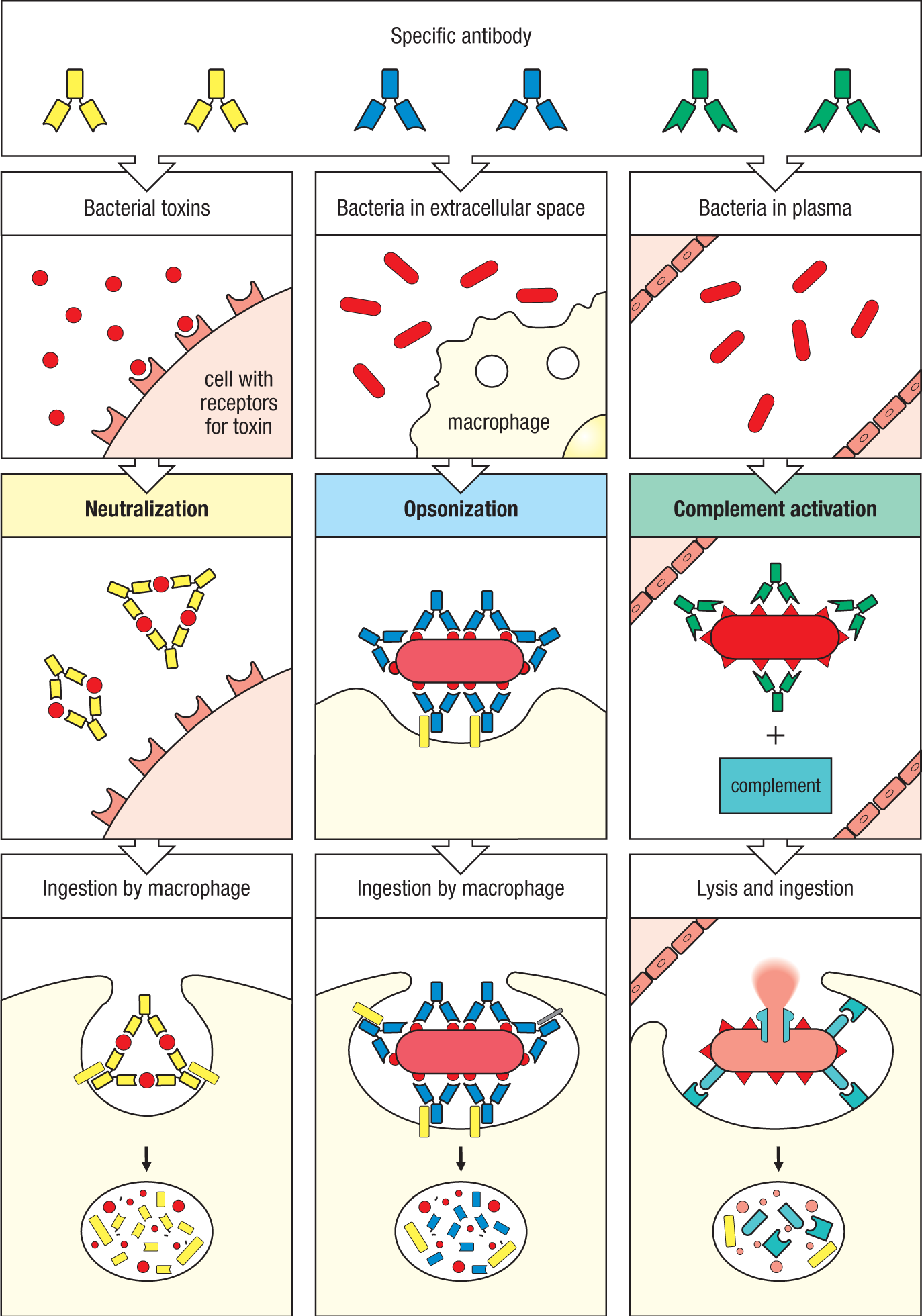
Antibodies coating an antigen render it recognizable as foreign by phagocytes (macrophages and neutrophils), which then ingest and destroy it; this is called opsonization. The center panels show opsonization and phagocytosis of a bacterial cell. Antibody first binds to antigens (red) on the bacterial cell through the variable regions. Then the antibody’s Fc region binds to Fc receptors (yellow) expressed by macrophages and other phagocytes, facilitating phagocytosis.
The right panels show activation of the complement system by antibodies coating a bacterial cell. Bound antibodies form a platform that activates the first protein in the complement system, which deposits complement proteins (blue) on the surface of the bacterium. This can lead in some cases to formation of a pore that lyses the bacterium directly. More generally, complement proteins on the bacterium can be recognized by complement receptors on phagocytes; this stimulates the phagocytes to ingest and destroy the bacterium. Thus, antibodies target pathogens and their toxic products for disposal by phagocytes.
For bacteria, however, binding by antibodies is not sufficient to stop their replication. In this case, a second function of antibodies is to enable a phagocytic cell such as a macrophage or a neutrophil to ingest and destroy the bacterium. Many bacteria evade the innate immune system because they have an outer coat that is not recognized by the pattern-recognition receptors of phagocytes. However, antigens in the coat can be recognized by antibodies, and phagocytes have receptors, called Fc receptors, that bind the constant region and facilitate phagocytosis of the bacterium (see Fig. 1.28, center panels). The coating of pathogens and foreign particles in this way is known as opsonization.
The third function of antibodies is complement activation. In Section 1-2 we briefly mentioned Bordet’s discovery of complement as a serum factor that ‘complements’ the activities of antibodies. Complement can be activated by microbial surfaces even without the help of antibodies, which leads to the covalent deposition of certain complement proteins onto the bacterial surface. But when an antibody binds first to the bacterial surface, its constant region provides a platform that is much more efficient in complement activation than microbial activation of complement alone. Thus, once antibodies are produced, complement activation against a pathogen can be substantially increased.
Certain complement components that are deposited on the bacterial surface can directly lyse the membranes of some bacteria, and this is important in a few bacterial infections (see Fig. 1.28, right panels). The major function of complement, however, is to enable phagocytes to engulf and destroy bacteria that the phagocytes would not otherwise recognize. Most phagocytes express receptors that bind certain complement proteins; called complement receptors, these receptors bind to the complement proteins deposited onto the bacterial surface and thus facilitate bacterial phagocytosis. Certain other complement proteins also enhance the phagocytes’ bactericidal capacity. The end result is that all pathogens and free molecules bound by antibody are eventually delivered to phagocytes for ingestion, degradation, and removal from the body (see Fig. 1.28, bottom panels). The complement system and the phagocytes that antibodies recruit are not themselves antigen-specific; they depend on antibody molecules to mark the particles as foreign.
1-21 T cells orchestrate cell-mediated immunity and regulate B-cell responses to most antigens.
Some bacteria and parasites, and all viruses, replicate inside cells, where they cannot be detected by antibodies, which access only the blood and extracellular space. The destruction of intracellular invaders is the function of the T lymphocytes, which are responsible for the cell-mediated immune responses of adaptive immunity. But T lymphocytes participate in responses to a wide variety of pathogens, including extracellular organisms, and so must exert a wide variety of effector activities.
T lymphocytes, of which there are several types, develop in the thymus. They are characterized by the type of T-cell receptors they express and by the expression of certain markers. The two main classes of T cells express either a cell-surface protein called CD8 or another called CD4. These are not just random markers, but are important for a T cell’s function, because they help to determine the interactions between the T cell and other cells. Recall from Section 1-10 that T cells detect peptides derived from foreign antigens that are displayed by MHC molecules on a cell’s surface. CD8 and CD4 function in antigen recognition by recognizing different regions of MHC molecules and by being involved in the signaling of the T-cell receptor that is engaged with its antigen. Thus, CD4 and CD8 are known as co-receptors and they provide a functional difference between CD8 and CD4 T cells.
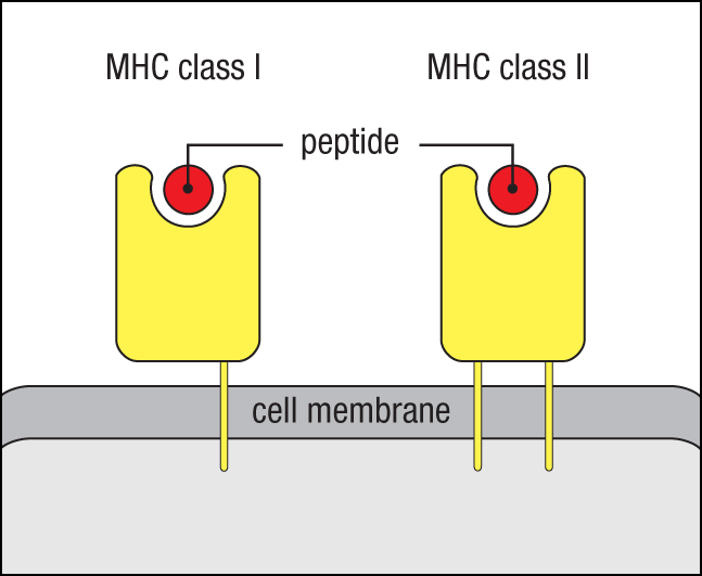
Importantly, there are two main types of MHC molecules, called MHC class I and MHC class II. These have slightly different structures, but both have an elongated groove on the outer surface that can bind a peptide (Fig. 1.29). The peptide becomes trapped in this groove during the synthesis and assembly of the MHC molecule inside the cell, and the peptide:MHC complex is then transported to the cell surface and displayed to T cells (Fig. 1.30). Because CD8 recognizes a region of the MHC class I protein while CD4 recognizes a region of the MHC class II protein, the two co-receptors functionally distinguish T cells. Therefore, CD8 T cells selectively recognize peptides that are presented by MHC class I molecules, while CD4 T cells selectively recognize peptides that are presented by MHC class II molecules.
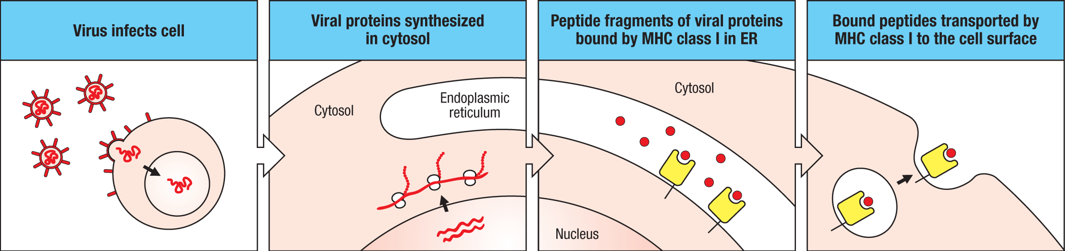
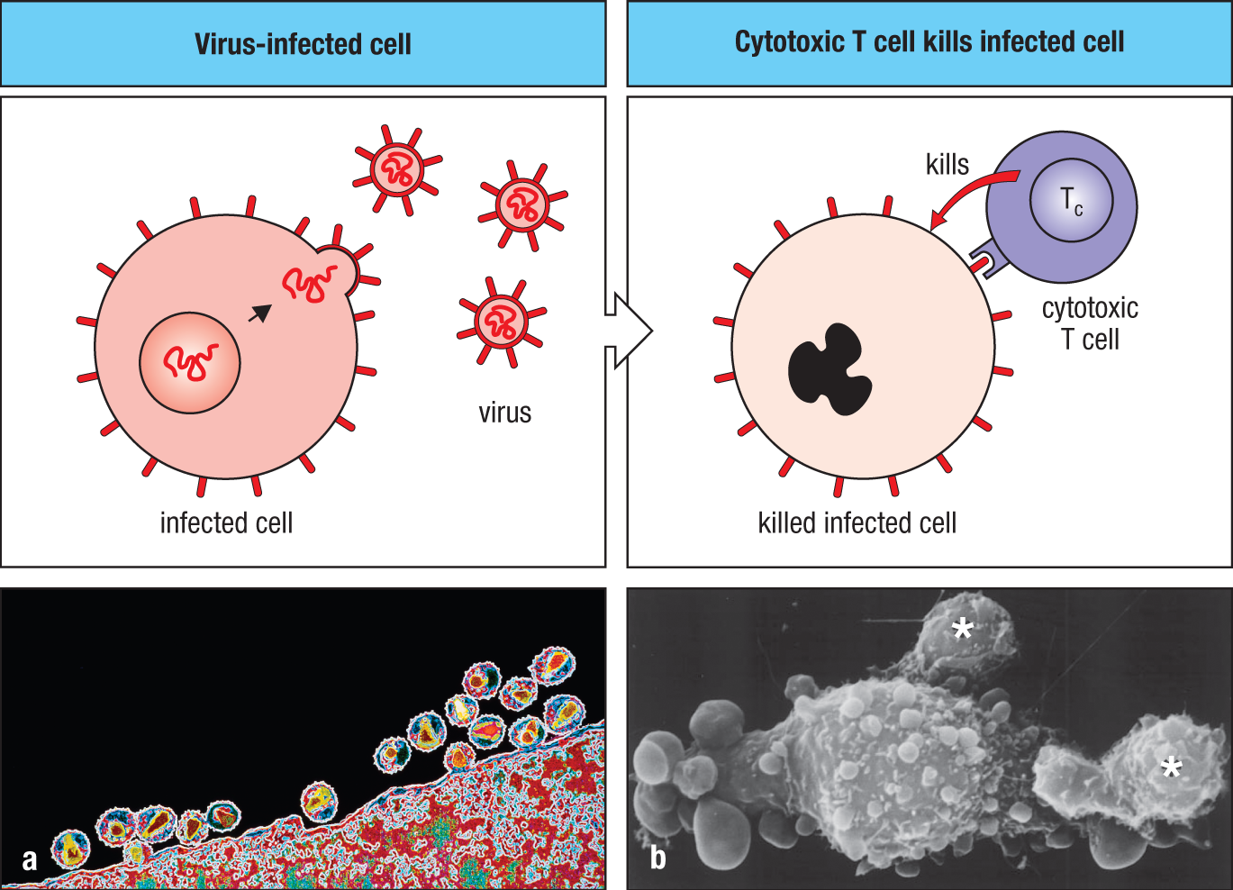
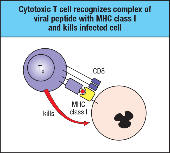
The most direct action of T cells is cytotoxicity. Cytotoxic T cells are effector T cells that act against cells infected with viruses. Antigens derived from the virus multiplying inside the infected cell are displayed on the cell’s surface, where they are recognized by the antigen receptors of cytotoxic T cells. These T cells can then control the infection by directly killing the infected cell before viral replication is complete and new viruses are released (Fig. 1.31). Cytotoxic T cells carry CD8, and so recognize antigen presented by MHC class I molecules. Because MHC class I molecules are expressed on most cells of the body, they serve as an important mechanism to defend against viral infections. MHC class I molecules bearing viral peptides are recognized by CD8-bearing cytotoxic T cells, which then kill the infected cell (Fig. 1.32).
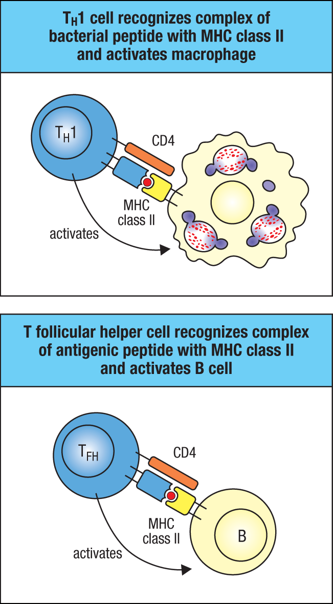
CD4 T cells recognize antigen presented by MHC class II molecules, which are expressed by the predominant antigen-presenting cells of the immune system: dendritic cells, macrophages, and B cells (Fig. 1.33). Thus CD4 T cells tend to recognize antigens taken up by phagocytosis from the extracellular environment. CD4 T cells are the helper T cells mentioned earlier in the chapter. They develop into a variety of different effector subsets, called TH1 (for T helper type 1), TH2, TH17, and so on, and they produce cytokines in patterns similar to those of the subsets of ILCs mentioned earlier that activate effector modules protective against different pathogens. These subsets act primarily at sites of infection or injury in peripheral tissues. In the lymphoid tissues, a subset of CD4 T cells, called the T follicular helper (TFH) cell, interacts with B cells to regulate antibody production during the immune response. The various T helper subsets are described in Chapter 9.
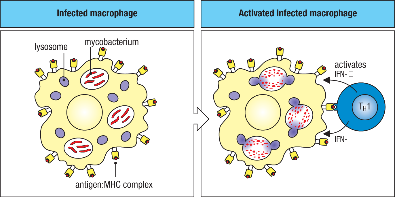
For example, the TH1 subset of CD4 T cells helps to control certain bacteria that take up residence in membrane-enclosed vesicles inside macrophages. They produce the same cytokine as ILC1 cells, IFN-γ, which activates macrophages to increase their intracellular killing power and destroy these bacteria. Important infections that are controlled by this function are tuberculosis and leprosy, which are caused by the bacteria Mycobacterium tuberculosis and M. leprae, respectively. Mycobacteria survive intracellularly because they prevent the vesicles they occupy from fusing with lysosomes, which contain a variety of degradative enzymes and antimicrobial substances (Fig. 1.34). However, on its surface, the infected macrophage presents mycobacteria-derived antigens that can be recognized by activated antigen-specific TH1 cells, which in turn secrete particular cytokines that induce the macrophage to overcome the block on vesicle fusion. TH2 and TH17 subsets produce cytokines that are specialized for promoting responses against parasites or extracellular bacteria and fungi, respectively. CD4 T cells, and their specialized subsets, play a pervasive role in adaptive immunity, and we will be returning to them many times in this book, including in Chapters 8, 9, 11, and 12.
1-22 Inherited and acquired defects in the immune system result in increased susceptibility to infection.
We tend to take for granted the ability of our immune systems to free our bodies of infection and prevent its recurrence. In some people, however, parts of the immune system fail. In the most severe of these immunodeficiency diseases, adaptive immunity is completely absent, and death occurs in infancy from overwhelming infection unless heroic measures are taken. Other less catastrophic failures lead to recurrent infections with particular types of pathogens, depending on the particular deficiency. Much has been learned about the functions of the different components of the human immune system through the study of these immunodeficiencies, many of which are caused by inherited genetic defects. Because understanding the features of immunodeficiencies requires a detailed knowledge of normal immune mechanisms, we have postponed discussion of most of these diseases until Chapter 13, where they can be considered together.
In the 1980s, a devastating form of immunodeficiency appeared, the acquired immune deficiency syndrome, or AIDS, which is caused by an infectious agent, the human immunodeficiency viruses HIV-1 and HIV-2. This disease destroys T cells, dendritic cells, and macrophages bearing CD4, leading to infections caused by intracellular bacteria and other pathogens normally controlled by such cells. These infections are the major cause of death from this increasingly prevalent immunodeficiency disease, which is discussed fully in Chapter 13 together with the inherited immunodeficiencies.
 Case Study: Acquired Immune Deficiency Syndrome (AIDS)
1-23 Understanding adaptive immune responses is important for the control of allergies, autoimmune disease, and the rejection of transplanted organs.
The main function of our immune system is to protect the human host from infectious agents. However, many medically important diseases are associated with a normal immune response directed against an inappropriate antigen, often in the absence of infectious disease. Immune responses directed at noninfectious antigens occur in allergy, in which the antigen is an innocuous foreign substance; in autoimmune disease, in which the response is to a self antigen; and in graft rejection, in which the antigen is borne by a transplanted foreign cell (discussed in Chapter 15). The major antigens provoking graft rejection are, in fact, the MHC molecules, as each of these is present in many different versions in the human population—that is, they are highly polymorphic—and most unrelated people differ in the set of MHC molecules they express, a property commonly known as their ‘tissue type.’ The MHC was originally recognized by the work of Peter Gorer in the 1930s as a gene locus in mice, the H-2 locus, that controlled the acceptance or rejection of transplanted tumors. Genes in this locus were later studied by George Snell, who examined their role in tissue transplantation by developing mouse strains differing only at these histocompatibility loci. The human MHC molecules were first discovered during the Second World War, when attempts were made to use skin grafts from donors to repair badly burned pilots and bomb victims. The patients rejected the grafts, which were recognized by their immune systems as being ‘foreign.’ What we call a successful immune response or a failure, and whether the response is considered harmful or beneficial to the host, depends not on the response itself but rather on the nature of the antigen and the circumstances in which the response occurs (Fig. 1.35). Snell was awarded the 1980 Nobel Prize (together with Baruj Benacerraf and Jean Dausset) for his work on the MHC.
|
Antigen |
Effect of response to antigen |
|
|
Normal response |
Deficient response |
|
|
Infectious agent |
Protective immunity |
Recurrent infection |
|
Innocuous substance |
Allergy |
No response |
|
Grafted organ |
Rejection |
Acceptance |
|
Self organ |
Autoimmunity |
Self tolerance |
|
Tumor |
Tumor immunity |
Cancer |
Fig. 1.35 Immune responses can be beneficial or harmful, depending on the nature of the antigen. Beneficial responses are shown in white boxes, harmful responses in red-shaded boxes. Where the response is beneficial, its absence is harmful.
Allergic diseases, which include asthma, are an increasingly common cause of disability in the developed world. Autoimmunity is also now recognized as the cause of many important diseases. An autoimmune response directed against pancreatic β cells is the leading cause of diabetes in the young. In allergies and autoimmune diseases, the powerful protective mechanisms of the adaptive immune response cause serious damage to the individual.
Immune responses to harmless antigens, to body tissues, or to organ grafts are, like all other immune responses, highly specific. At present, the usual way to treat these responses is with immunosuppressive drugs, which inhibit all immune responses, desirable and undesirable alike. If it were possible to suppress only those lymphocyte clones responsible for the unwanted response, the disease could be cured or the grafted organ protected without impeding protective immune responses. At present, antigen-specific immunoregulation is outside the reach of clinical treatment. But as we shall see in Chapter 16, many new drugs have been developed recently that offer more selective immune suppression to control autoimmune and other unwanted immune responses. Among these, therapies using highly specific monoclonal antibodies were made possible by Georges Köhler and César Milstein, who shared the 1984 Nobel Prize along with Niels K. Jerne. Köhler and Milstein received the prize for their “discovery of the principle for production of monoclonal antibodies.” We shall discuss the current state of understanding of allergies, autoimmune disease, graft rejection, and immunosuppressive drugs and monoclonal antibodies in Chapters 14–16, and we shall see in Chapter 15 how the mechanisms of immune regulation are beginning to emerge from a better understanding of the functional subsets of lymphocytes and the cytokines that control them.
1-24 Vaccination is the most effective means of controlling infectious diseases.
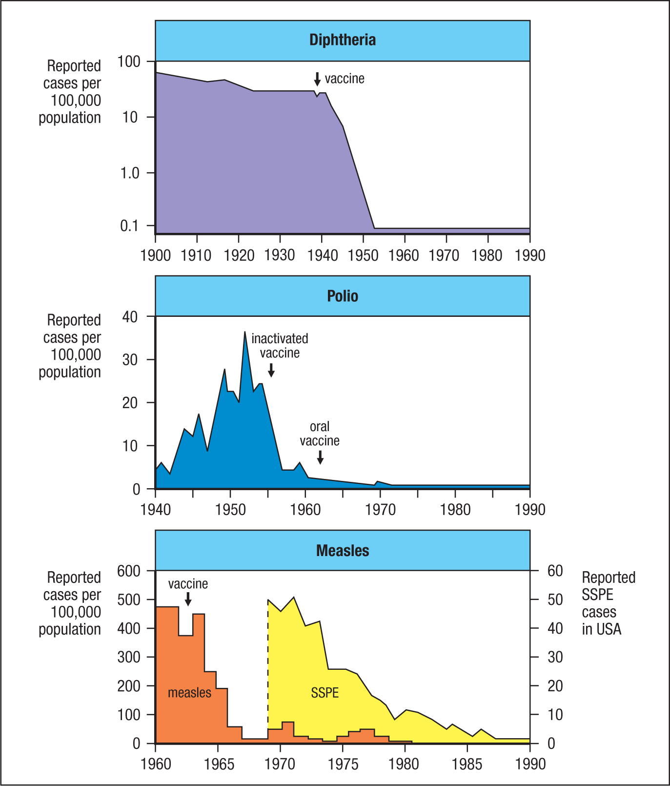
The deliberate stimulation of an immune response by immunization, or vaccination, has achieved many successes in the two centuries since Jenner’s pioneering experiment. Mass immunization programs have led to the virtual eradication of several diseases that used to be associated with significant morbidity (illness) and mortality (Fig. 1.36). Immunization is considered so safe and so important that most states in the United States require children to be immunized against up to seven common childhood diseases. Impressive as these accomplishments are, there are still many diseases for which we lack effective vaccines. And even though vaccines for diseases such as measles can be used effectively in developed countries, technical and economic problems can prevent their widespread use in developing countries, where mortality from these diseases is still high.
The tools of modern immunology and molecular biology are being applied to develop new vaccines and improve old ones, and we discuss these advances in Chapter 16. The prospect of controlling these important diseases is tremendously exciting. The guarantee of good health is a critical step toward population control and economic development. At a cost of pennies per person, great hardship and suffering can be alleviated.
Many serious pathogens have resisted efforts to develop vaccines against them, often because they can evade or subvert the protective mechanisms of an adaptive immune response. We examine some of the evasive strategies used by successful pathogens in Chapter 13. The conquest of many of the world’s leading diseases, including malaria and diarrheal diseases (the leading killers of children) as well as the more recent threat from AIDS, depends on a better understanding of the pathogens that cause them and their interactions with the cells of the immune system.
1-25 Emerging pathogens present new challenges for the immune system.
Pathogenic microbes have been besieging humans for centuries, if not millennia. Reports of the bubonic plague, caused by the bacterium Yersinia pestis, date back to the Philistines in 1320 BCE. Influenza virus has been causing human disease for centuries, with the earliest report of an influenza-like illness by Hippocrates, the Greek physician, in ~410 BCE. Current estimates indicate that Mycobacterium tuberculosis, the causative agent of tuberculosis, has existed for more than 70,000 years. However, as recent history has shown us, new pathogenic organisms occasionally arise and wreak devastation on the human population. Frequently, these emerging pathogens are viruses with relatively small genomes and high mutation rates, ideal conditions for the development of human-to-human transmission capabilities.
The early 21st century witnessed the emergence of several novel coronaviruses that cause respiratory infections with a high rate of transmission. These viruses are believed to have crossed into humans from bats, their natural reservoir. Two of these viruses caused epidemics with a high fatality rate, SARS-CoV in 2002–2003 and MERS-CoV in 2012, but ultimately were responsible for fewer than 1000 deaths each. A third coronavirus, SARS-CoV-2, emerged in late 2019 and rapidly spread across the globe, causing a worldwide pandemic that disrupted societies and economies and by June 2020 was responsible for more than 4000 deaths a day. Studies of the immune response to SARS-CoV-2 are proceeding at a remarkable pace, and by early 2021 multiple effective vaccines had been developed and introduced to the public.
Summary.
The responses to infection can be organized into several effector modules that target the various types of pathogen lifestyles. Innate sensor cells that detect infection generate mediators that activate innate lymphoid cells (ILCs) and T cells, which amplify the immune response and also activate various effector modules. ILCs include subsets that produce different cytokines and activate distinct effector modules. T cells fall into two major classes that are based on the expression of the co-receptors CD8 and CD4. CD8 and CD4 T cells recognize antigen presented by MHC class I or MHC class II proteins, respectively. These subsets of T cells, like their ILC counterparts, also promote the actions of distinct effector modules. NK cells and CD8 T cells can exert cytotoxic activity to target intracellular infections such as viruses. Other subsets of innate lymphoid and helper T cells can secrete mediators that activate other effector functions, ones that target intracellular bacteria, extracellular bacteria and fungi, and parasites. T cells also provide signals that help regulate B cells and stimulate them to produce antibodies. Specific antibodies mediate the clearance and elimination of soluble toxins and extracellular pathogens. They interact not only with the toxins or the antigens on microbes but also with the Fc region of specific receptors that are expressed by many types of phagocytes. Phagocytes also express receptors for complement proteins that are deposited on microbial surfaces, particularly in the presence of antibody.
Failures of immunity can be caused by genetic defects or by infections that target important components of the immune system. Misdirected immune responses can damage host tissues, as in autoimmune diseases or allergy, or lead to the failure of transplanted organs. While vaccination is still the greatest tool of immunology to fight diseases, modern approaches have added new tools, such as monoclonal antibodies, that have become progressively more important in the clinic since their initial use in the mid-1980s.
Summary to Chapter 1.
The immune system defends the host against infection. Innate immunity serves as a first line of defense but lacks the ability to recognize certain pathogens and to provide the specific protective immunity that prevents reinfection. Adaptive immunity is based on clonal selection from a repertoire of lymphocytes bearing highly diverse antigen-specific receptors that enable the immune system to recognize any foreign antigen. In the adaptive immune response, antigen-specific lymphocytes proliferate and differentiate into clones of effector lymphocytes that eliminate the pathogen. Figure 1.7 summarizes the phases of the immune response and their approximate timings. Host defense requires different recognition systems and a wide variety of effector mechanisms to seek out and destroy the wide variety of pathogens in their various habitats within the body and at its external and internal surfaces. Not only can the adaptive immune response eliminate a pathogen, but, in the process, it also generates increased numbers of differentiated memory lymphocytes through clonal selection, and this allows a more rapid and effective response upon reinfection. The regulation of immune responses, whether to suppress them when unwanted or to stimulate them in the prevention of infectious disease, is the major medical goal of research in immunology.
Glossary
- effector module
- A collection of cell-mediated and humoral mechanisms that involve components of both the innate and adaptive immune systems. These mechanisms act together to achieve elimination of a particular category of pathogen.
- type 1 immunity
- Class of effector activities aimed at elimination of intracellular pathogens.
- ILC1
- A subset of innate lymphoid cells characterized by production of IFN-γ.
- type 2 immunity
- Class of effector activities aimed at elimination of parasites and promoting barrier and mucosal immunity.
- type 3 immunity
- Class of effector activities aimed at elimination of extracellular pathogens such as bacteria and fungi.
- classes
- The class of an antibody is defined by the type of heavy chain it contains. There are five main antibody classes: IgA, IgD, IgM, IgG, and IgE, containing heavy chains α, δ, μ, γ, and ε, respectively. The IgG class has several subclasses. See also isotypes.
- isotype
- The designation of an immunoglobulin chain with respect to the type of constant region it has. Light chains can be of either κ or λ isotype. Heavy chains can be of μ, δ, γ, α, or ε isotype. The different heavy-chain isotypes have different effector functions and determine the class and functional properties of antibodies (IgM, IgD, IgG, IgA, and IgE, respectively).
- neutralization
- Inhibition of the infectivity of a virus or the toxicity of a toxin molecule by the binding of antibodies.
- Fc receptors
- Family of cell-surface receptors that bind the Fc portions of different immunoglobulins: Fcγ receptors bind IgG, for example, and Fcε receptors bind IgE.
- opsonization
- The coating of the surface of a pathogen by antibody and/or complement that makes it more easily ingested by phagocytes.
- complement activation
- The activation of the normally inactive proteins of the complement system that occurs on infection. See classical pathway, alternative pathway, lectin pathway.
- cell-mediated immune responses
- An adaptive immune response in which antigen-specific effector T cells have the main role. The immunity to infection conferred by such a response is called cell-mediated immunity. A primary cell-mediated immune response is the T-cell response that occurs the first time a particular antigen is encountered.
- CD8
- The co-receptor for T-cell receptors that recognize peptide antigens bound to MHC class I molecules. It binds to the lateral face of the MHC molecule.
- CD4
- The co-receptor for T-cell receptors that recognize peptide antigens bound to MHC class II molecules. It binds to the lateral face of the MHC molecule.
- co-receptor
- Cell-surface protein that increases the sensitivity of a receptor to its ligand by binding to associated ligands and participating in signaling. The antigen receptors on T cells and B cells act in conjunction with co-receptors, which are either CD4 or CD8 on T cells, and a co-receptor complex of three proteins, one of which is the complement receptor CR2, on B cells.
- MHC class I
- Polymorphic cell-surface proteins encoded in the MHC locus and expressed on most cells. They present antigenic peptides generated in the cytosol to CD8 T cells and also bind the co-receptor CD8.
- MHC class II
- Polymorphic cell-surface proteins encoded in the MHC locus and expressed primarily on specialized antigen-presenting cells. They present antigenic peptides derived from internalized extracellular pathogens to CD4 T cells and also bind the co-receptor CD4.
- T follicular helper (TFH)
- An effector T cell found in lymphoid follicles that provides help to B cells for antibody production and class switching.
- immunodeficiency diseases
- Any inherited or acquired disorder in which some aspect or aspects of host defense are absent or functionally defective.
- acquired immune deficiency syndrome (AIDS)
- A disease caused by infection with the human immunodeficiency virus (HIV-1 or HIV-2). AIDS occurs when an infected individual has lost most of his or her CD4 T cells, so that infections with opportunistic pathogens occur.
- AIDS
- A disease caused by infection with the human immunodeficiency virus (HIV-1 or HIV-2). AIDS occurs when an infected individual has lost most of his or her CD4 T cells, so that infections with opportunistic pathogens occur.
- allergy
- The state in which a symptomatic immune reaction is made to a normally innocuous environmental antigen. It involves the interaction between the antigen and antibody or primed T cells produced by earlier exposure to the same antigen.
- autoimmune disease
- Disease in which the pathology is caused by adaptive immune responses to self antigens.
- graft rejection
- The immunologically mediated rejection of grafted tissues or organs from a genetically nonidentical donor. It is due chiefly to recognition of nonself MHC molecules on the graft.
- polymorphic
- Existing in a variety of different forms; applied to a gene, occurring in a variety of different alleles.
- monoclonal antibodies
- Antibodies produced by a single clone of B lymphocytes, so that they are all identical.
- IFN-γ
- A cytokine of the interferon structural family produced by effector CD4 TH1 cells, CD8 T cells, and NK cells. Its primary function is the activation of macrophages, and it acts through a different receptor from that of the type I interferons.
- humoral immunity, humoral immune response
- Immunity due to proteins circulating in the blood, such as antibodies (in adaptive immunity) or complement (in innate immunity). Adaptive humoral immunity can be transferred to unimmunized recipients by the transfer of serum containing specific antibody.
- TH1
- A subset of effector CD4 T cells characterized by the cytokines they produce, notably IFN-γ and TNF-α. They are mainly involved in activating macrophages and promoting inflammation.
- TH2
- A subset of effector CD4 T cells that are characterized by the cytokines they produce, notably IL-4, IL-5, and IL-13. They are involved in recruiting eosinophils and activating mast cells and basophils, and in promoting enhanced barrier protection.
- TH17
- A subset of CD4 T cells that are characterized by production of the cytokine IL-17. They help recruit neutrophils to sites of infection.
- H-2 locus, H-2 genes
- The major histocompatibility complex of the mouse. Haplotypes are designated by a lowercase superscript, as in H-2b.