Consequences of innate immune activation.
We will now examine the responses of innate immunity induced as an immediate consequence of pathogen recognition by the sensors described in the first half of this chapter. We will focus on the major phagocytes—neutrophils, macrophages, and dendritic cells—and the cytokines they produce that induce and maintain inflammation. First, we will introduce the families of cytokines and chemokines that coordinate many cellular responses, such as the recruitment of neutrophils and other immune cells to sites of infection. We will discuss the various adhesion molecules that are induced on immune cells circulating in the blood and on endothelial cells of blood vessels to coordinate movement of cells out of the blood and into infected tissues. We will consider in some detail how macrophage-derived chemokines and cytokines promote the continued destruction of infecting microbes. This is achieved both by stimulating the production and recruitment of fresh phagocytes and by inducing another phase of the innate immune response—the acute-phase response—in which the liver produces proteins that act as opsonizing molecules, helping to augment the actions of complement. We will also look at the mechanism of action of antiviral interferons, the type I interferons, and finally examine the growing class of innate lymphoid cells, or ILCs, which include the NK cells that contribute to innate immune defense against viruses, other intracellular pathogens, and even cancerous cells. ILCs exert a diverse array of effector functions that contribute to a rapid innate immune response to infection. They respond to early cytokine signals provided by innate sensor cells and amplify the response by producing various types of effector cytokines. Some time after induction of the innate response, an adaptive response will ensue that uses many of the same effector mechanisms used by the innate immune system but targets them with much greater precision. The effector mechanisms described here therefore serve as a primer for the focus on adaptive immunity in the later parts of this book.
3-15 Cytokines and their receptors fall into distinct families of structurally related proteins.
Cytokines are small proteins (about 25 kDa) that are released by various cells in the body, usually in response to an activating stimulus, and that induce responses through binding to specific receptors. Cytokines can act in an autocrine manner, affecting the behavior of the cell that releases the cytokine, or in a paracrine manner, affecting adjacent cells. Some cytokines are even stable enough to act in an endocrine manner, affecting distant cells, although this depends on their ability to enter the circulation and on their half-life in the blood. In an attempt to develop a standardized nomenclature for molecules secreted by, and acting on, leukocytes, many cytokines are called by the name interleukin (IL) followed by a number (for example, IL-1 or IL-2). However, not all cytokines are included in this system; thus, students of immunology are still faced with a somewhat confusing and difficult task. The cytokines are listed alphabetically, together with their receptors, in eAppendix III.
Cytokines can be grouped by structure into families—the IL-1 family, the hematopoietin superfamily, the interferons (described in Section 3-7), the TNF family, and chemokines—and their receptors can likewise be grouped (Fig. 3.24). The IL-1 family contains 11 members, notably IL-1α, IL-1β, IL-18, and IL-33. Most members of this family are produced as inactive proproteins that are cleaved (removing an amino-terminal peptide) to produce the mature cytokine. The exception to this rule is IL-1α, for which both the proprotein and its cleaved forms are biologically active. As discussed earlier (see Section 3-11), mature IL-1β and IL-18 are produced by macrophages through the action of caspase 1 in response to TLR signaling and inflammasome activation. The IL-1 family receptors have TIR domains in their cytoplasmic tails and signal by the NFΚB pathway described earlier for TLRs. The IL-1 receptor functions in concert with a second transmembrane protein, the IL-1 receptor accessory protein (IL1RAP), which is required for IL-1 signal transduction.
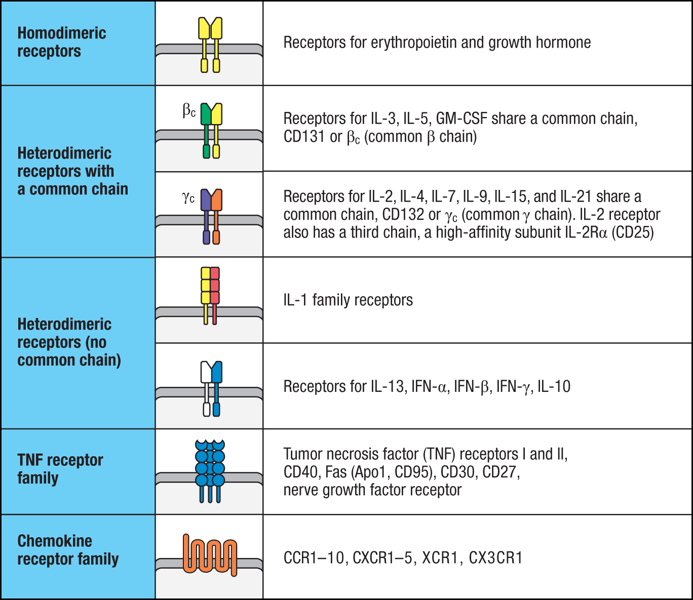
The hematopoietin superfamily of cytokines is quite large and includes non-immune-system growth and differentiation factors, such as erythropoietin (which stimulates red blood cell development), as well as interleukins with roles in innate immunity and adaptive immunity. IL-6 is a member of this superfamily, as is the cytokine GM-CSF, which stimulates the production of new monocytes and granulocytes in the bone marrow. Many of the soluble cytokines made by activated T cells are members of the hematopoietin superfamily. The receptors for the hematopoietin cytokines are tyrosine kinase–associated receptors that form dimers when their cytokine ligand binds. Dimerization initiates intracellular signaling from the tyrosine kinases associated with the cytoplasmic domains of the receptor. Some types of cytokine receptors are composed of two identical subunits, but others have two different subunits. An important feature of cytokine signaling is the large variety of different receptor subunit combinations that occur.
These cytokines and their receptors can also be further divided into subfamilies characterized by functional similarities and genetic linkage. For instance, IL-3, IL-4, IL-5, IL-13, and GM-CSF are related structurally, their genes are closely linked in the genome, and they are often produced together by the same kinds of cells. In addition, they bind to closely related receptors, which belong to the family of class I cytokine receptors. The IL-3, IL-5, and GM-CSF receptors form a subgroup that shares a common β chain (βc). Another subgroup of class I cytokine receptors is defined by a common γ chain (γc) of the IL-2 receptor. This chain is shared by receptors for the cytokines IL-2, IL-4, IL-7, IL-9, IL-15, and IL-21 and is encoded by a gene located on the X chromosome. Mutations that inactivate γc cause an X-linked severe combined immune deficiency (X-linked SCID) because of inactivation of the signaling pathways for several cytokines—IL-7, IL-15, and IL-2—that are required for normal lymphocyte development (see Section 13-2). More distantly related, the receptor for IFN-γ is a member of a small family of heterodimeric cytokine receptors with some similarities to the hematopoietin receptor superfamily. These so-called class II cytokine receptors (also known as interferon receptors) include the receptors for IFN-α and IFN-β and the IL-10 receptor. The hematopoietin and interferon receptors all signal through the JAK–STAT pathway described in the next section and activate different combinations of STATs with different effects.
 Case Study: X-linked Severe Combined Immune Deficiency
The TNF family, of which TNF-α is the prototype, contains more than 17 cytokines with important functions in adaptive immunity and innate immunity. Unlike most of the other immunologically important cytokines, many members of the TNF family are transmembrane proteins, a characteristic that gives them distinct properties and limits their range of action. Some, however, can be released from the membrane in some circumstances. They are usually found as homotrimers of a membrane-bound subunit, although some heterotrimers consisting of different subunits also occur. TNF-α (sometimes called simply TNF) is initially expressed as a trimeric membrane-bound cytokine but can be released from the membrane. The effects of TNF-α are mediated by either of two TNF receptors. TNF receptor I (TNFR-I) is expressed on a wide range of cells, including endothelial cells and macrophages, whereas TNFR-II is expressed largely by lymphocytes. The receptors for cytokines of the TNF family are structurally unrelated to the receptors described above and also have to cluster to become activated. Because TNF family cytokines are produced as trimers, the binding of these cytokines induces the clustering of three identical receptor subunits. The signaling pathway activated by these receptors is described in Chapter 7, where we see that signaling uses members of the TRAF family to activate the so-called non-canonical NFΚB pathway.
Members of the chemokine receptor family are listed in eAppendix IV, along with the chemokines they recognize. These receptors have a seven-transmembrane structure and signal by interacting with G proteins as described in Section 3-2.
3-16 Cytokine receptors of the hematopoietin superfamily are associated with the JAK family of tyrosine kinases, which activate STAT transcription factors.
The signaling chains of the hematopoietin superfamily of cytokine receptors are noncovalently associated with protein tyrosine kinases of the Janus kinase (JAK) family—so called because they have two tandem kinase-like domains and thus resemble the two-headed mythical Roman god Janus. There are four members of the JAK family: Jak1, Jak2, Jak3, and Tyk2. Because mice deficient for individual JAK family members show different phenotypes, each kinase must have a distinct function. For example, Jak3 is used by γc for signaling by several of the cytokines described above. Mutations that inactivate Jak3 cause a form of SCID that is not X-linked.
 3.7 Cytokine Signaling
The dimerization or clustering of receptor signaling chains brings the JAKs into close proximity, causing phosphorylation of each JAK on a tyrosine residue that stimulates its kinase activity. The activated JAKs then phosphorylate their associated receptors on specific tyrosine residues. This phosphotyrosine, and the specific amino acid sequence surrounding it, creates a binding site that is recognized by SH2 domains found in other proteins, in particular members of a family of transcription factors known as signal transducers and activators of transcription (STATs) (Fig. 3.25).
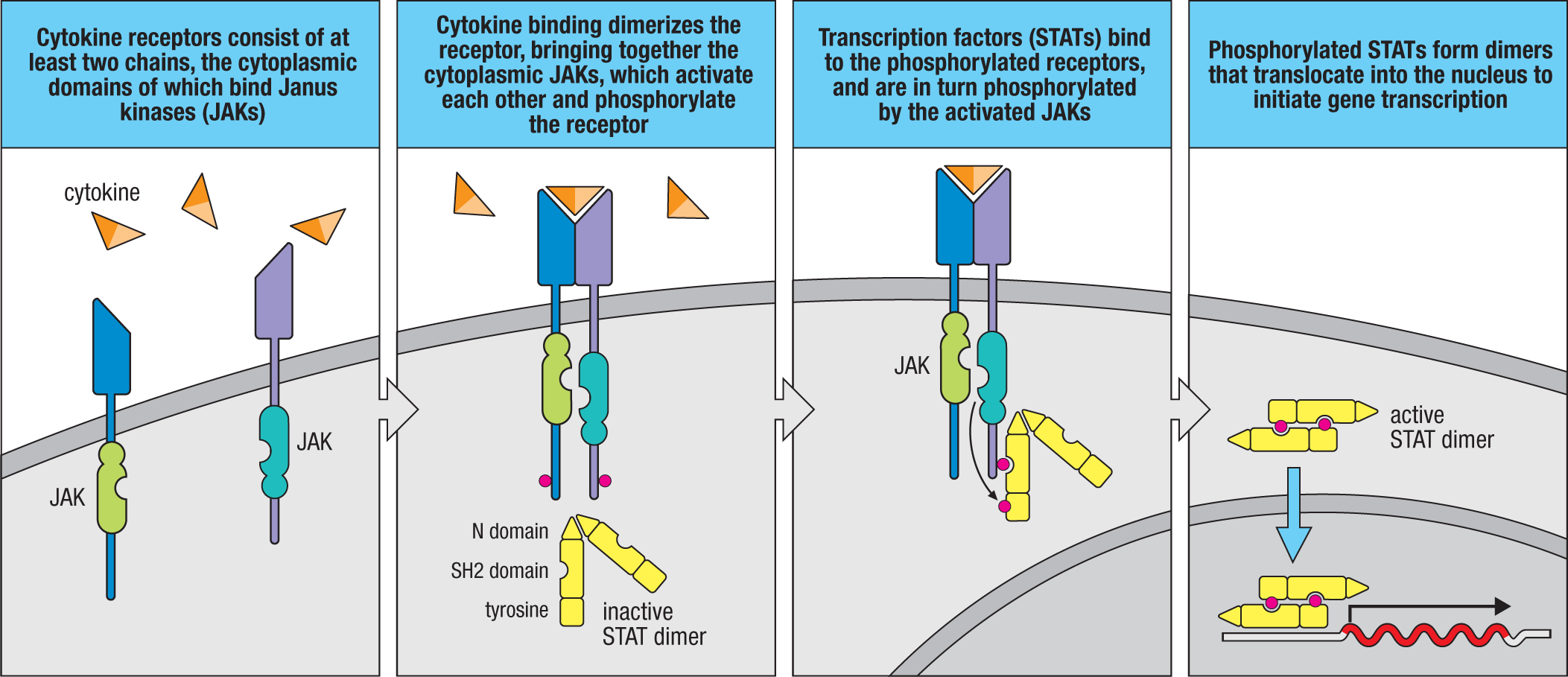
There are seven STATs (1–4, 5a, 5b, and 6), which reside in the cytoplasm in an inactive form until activated by cytokine receptors. Before activation, most STATs form homodimers, due to a specific homotypic interaction between domains present at the amino termini of the individual STAT monomers. The receptor specificity of each STAT is determined by the recognition of the distinctive phosphotyrosine sequence on each activated receptor by the different SH2 domains within the various STAT proteins. Recruitment of a STAT to the activated receptor brings the STAT close to an activated JAK, which can then phosphorylate the conserved tyrosine residue in the carboxyl terminus of each of the STATs. This leads to a rearrangement of the STAT dimer, in which the phosphotyrosine of each STAT monomer binds to the SH2 domain of the other STAT monomer, forming a configuration that can bind DNA with high affinity. Activated STATs predominantly form homodimers, with a cytokine typically activating one type of STAT. For example, IFN-γ activates STAT1 and generates STAT1 homodimers, whereas IL-4 activates STAT6, generating STAT6 homodimers. Other cytokine receptors can activate several STATs, and some STAT heterodimers can be formed, such as STAT1–STAT2 heterodimers formed downstream of the receptor for IFN-α and IFN-β. The phosphorylated STAT dimer enters the nucleus, where it acts as a transcription factor to initiate the expression of selected genes that can regulate growth, differentiation, and/or survival of particular subsets of lymphocytes.
Because signaling by these receptors depends on tyrosine phosphorylation, dephosphorylation of the receptor complex by tyrosine phosphatases is one way that cells can terminate signaling. A variety of tyrosine phosphatases have been implicated in the dephosphorylation of cytokine receptors, JAKs, and STATs. These include the nonreceptor tyrosine phosphatases SHP-1 and SHP-2 (encoded by PTPN6 and PTPN11) and the transmembrane receptor tyrosine phosphatase CD45, which is expressed as multiple isoforms on many hematopoietic cells. Cytokine signaling can also be terminated by negative feedback involving specific inhibitors that are induced by cytokine activation. The suppressor of cytokine signaling (SOCS) proteins are a class of inhibitors that terminate the signaling of many cytokine and hormone receptors. SOCS proteins contain an SH2 domain that can recruit them to the phosphorylated JAK protein or receptor, and they can inhibit JAK activity directly, or compete for the receptor, and direct the ubiquitination and subsequent degradation of JAKs and STATs. SOCS proteins are induced by STAT activation and thus inhibit receptor signaling after the cytokine has had its effect. Their importance can be seen in SOCS1-deficient mice, which develop a multiorgan inflammatory infiltrate caused by increased signaling from interferon receptors, γc-containing receptors, and TLRs. Another class of inhibitory proteins, the protein inhibitors of activated STAT (PIAS), regulate the response to cytokines, as well as other biological pathways, by promoting the degradation of pathway components.
3-17 Chemokines released by macrophages and dendritic cells recruit effector cells to sites of infection.
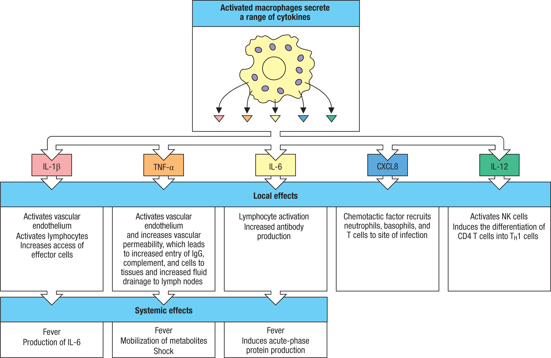
All the cytokines produced by macrophages in innate immune responses have important local and systemic effects that contribute to both innate and adaptive immunity, and these are summarized in Fig. 3.26. The recognition of different classes of pathogens by phagocytes and dendritic cells may involve signaling through different receptors, such as the various TLRs, and can result in some variation in the cytokines expressed by stimulated macrophages and dendritic cells. This is one way in which appropriate immune responses can be selectively activated, as the released cytokines orchestrate the next phase of host defense. In response to activation by PRRs, macrophages and dendritic cells secrete a diverse group of cytokines that includes IL-1β, IL-6, IL-12, TNF-α, and the chemokine CXCL8 (formerly known as IL-8).
3.8 Neutrophil Chase
Among the cytokines released by tissues in the earliest phases of infection are members of a family of chemoattractant cytokines known as chemokines. These small proteins induce directed chemotaxis in nearby responsive cells, resulting in the movement of the cells toward the source of the chemokine. Because chemokines were first detected in functional assays, they were initially given a variety of names, which are listed along with their standardized nomenclature in eAppendix IV. All the chemokines are related in amino acid sequence, and their receptors are G protein–coupled receptors (see Section 3-2). The signaling pathway stimulated by chemokines causes changes in cell adhesiveness and changes in the cell’s cytoskeleton that lead to directed migration. Chemokines can be produced and released by many different types of cells, not only those of the immune system. In the immune system they function mainly as chemoattractants for leukocytes, recruiting monocytes, neutrophils, and other effector cells of innate immunity from the blood into sites of infection. They also guide lymphocytes in adaptive immunity, as we will learn in Chapters 9–11. Some chemokines also function in lymphocyte development and migration and in angiogenesis (the growth of new blood vessels). There are more than 50 known chemokines, and this striking multiplicity may reflect their importance in delivering cells to their correct locations, which seems to be their main function in the case of lymphocytes. Some of the chemokines that are produced by or that affect human innate immune cells are listed in Fig. 3.27, along with their properties.
|
Class |
Chemokine |
Produced by |
Receptors |
Cells attracted |
Major effects |
|
CXC |
CXCL8 (IL-8) |
Monocytes Macrophages Fibroblasts Epithelial cells Endothelial cells |
CXCR1 CXCR2 |
Neutrophils Naive T cells |
Mobilizes, activates, and degranulates neutrophils Angiogenesis |
|
CXCL7 (PBP, β-TG, NAP-2) |
Platelets |
CXCR2 |
Neutrophils |
Activates neutrophils Clot resorption Angiogenesis |
|
|
CXCL1 (GROα) CXCL2 (GROβ) CXCL3 (GROγ) |
Monocytes Fibroblasts Endothelium |
CXCR2 |
Neutrophils Naive T cells Fibroblasts |
Activates neutrophils Fibroplasia Angiogenesis |
|
|
CC |
CCL3 (MIP-1α) |
Monocytes T cells Mast cells Fibroblasts |
CCR1, 3, 5 |
Monocytes NK and T cells Basophils Dendritic cells |
Competes with HIV-1 Antiviral defense Promotes TH1 immunity |
|
CCL4 (MIP-1β) |
Monocytes Macrophages Neutrophils Endothelium |
CCR1, 3, 5 |
Monocytes NK and T cells Dendritic cells |
Competes with HIV-1 |
|
|
CCL2 (MCP-1) |
Monocytes Macrophages Fibroblasts Keratinocytes |
CCR2B |
Monocytes NK and T cells Basophils Dendritic cells |
Activates macrophages Basophil histamine release Promotes TH2 immunity |
|
|
CCL5 (RANTES) |
T cells Endothelium Platelets |
CCR1, 3, 5 |
Monocytes NK and T cells Basophils Eosinophils Dendritic cells |
Degranulates basophils Activates T cells Chronic inflammation |
|
|
CXXXC (CX3C) |
CX3CL1 (Fractalkine) |
Monocytes Endothelium Microglial cells |
CX3CR1 |
Monocytes T cells |
Leukocyte–endothelial adhesion Brain inflammation |
Fig. 3.27 Properties of selected human chemokines. Chemokines fall mainly into two related but distinct groups: the CC chemokines, which have two adjacent cysteine residues near the amino terminus; and the CXC chemokines, in which the equivalent cysteine residues are separated by a single amino acid. In humans, the genes for CC chemokines are mostly clustered in one region of chromosome 4. Genes for CXC chemokines are found mainly in a cluster on chromosome 17. The two groups of chemokines act on different sets of receptors, all of which are G protein–coupled receptors. CC chemokines bind to receptors designated CCR1–10. CXC chemokines bind to receptors designated CXCR1–7. Different receptors are expressed on different cell types, and so a particular chemokine can be used to attract a particular cell type. In general, CXC chemokines with a Glu-Leu-Arg tripeptide motif immediately before the first cysteine promote the migration of neutrophils. CXCL8 is an example of this type. Most of the other CXC chemokines, including those that interact with receptors CXCR3, 4, and 5, lack this motif. Fractalkine is unusual in several respects: it has three amino acid residues between the two cysteines, and it exists in two forms, one that is tethered to the membrane of the endothelial and epithelial cells that express it, where it serves as an adhesion protein, and a soluble form that is released from the cell surface and acts as a chemoattractant for a wide range of cell types. A more comprehensive list of chemokines and their receptors is given in eAppendix IV.
Chemokines fall mainly into two related but distinct groups. CC chemokines have two adjacent cysteine residues near the amino terminus, whereas in CXC chemokines the corresponding two cysteine residues are separated by a single amino acid. The CC chemokines promote the migration of monocytes, lymphocytes, and other cell types. One example relevant to innate immunity is CCL2, which attracts monocytes through the receptor CCR2B, inducing their migration from the bloodstream to become tissue macrophages. In contrast, neutrophil migration is promoted by CXC chemokines. CXCL8, acting through CXCR2, mobilizes neutrophils from bone marrow and induces them to leave the blood and migrate into the surrounding tissues. CCL2 and CXCL8 therefore have similar but complementary functions in the innate immune response, attracting monocytes and neutrophils, respectively.
The role of chemokines in cell recruitment is twofold. First, they act on the leukocyte as it rolls along endothelial cells at sites of inflammation, converting this rolling into stable binding by triggering a change of conformation in the adhesion molecules known as leukocyte integrins. These conformational changes enable integrins to bind strongly to their ligands on the endothelial cells, which allows the leukocyte to cross the blood vessel walls by squeezing between the endothelial cells. Second, the chemokine directs the migration of the leukocyte along a gradient of chemokine molecules bound to the extracellular matrix and the surfaces of endothelial cells. This gradient increases in concentration toward the site of infection.
3.9 Immune-Cell Homing
Chemokines are produced by a wide variety of cell types in response to bacterial products, viruses, and agents that cause physical damage, such as silica, alum, or the urate crystals that occur in gout. Complement fragments, such as C3a and C5a, and fMLF bacterial peptides also act as chemoattractants for neutrophils. Thus, infection or physical damage to tissues induces the production of chemokine gradients that can direct phagocytes to the sites where they are needed. Neutrophils arrive rapidly in large numbers at a site of infection. The recruitment of monocytes occurs simultaneously, but they accumulate more slowly at the site of infection, perhaps because they are less abundant in the circulation. The complement fragment C5a and the chemokines CXCL8 and CCL2 activate their respective target cells, so that not only are neutrophils and monocytes brought to potential sites of infection, but also, in the process, they are armed to deal with the pathogens they encounter there. In particular, the signaling induced by C5a or CXCL8 in neutrophils serves to augment the respiratory burst that generates oxygen radicals and nitric oxide and to induce the neutrophils to release their stored antimicrobial granule contents (see Section 3-2).
Chemokines do not act alone in cell recruitment. They require the action of vasoactive mediators that bring leukocytes close to the blood vessel wall (see Section 3-3) and cytokines such as TNF-α to induce the necessary adhesion molecules on endothelial cells. We will return to the chemokines in later chapters, where they are discussed in the context of the adaptive immune response. Now, however, we turn to the molecules that enable leukocytes to adhere to the endothelium, and we shall then describe step by step the extravasation process by which monocytes and neutrophils enter infected sites.
3-18 Cell-adhesion molecules control interactions between leukocytes and endothelial cells during an inflammatory response.
The recruitment of activated phagocytes to sites of infection is one of the most important functions of innate immunity. Recruitment occurs as part of the inflammatory response and is mediated by cell-adhesion molecules that are induced on the surface of the endothelial cells of local blood vessels. Here we consider those functions that participate in the recruitment of inflammatory cells in the hours to days after the establishment of an infection.
3.10 Leukocyte Rolling
As with the complement components, a significant barrier to understanding the functions of cell-adhesion molecules is their nomenclature. Most adhesion molecules, especially those on leukocytes, which are relatively easy to analyze functionally, were originally named after the effects of specific monoclonal antibodies directed against them. Their names therefore bear no relation to their structural class. For instance, the leukocyte functional antigens LFA-1, LFA-2, and LFA-3 are actually members of two different protein families. In Fig. 3.28, the adhesion molecules relevant to innate immunity are grouped according to their molecular structure, which is shown in schematic form alongside their different names, sites of expression, and ligands. Three structural families of adhesion molecules are important for leukocyte recruitment. The selectins are membrane glycoproteins with a distal lectin-like domain that binds specific carbohydrate groups. Members of this family are induced on activated endothelium and initiate endothelium–leukocyte interactions by binding to fucosylated oligosaccharide ligands on passing leukocytes (see Fig. 3.28).
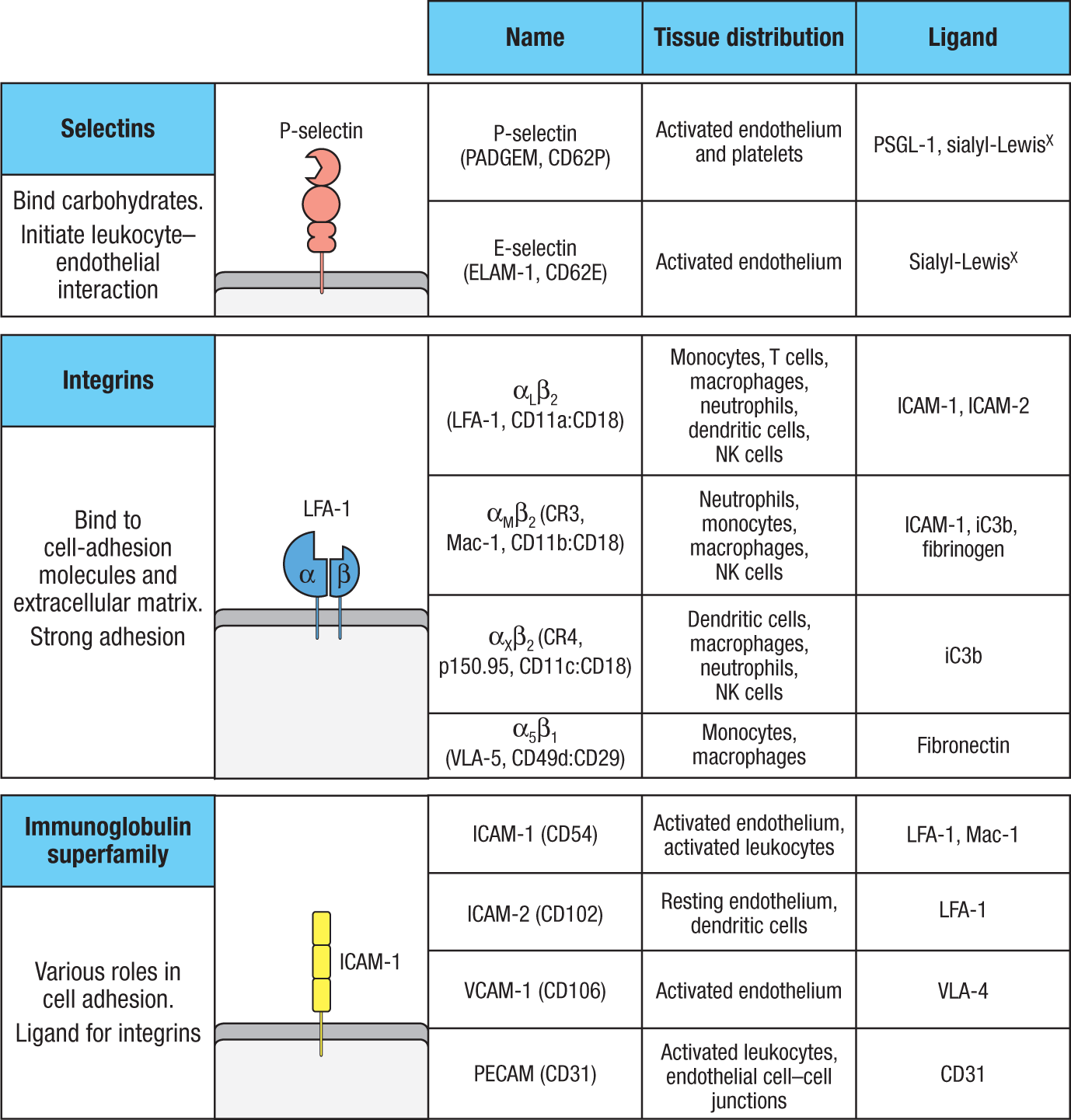
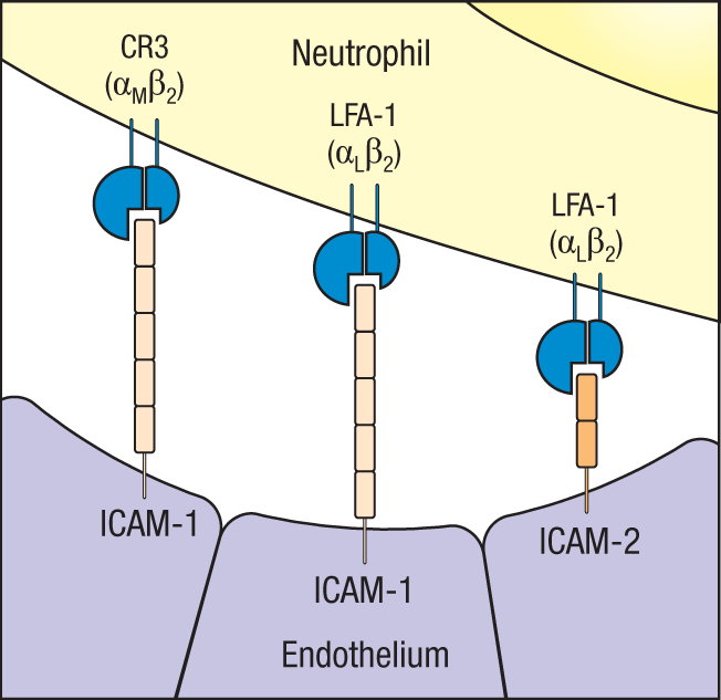
The next step in leukocyte recruitment depends on tighter adhesion, which is due to the binding of intercellular adhesion molecules (ICAMs) on the endothelium to heterodimeric proteins of the integrin family on leukocytes. ICAMs are single-pass membrane proteins that belong to the large superfamily of immunoglobulin-like proteins, which contain protein domains similar to those of immunoglobulins. The extracellular regions of ICAMs are composed of several immunoglobulin-like domains. An integrin molecule is composed of two transmembrane protein chains, α and β, of which there are numerous different types. Subsets of integrins have a common β chain partnered with different α chains. The leukocyte integrins important for extravasation are LFA-1 (αLβ2, also known as CD11a:CD18) and CR3 (αMβ2, complement receptor 3, also known as CD11b:CD18 or Mac-1). We described CR3 in Section 2-13 as a receptor for iC3b, but it also binds other ligands. Both LFA-1 and CR3 bind to ICAM-1 (Fig. 3.29). Even in the absence of infection, circulating monocytes are continually leaving the blood and entering certain tissues, such as the intestine, where they become resident macrophages. LFA-1 also binds to ICAM-2. To navigate out of the blood vessel, monocytes may adhere to ICAM-2, which is expressed at low levels by unactivated endothelium. CR3 also binds to fibrinogen and factor X, both substrates of the coagulation cascade.
Strong adhesion between leukocytes and endothelial cells is promoted by the induction of ICAM-1 on inflamed endothelium together with a conformational change in LFA-1 and CR3 that occurs on the leukocyte. Integrins can switch between an ‘active’ state, in which they bind strongly to their ligands, and an ‘inactive’ state, in which binding is easily broken. This enables cells to make and break integrin-mediated adhesions in response to signals received by the cell either through the integrin itself or through other receptors. In the activated state, an integrin molecule is linked via the intracellular protein talin to the actin cytoskeleton. In the case of migrating leukocytes, chemokines binding to their receptors on the leukocyte generate intracellular signals that cause talin to bind to the cytoplasmic tails of the β chains of LFA-1 and CR3, forcing the integrin extracellular regions to assume an active binding conformation. The importance of leukocyte integrin function in inflammatory-cell recruitment is illustrated by leukocyte adhesion deficiencies (LADs), which can be caused by defects in the integrins themselves or in the proteins required for modulating adhesion. People with these diseases suffer from recurrent bacterial infections and impaired healing of wounds.
Endothelial activation is driven by macrophage-produced cytokines, particularly TNF-α, which induce the rapid externalization of granules called Weibel–Palade bodies in the endothelial cells. These granules contain preformed P-selectin, which appears on the surfaces of local endothelial cells just minutes after macrophages have responded to the presence of microbes by producing TNF-α. Shortly after P-selectin gets to the cell surface, mRNA encoding E-selectin is synthesized, and within 2 hours the endothelial cells are expressing mainly E-selectin. Both P-selectin and E-selectin interact with sulfated sialyl-LewisX, a sulfated form of a carbohydrate structure that is also an important blood group antigen. Sulfated sialyl-LewisX is present on the surface of neutrophils, and its interactions with P-selectin and E-selectin are important for neutrophil rolling on the endothelium. Mutations in enzymes involved in its synthesis, such as fucosyltransferase, cause defective sialyl-LewisX expression that results in an immunodeficiency, leukocyte adhesion deficiency type 2.
Integrins are also convenient cell-surface markers for distinguishing different cell types. Dendritic cells, macrophages, and monocytes express different integrin α chains and thus display distinct β2 integrins on their surface. The predominant leukocyte integrin on conventional dendritic cells is αXβ2, also known as CD11c:CD18 or complement receptor 4 (CR4) (see Fig. 3.28). This integrin is a receptor for the complement C3 cleavage product iC3b, fibrinogen, and ICAM-1. In contrast to conventional dendritic cells, most monocytes and macrophages express low levels of CD11c and predominantly express the integrin αMβ2 (CD11b:CD18; CR3). However, patterns of integrin expression can vary, with some tissue macrophages, such as those in the lung, expressing high levels of CD11c:CD18. In the mouse, the two major branches of conventional dendritic cells can be distinguished by expression of CD11b:CD18: one branch characterized by high expression of CD11b:CD18, and a second branch that lacks CD11b:CD18. Plasmacytoid dendritic cells (pDCs) express lower levels of CD11c but can be distinguished from conventional dendritic cells using other markers; human pDCs express the C-type lectin BDCA-2 (blood dendritic cell antigen 2), and mouse pDCs express BST2 (bone marrow stromal antigen 2), neither of which is expressed by conventional dendritic cells.
3-19 Neutrophils make up the first wave of cells that cross the blood vessel wall to enter an inflamed tissue.
The migration of leukocytes out of blood vessels, the process known as extravasation, occurs in response to signals generated at sites of infection. Under normal conditions, leukocytes travel in the center of small blood vessels, where blood flow is fastest. Within sites of inflammation, the vessels are dilated and the consequent slower blood flow allows leukocytes to interact in large numbers with the vascular endothelium. During an inflammatory response, the induction of adhesion molecules on the endothelial cells of blood vessels within the infected tissue, as well as changes in the adhesion molecules expressed on leukocytes, recruits large numbers of circulating leukocytes to the site of infection. We will describe this process with regard to monocytes and neutrophils (Fig. 3.30).
3.11 Rolling Adhesion
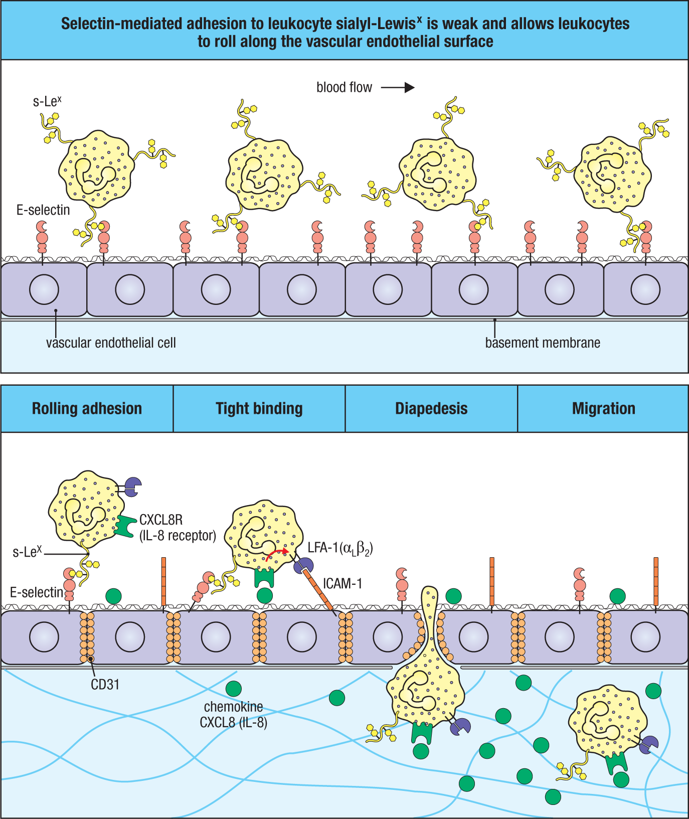
Extravasation proceeds in four stages. In the first, induction of selectins enables leukocyte rolling along the endothelium. P-selectin appears on endothelial cell surfaces within a few minutes of exposure to leukotriene B4, C5a, or histamine, which is released from mast cells in response to C5a. P-selectin can also be induced by TNF-α or LPS, and both of these induce synthesis of E-selectin, which appears on the endothelial cell surface a few hours later. When the sulfated sialyl-LewisX on monocytes and neutrophils contacts these exposed P- and E-selectins, these cells adhere reversibly to the vessel wall and begin to ‘roll’ along endothelium (see Fig. 3.30, top panel), permitting stronger interactions of the next step in leukocyte migration. Neutrophils are particularly efficient at rolling along endothelium, even under flow rates that prevent rolling by other cells. Such shear-resistant rolling by neutrophils uses long extensions of plasma membrane, termed slings, that bind the endothelium and wrap around the cell as it rolls, serving to tether the cell firmly to the endothelium and to promote rapid entry to sites of infection.
3.12 Neutrophil Rolling Using Slings
The second step depends on interactions between the leukocyte integrins LFA-1 and CR3 and adhesion molecules such as ICAM-1 (which can be induced on endothelial cells by TNF-α) or ICAM-2 on endothelium (see Fig. 3.30, bottom panel). LFA-1 and CR3 normally bind their ligands only weakly, but CXCL8 (or other chemokines), bound to proteoglycans on the surface of endothelial cells, binds to specific chemokine receptors on the leukocyte and signals the cell to trigger a conformational change in LFA-1 and CR3 on the rolling leukocyte; this greatly increases the adhesive properties of the leukocyte, as discussed in Section 3-18. The cell then attaches firmly to the endothelium, and its rolling is arrested.
3.13 Extravasation
In the third step the leukocyte extravasates, or crosses the endothelial wall. This step also involves LFA-1 and CR3, as well as a further adhesive interaction involving an immunoglobulin-related molecule called PECAM, or CD31, which is expressed both on the leukocyte and at the intercellular junctions of endothelial cells. These interactions enable cells to squeeze between the endothelial cells. It then penetrates the basement membrane with the aid of enzymes that break down the extracellular matrix proteins of the basement membrane. The movement through the basement membrane is known as diapedesis, and it enables cells to enter the subendothelial tissues.
The fourth and final step in extravasation is the migration of leukocytes through the tissues under the influence of chemokines. Chemokines such as CXCL8 and CCL2 (see Section 3-17) are produced at the site of infection and bind to proteoglycans in the extracellular matrix and on endothelial cell surfaces. In this way, a matrix-associated concentration gradient of chemokines is formed on a solid surface along which the leukocyte can migrate to the focus of infection (see Fig. 3.30). CXCL8 is released by the macrophages that first encounter pathogens; it recruits neutrophils, which enter the infected tissue in large numbers in the early part of the induced response. Their influx usually peaks within the first 6 hours of an inflammatory response. Monocytes are recruited through the action of CCL2 and accumulate more slowly than neutrophils. Once in the inflamed tissue, neutrophils are able to eliminate many pathogens by phagocytosis. In an innate immune response, neutrophils use their complement receptors and the direct pattern-recognition receptors discussed earlier in this chapter (see Section 3-1) to recognize and phagocytose pathogens or pathogen components directly or after opsonization with complement (see Section 2-13). In addition, as we will see in Chapter 10, neutrophils act as phagocytic effectors in humoral adaptive immunity, taking up antibody-coated microbes by means of specific receptors.
The importance of neutrophils in immune defense is dramatically illustrated by diseases or medical treatments that severely reduce neutrophil numbers. Individuals suffering this affliction are said to have neutropenia, and they are highly susceptible to deadly infection with a wide range of pathogens and commensal organisms. Restoring neutrophil levels in such individuals by transfusion of neutrophil-rich blood fractions or by stimulating their production with specific growth factors largely corrects this susceptibility.
3-20 TNF-α is an important cytokine that triggers local containment of infection but induces shock when released systemically.
TNF-α acting on endothelial cells stimulates the expression of adhesion molecules and aids the extravasation of cells such as monocytes and neutrophils. Another important action of TNF-α is to stimulate endothelial cells to express proteins that trigger blood clotting in the local small vessels, occluding them and cutting off blood flow. This can be important in preventing the pathogen from entering the bloodstream and spreading through the blood to organs all over the body. The importance of TNF-α in the containment of local infection is illustrated by experiments in which rabbits were infected locally with a bacterium. Normally, the infection would be contained at the site of the inoculation; if, however, an injection of anti-TNF-α antibody was also given to block the action of TNF-α, the infection spread via the blood to other organs. In parallel, the fluid that has leaked into the tissue in the early phases of an infection carries the pathogen, usually enclosed in dendritic cells, via the lymph to the regional lymph nodes, where an adaptive immune response can be initiated.
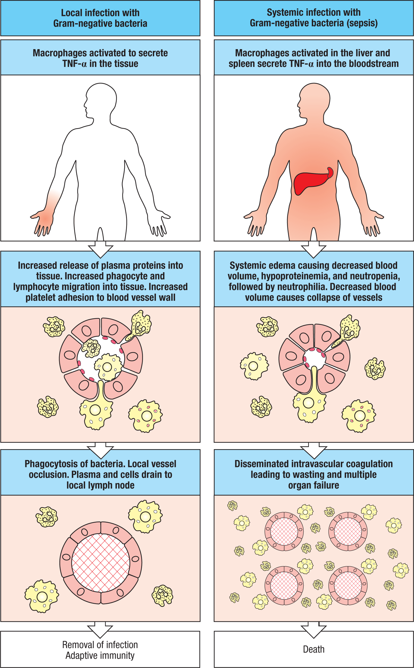
Once an infection has spread to the bloodstream, however, the same mechanisms by which TNF-α so effectively contains local infection instead become catastrophic (Fig. 3.31). Although produced as a membrane-associated cytokine, TNF-α can be cleaved by a protease, TACE (TNF–α–converting enzyme, which is encoded by the ADAM17 gene), and released from the membrane as a soluble cytokine. The presence of infection in the bloodstream, or sepsis, is accompanied by a massive release of soluble TNF-α from macrophages in the liver, spleen, and other sites throughout the body. The systemic release of TNF-α into the bloodstream causes vasodilation, which leads to a loss of blood pressure and increased vascular permeability; this in turn leads to a loss of plasma volume and eventually to shock, known in this case as septic shock because the underlying cause is a bacterial infection. The TNF-α released in septic shock also triggers blood clotting in small vessels throughout the body—known as disseminated intravascular coagulation (DIC)—which leads to the massive consumption of clotting proteins, so that the individual’s blood cannot clot appropriately. Disseminated intravascular coagulation frequently leads to the failure of vital organs such as the kidneys, liver, heart, and lungs, which are quickly compromised by the failure of normal blood perfusion; consequently, septic shock has a very high mortality rate.
Blockade of TNF-α activity, either with specific antibodies or with soluble proteins that mimic the receptor, is a successful treatment for several inflammatory disorders, including rheumatoid arthritis. However, these treatments have been found to reactivate tuberculosis in some apparently well individuals with evidence of previous infection (as demonstrated by skin test), which is a direct demonstration of the importance of TNF-α in keeping infection local and in check. The efficacy of TNF-α blockade as a treatment for sepsis remains unclear.
3-21 Cytokines made by macrophages and dendritic cells induce a systemic reaction known as the acute-phase response.
As well as their important local effects, the cytokines produced by macrophages and dendritic cells have long-range effects that contribute to host defense. One of these is the elevation of body temperature, which is caused mainly by TNF-α, IL-1β, and IL-6. These cytokines are termed endogenous pyrogens because they cause fever and derive from an endogenous source rather than from bacterial components such as LPS, which also induces fever and is an exogenous pyrogen. Endogenous pyrogens cause fever by inducing the synthesis of prostaglandin E2 by the enzyme cyclooxygenase-2, the expression of which is induced by these cytokines. Prostaglandin E2 then acts on the hypothalamus, resulting in an increase in both heat production from the catabolism of brown fat and heat retention from vasoconstriction, which decreases the loss of excess heat through the skin. Exogenous pyrogens are able to induce fever by promoting the production of the endogenous pyrogens and also by directly inducing cyclooxygenase-2 as a consequence of signaling through TLR-4, leading to the production of prostaglandin E2. Fever is generally considered beneficial to host defense, although the precise mechanisms by which fever facilitates pathogen clearance remain unclear.
The effects of TNF-α, IL-1β, and IL-6 are summarized in Fig. 3.32. One of the most important of these occurs in the liver and is the initiation of a response known as the acute-phase response (Fig. 3.33). The cytokines act on hepatocytes, which respond by changing the profile of proteins that they synthesize and secrete into the blood. In the acute-phase response, blood levels of some proteins go down, whereas levels of others increase markedly. The proteins induced by TNF-α, IL-1β, and IL-6 are called the acute-phase proteins. Several of these are of particular interest because they mimic the action of antibodies, but unlike antibodies they have broad specificity for pathogen-associated molecular patterns and depend only on the presence of cytokines for their production.
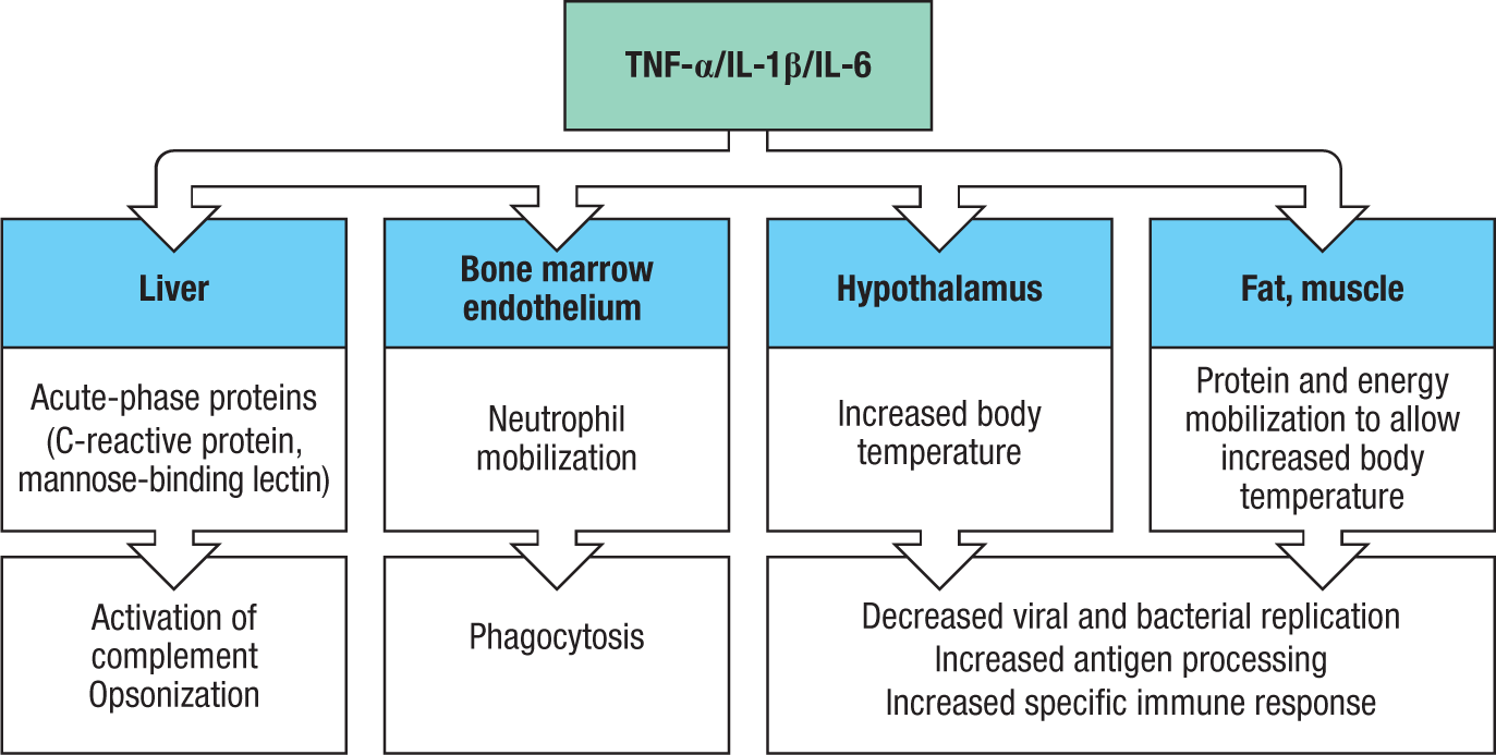
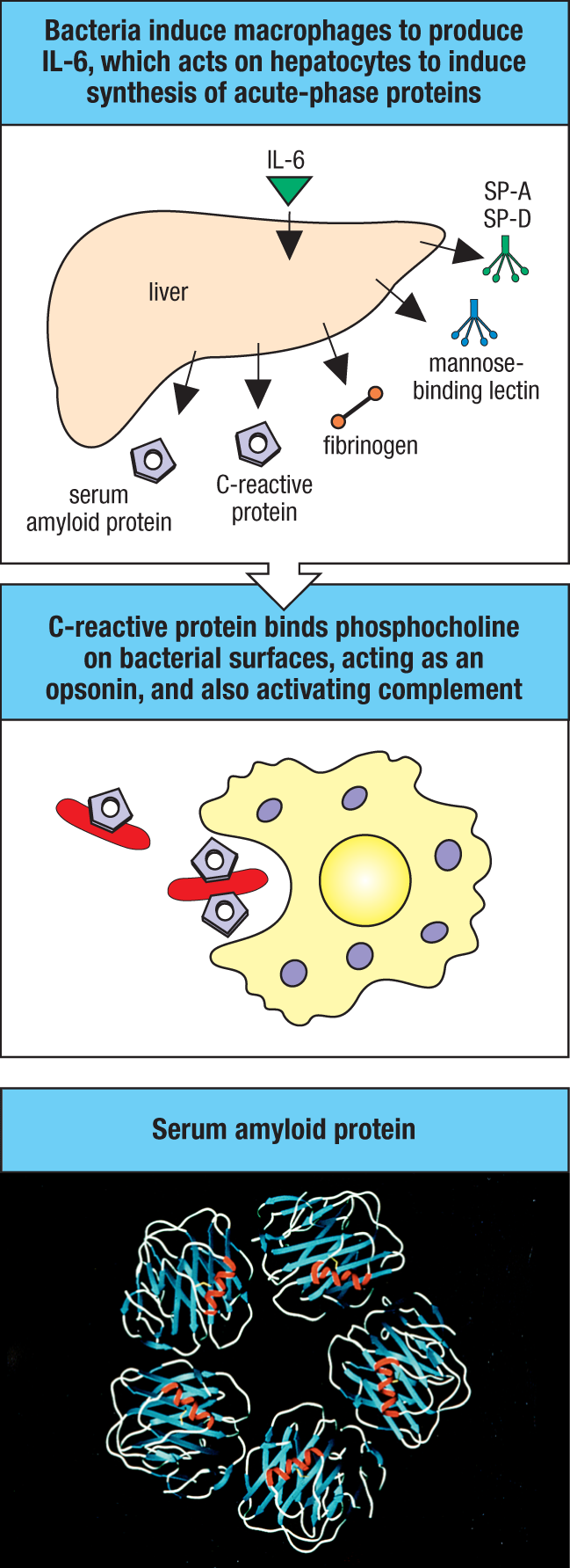
One acute-phase protein, the C-reactive protein, is a member of the pentraxin protein family, so called because the proteins are formed from five identical subunits. C-reactive protein is yet another example of a multipronged pathogen-recognition molecule, and it binds to the phosphocholine portion of certain bacterial and fungal cell-wall lipopolysaccharides. Phosphocholine is also found in mammalian cell-membrane phospholipids, but it cannot be bound by C-reactive protein. When C-reactive protein binds to a bacterium, it not only is able to opsonize the bacterium but can also activate the complement cascade by binding to C1q, the first component of the classical pathway of complement activation (see Section 2-7). The interaction with C1q involves the collagen-like parts of C1q rather than the globular heads that make contact with pathogen surfaces, but the same cascade of reactions is initiated.
Mannose-binding lectin (MBL) is another acute-phase protein; it serves as an innate recognition molecule that can activate the lectin pathway of complement (see Section 2-6). MBL is present at low levels in the blood of healthy individuals, but it is produced in increased amounts during the acute-phase response. By recognizing mannose residues on microbial surfaces, MBL can act as an opsonin that is recognized by monocytes, which do not express the macrophage mannose receptor. Two other proteins with opsonizing properties that are also produced in increased amounts during an acute-phase response are the surfactant proteins A and D (SP-A and SP-D). These are produced by the liver and a variety of epithelia. They are, for example, found along with macrophages in the alveolar fluid of the lung, where they are secreted by pneumocytes, and are important in promoting the phagocytosis of opportunistic respiratory pathogens such as Pneumocystis jirovecii (formerly known as P. carinii), one of the main causes of pneumonia in individuals with AIDS.
Thus, within a day or two, the acute-phase response provides the host with several proteins with the functional properties of antibodies but able to bind a broad range of pathogens. However, unlike antibodies, which we describe in Chapters 4 and 10, acute-phase proteins have no structural diversity and are made in response to any stimulus that triggers the release of TNF-α, IL-1β, and IL-6. Therefore, unlike antibodies, their synthesis is not specifically induced and targeted.
A final, distant effect of the cytokines produced by macrophages is to induce leukocytosis, an increase in the numbers of circulating neutrophils. The neutrophils come from two sources: the bone marrow, from which mature leukocytes are released in increased numbers; and sites in blood vessels, where they are attached loosely to endothelial cells. Thus, the effects of these cytokines contribute to the control of infection while the adaptive immune response is being developed.
3-22 Interferons induced by viral infection make several contributions to host defense.
Viral infection induces the production of interferons, originally named because of their ability to interfere with viral replication in previously uninfected tissue culture cells. Interferons have a similar role in vivo, blocking the spread of viruses to uninfected cells. There are numerous genes encoding antiviral, or type I, interferons. Best understood are the IFN-α family of 12 closely related human genes and IFN-β, the product of a single gene; less well studied are IFN-Κ, IFN-ϵ, and IFN-ω. IFN-γ is the sole type II interferon.
Type III interferons are a newly classified IFN family composed of the products of three IFN-λ genes, also known as IL-28A, IL-28B, and IL-29, which bind a heterodimeric IFN-λ receptor composed of a unique IL-28Rα subunit and the β subunit of the IL-10 receptor. While receptors for type I interferons and IFN-γ are widespread in their tissue distribution, type III receptors are more restricted. For example, they are not expressed by fibroblasts but are expressed on epithelial cells.
Type I interferons are inducible and are synthesized by many cell types after infection by diverse viruses. Almost all types of cells can produce IFN-α and IFN-β in response to activation of several innate sensors. For example, type I interferons are induced by RLRs and cGAS–STING signaling (see Sections 3-8 and 3-9). However, some immune cells seem to be specialized for this task. In Section 3-1 we introduced the plasmacytoid dendritic cell (pDC). Also called interferon-producing cells (IPCs) or natural interferon-producing cells, human plasmacytoid dendritic cells were initially recognized as rare peripheral blood cells that accumulate in peripheral lymphoid tissues during a viral infection and make abundant type I interferons (IFN-α and IFN-β)—up to 1000 times more than that made by other cell types. This abundant production of type I interferons may result from the efficient coupling of viral recognition by TLRs to the pathways of interferon production (see Section 3-7). Plasmacytoid dendritic cells express a subset of TLRs that includes TLR-7 and TLR-9, which are endosomal sensors of viral RNA and DNA (see Fig. 3.11). The requirement for TLR-9 in sensing infections caused by DNA viruses has been demonstrated, for example, by the inability of TLR-9–deficient plasmacytoid dendritic cells to generate type I interferons in response to herpes simplex virus.
Interferons help defend against viral infection in several ways (Fig. 3.34). IFN-β induces an antiviral state in cells, which includes making cells more likely to produce IFN-α in response to PRR triggering, thus amplifying the interferon response. IFN-α and IFN-β bind to a common cell-surface receptor, known as the interferon-a receptor (IFNAR), which uses the JAK and STAT pathways described in Section 3-16. IFNAR uses the kinases Tyk2 and Jak1 to activate the factors STAT1 and STAT2, which can interact with IRF9 and form a complex called ISGF3, which binds to the promoters of many interferon-stimulated genes (ISGs). The induction of ISGs induces a state of resistance to viral replication in all cells. Many of the genes involved in nucleic-acid sensing and signaling are ISGs, including the genes for RIG-I, MDA-5, IRF7, and STING, such that cells exposed to interferons have a heightened ability to recognize viral nucleic acids.
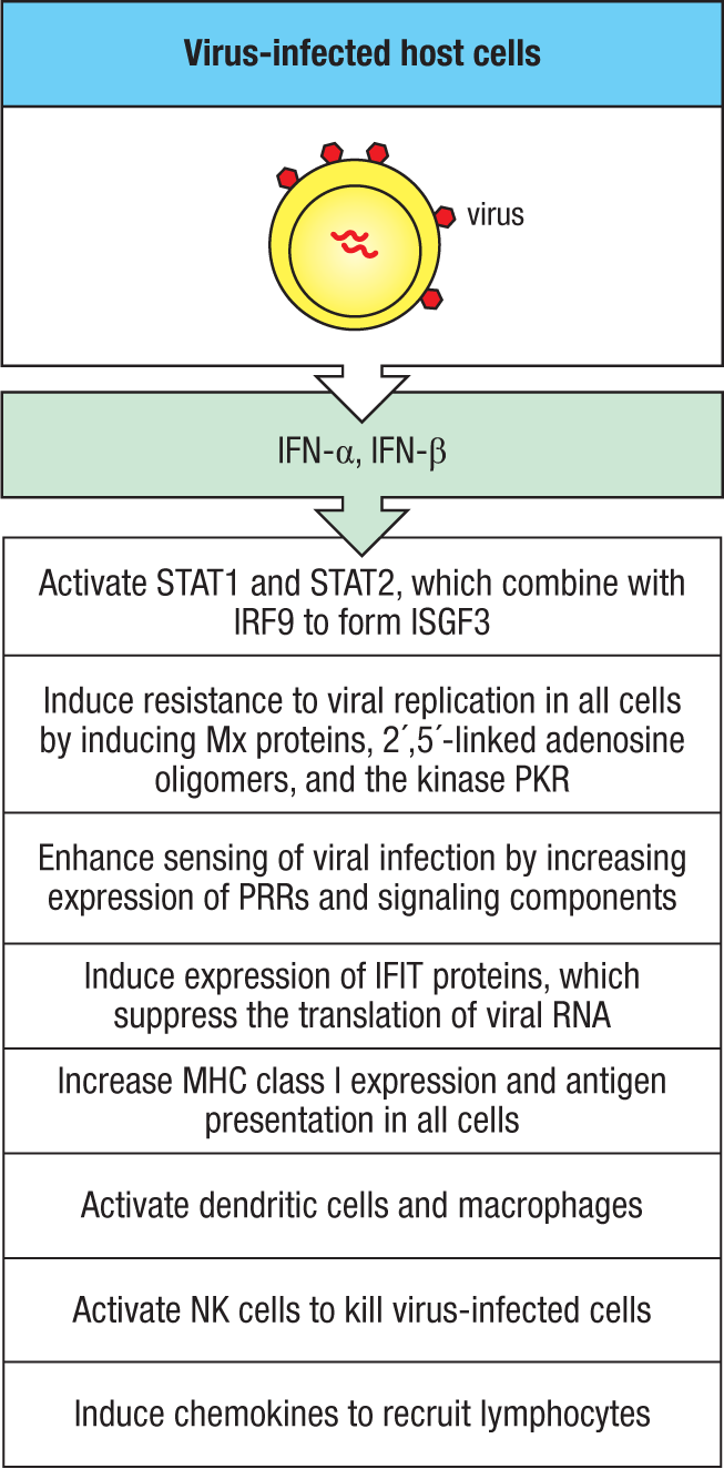
ISGs exert antiviral activities through a variety of mechanisms. One ISG encodes the enzyme oligoadenylate synthetase, which polymerizes ATP into 2′,5′-linked oligomers (whereas nucleotides in nucleic acids are normally linked 3′,5′). These 2′,5′-linked oligomers serve as second messengers and activate an endoribonuclease that then degrades viral RNA (note the parallels to cGAMP activation of STING, described in Section 3-9). Another ISG-encoded protein induced by IFN-α and IFN-β is a dsRNA-dependent protein kinase called PKR. This serine/threonine kinase phosphorylates the α subunit of eukaryotic initiation factor 2 (eIF2), thus suppressing protein translation and contributing to the inhibition of viral replication. Mx (myxoma resistant) proteins are also ISG-encoded proteins induced by type I interferons. Humans and wild mice have two highly similar proteins, Mx1 and Mx2, which are GTPases belonging to the dynamin protein family, but how they interfere with viral replication is not understood.
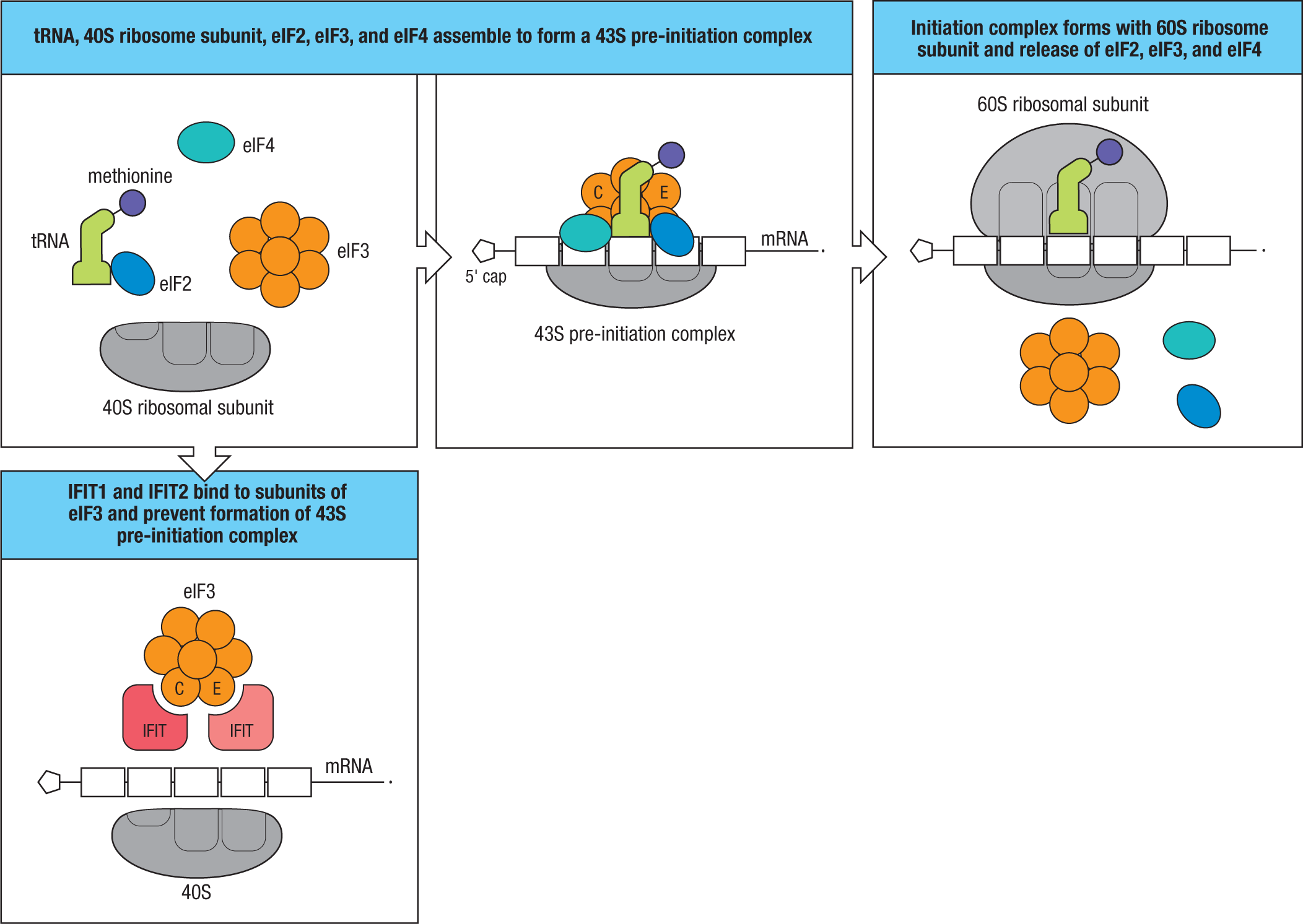
The IFIT (IFN-induced protein with tetratricoid repeats) family of ISG-encoded proteins contains four human and three mouse proteins that function in restraining the translation of viral RNA into proteins. IFIT1 and IFIT2 can both suppress the translation of normal capped mRNAs by binding to subunits of the eukaryotic initiation factor 3 (eIF3) complex, which prevents eIF3 from interacting with eIF2 to form the 43S pre-initiation complex (Fig. 3.35). This action may be responsible in part for the reduction in cellular proliferation induced by type I interferons. Mice lacking IFIT1 or IFIT2 show increased susceptibility to infection by certain viruses, such as vesicular stomatitis virus.
Another function of IFIT1 is to suppress translation of viral RNA that lacks a normal host modification of the 5′ cap. Recall that the normal mammalian 5′ cap is initiated by linking a 7-methylguanosine nucleotide to the first ribose sugar of the mRNA by a 5′,5′-linked triphosphate bridge to produce a structure called cap-0. This structure is further modified by cytoplasmic methylation of the 2′ hydroxyl groups on the first and second ribose sugars of the RNA. Methylation of the first ribose sugar produces a structure called cap-1; methylation of the second generates cap-2. IFIT1 has a high affinity for cap-0 but much lower affinity for cap-1 and cap-2. Some viruses, such as Sindbis virus (family Togaviridae), lack 2′-O-methylation and therefore are restricted by this action of IFIT1. Many viruses, such as West Nile virus and SARS coronavirus, have acquired a 2′-O-methyltransferase (MTase) that produces cap-1 or cap-2 on their viral transcripts. These viruses can thus evade restriction by IFIT1.
Members of the interferon-induced transmembrane protein (IFITM) family are expressed at a basal level on many types of tissues but are strongly induced by type I interferons. There are four functional IFITM genes in humans and in mice, and these encode proteins that have two transmembrane domains and are localized to various vesicular compartments of the cell. IFITM proteins act to inhibit, or restrict, viruses at early steps of infection. Although the molecular details are unclear, IFITM1 appears to interfere with the fusion of viral membranes with the membrane of the lysosome, which is required for introducing some viral genomes into the cytoplasm. Viruses that must undergo this fusion event in lysosomes, such as the Ebola virus, are restricted by IFITM1. Similarly, IFITM3 interferes with membrane fusion in late endosomes, and so restricts the influenza A virus, which undergoes fusion there. The importance of this mechanism is demonstrated by the increased viral load and higher mortality in mice lacking IFITM3 that are infected with the influenza A virus.
Interferons also stimulate production of the chemokines CXCL9, CXCL10, and CXCL11, which recruit lymphocytes to sites of infection. They also increase the expression of MHC class I molecules on all types of cells, which facilitates recognition of virally infected cells by cytotoxic T lymphocytes via the display of viral peptides complexed to MHC class I molecules on the infected cell surface (see Fig. 1.30). Through these effects, interferons indirectly help promote the killing of virus-infected cells by CD8 cytotoxic T cells. Another way in which interferons act is to activate populations of innate immune cells, such as NK cells, that can kill virus-infected cells, as described below.
3-23 Several types of innate lymphoid cells provide protection in early infection.
A defining feature of adaptive immunity is the clonal expression of antigen receptors, produced by somatic gene rearrangements, that provide the extraordinarily diverse specificities of T and B lymphocytes (see Section 1-11). However, for several decades, immunologists have recognized cells with lymphoid characteristics that lack specific antigen receptors. Natural killer (NK) cells have been known the longest, but in the past several years other distinct groups of such cells have been identified. Collectively, these are now called innate lymphoid cells (ILCs) and include NK cells (Fig. 3.36). ILCs develop in the bone marrow from the same common lymphoid progenitor (CLP) that gives rise to B and T cells. Expression of the transcription factor Id2 (inhibitor of DNA binding 2) in the CLP represses B- and T-cell fates and is required for the development of all ILCs. ILCs are identified by the absence of T- and B-cell antigen receptor and co-receptor complexes, but they express receptors for common γ-chain cytokines. They migrate from the bone marrow and take up residence in lymphoid tissues and peripheral organs, notably the dermis, liver, small intestine, adipose tissue, and lung.
|
The major categories of innate lymphoid cells (ILCs) and their properties |
|||
|
Innate lymphoid subgroup |
Inducing cytokines |
Effector molecules produced |
Function |
|
NK cells |
IL-12, IL-18, type I IFN |
IFN-γ, perforin, granzyme |
Immunity against viruses, intracellular pathogens |
|
ILC1 |
IL-12, IL-18 |
IFN-γ |
Defense against viruses, intracellular pathogens |
|
ILC2 |
IL-25, IL-33, TSLP |
IL-5, IL-13, amphiregulin, IL-4 |
Expulsion of extracellular parasites, tissue repair |
|
ILC3 (LTi cells) |
IL-1β, IL-23 |
IL-17, IL-22 |
Immunity to extracellular bacteria |
Fig. 3.36 The major categories of innate lymphoid cells (ILCs) and their properties.
ILCs function in innate immunity as effector cells that amplify the signals delivered by innate recognition. They are stimulated by cytokines produced by other innate cells, such as macrophages or dendritic cells, or by nonimmune cells, such as epithelial cells, that have been activated by innate sensors of microbial infection or cellular damage. Three major subgroups of ILCs are defined, largely on the basis of the types of cytokines that each produces. Group 1 ILCs (ILC1s) generate IFN-γ in response to activation by certain cytokines, in particular IL-12 and IL-18, made by dendritic cells and macrophages, and they function in protection against infection by viruses or intracellular pathogens. NK cells are now considered to be a type of ILC. ILC1s and NK cells are closely related but have distinct functional properties and differ in the cytokines and transcription factors required for their development. For example, both NK and ILC1 cells require the transcription factors Id2 and Nfil3 for their development, but ILC1s also require the transcription factor PLZF whereas NK cells do not. NK cells require the cytokine IL-15, while liver ILC1 cells require the cytokine IL-7 and the transcription factor Tbet. Functionally, NK cells are more similar to CD8 T cells, while ILC1s resemble more closely the TH1 subset of CD4 T cells (see Section 3-24). NK cells can be found within tissues, but they also circulate through the blood, while ILC1 cells appear to be largely noncirculating tissue-resident cells. In the mouse, conventional NK cells express the integrin α2 (CD49b), while ILC1 cells, for example in the liver, lack CD49b but express the surface protein CD49a.
Group 2 ILCs (ILC2s) produce the cytokines IL-4, IL-5, and IL-13 in response to various cytokines, particularly thymic stromal lymphopoietin (TSLP), IL-25, and IL-33. ILC2 cytokines function in promoting mucosal and barrier immunity and aid in protection against parasites. ILC2s also produce amphiregulin, an EGF family member that induces epithelial cell proliferation and differentiation, which facilitates repair of damaged tissues.
Group 3 ILCs (ILC3s) respond to the cytokines IL-1β and IL-23 and produce several cytokines, including IL-17 and IL-22, which increase defenses against extracellular bacteria and fungi. IL-17 functions by stimulating the production of chemokines that recruit neutrophils, while IL-22 acts directly on epithelial cells to stimulate the production of antimicrobial peptides such as RegIIIγ (see Section 2-4).
The classification of ILC subtypes and the analysis of their development and function is still an active area, and studies to define the relative importance of these cells in immune responses are ongoing. The ILC subgroups identified so far appear to be highly parallel in structure to the subsets of effector CD8 and CD4 T cells that were defined over the past three decades. The transcription factors that control the development of different ILC subsets seem, for now at least, to be the same as those that control the corresponding T-cell subsets. Because of these similarities, we will postpone a detailed description of ILC development until Chapter 9, where we will cover this topic along with the development of T-cell subsets.
3-24 NK cells are activated by type I interferon and macrophage-derived cytokines.
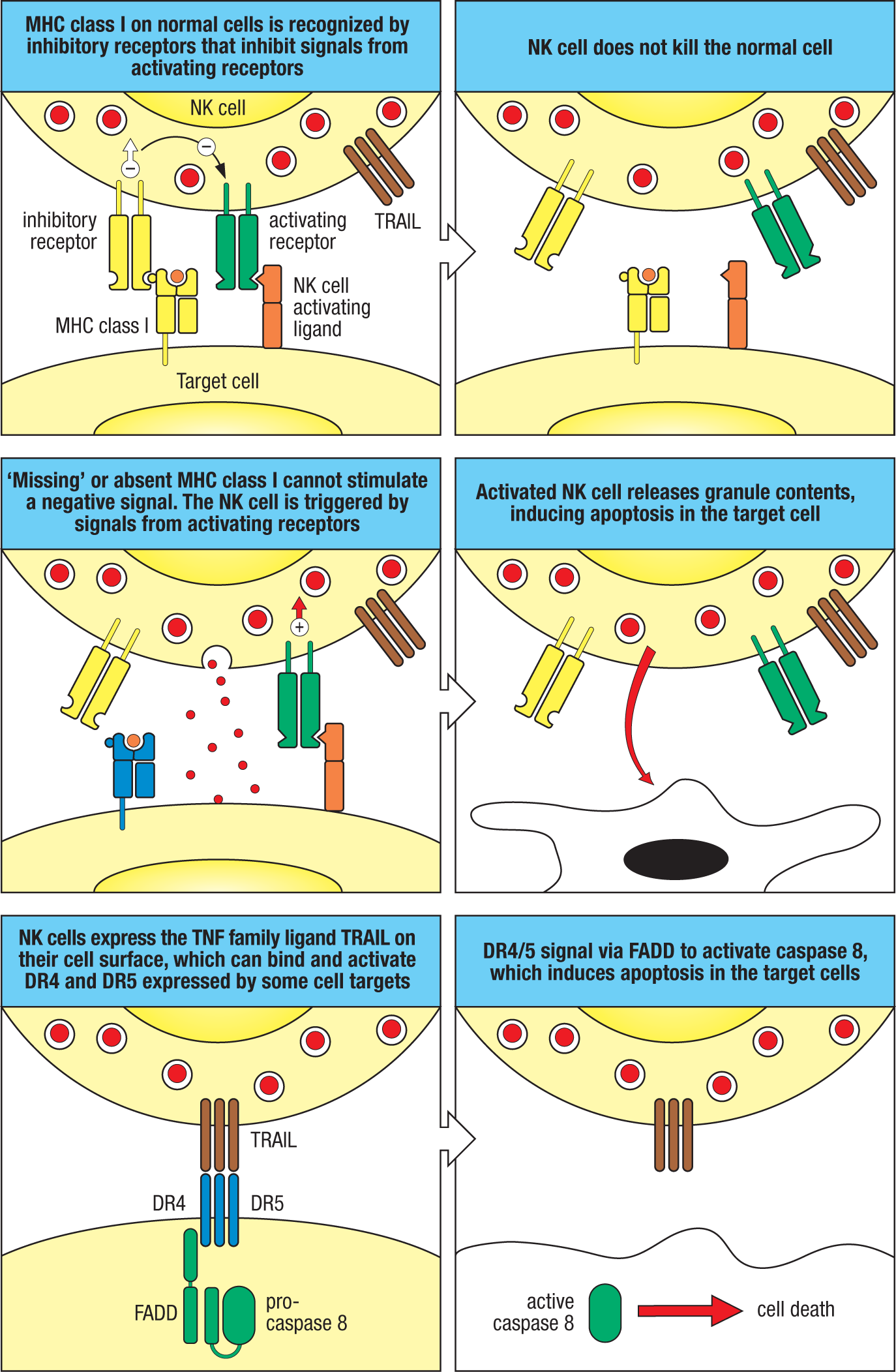
NK cells are larger than naive T and B cells, have distinctive cytoplasmic granules containing cytotoxic proteins, and were functionally identified by their ability to kill certain tumor cell lines in vitro without the need for prior immunization. NK cells kill cells by releasing their cytotoxic granules, which are similar to those of cytotoxic T cells and have the same effects (discussed in Chapter 9). In brief, the contents of cytotoxic granules, which contain granzymes and the pore-forming protein perforin, are released onto the surface of the target cell and penetrate the cell membrane and induce programmed cell death. However, unlike T cells, killing by NK cells is triggered by germline-encoded receptors that recognize molecules on the surface of infected or malignantly transformed cells. A second pathway used by NK cells to kill target cells involves the TNF family member known as TRAIL (tumor necrosis factor–related apoptosis-inducing ligand). NK cells express TRAIL on their cell surface. TRAIL interacts with two TNFR superfamily ‘death’ receptors, DR4 and DR5 (encoded by TNFSF10A and TNFSF10B), that are expressed by many types of cells. When NK cells recognize a target cell, TRAIL stimulates DR4 and DR5 to activate the pro-enzyme caspase 8, which leads to apoptosis. In contrast to pyroptosis, induced by caspase 1 after inflammasome activation (see Section 3-11), apoptosis is not associated with production of inflammatory cytokines. We will discuss more details of the mechanisms of caspase-induced apoptosis when we discuss killing by cytotoxic T cells in Chapter 9. Finally, NK cells express Fc receptors (see Section 1-20); binding of antibodies to these receptors activates NK cells to release their cytotoxic granules, a process known as antibody-dependent cell-mediated cytotoxicity, or ADCC, to which we will return in Chapter 10.
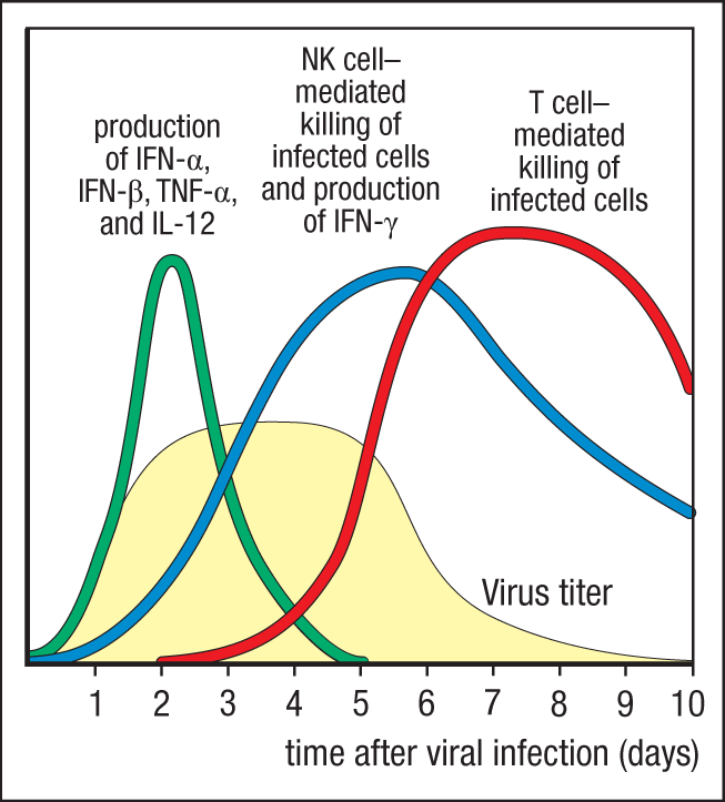
The ability of NK cells to kill target cells can be enhanced by interferons or certain cytokines. NK cells that can kill sensitive targets can be isolated from uninfected individuals, but this activity is increased 20- to 100-fold when NK cells are exposed to IFN-α and IFN-β or to IL-12, a cytokine produced by dendritic cells and macrophages during infection by many types of pathogens. Activated NK cells serve to contain virus infections while the adaptive immune response is generating antigen-specific cytotoxic T cells and neutralizing antibodies that can clear the infection (Fig. 3.37). A clue to the physiological function of NK cells in humans comes from rare individuals deficient in these cells, who are frequently susceptible to herpesvirus infection. For example, a selective NK-cell deficiency results from mutations in the human MCM4 (minichromosome maintenance-deficient 4) protein, which is associated with predisposition to viral infections. Similarly, depletion of NK cells in mice, either by antibody or genetically, results in susceptibility to a number of viruses, including herpesviruses.
IL-12, produced by activated macrophages and dendritic cells, acts in synergy with the cytokine IL-18 to stimulate NK cells to secrete large amounts of IFN-γ. This IFN-γ is crucial in controlling certain infections because it precedes the production of IFN-γ by activated CD8 cytotoxic T cells. IFN-γ, whose receptor activates only the STAT1 transcription factor, is quite distinct functionally from the antiviral type I interferons IFN-α and IFN-β and is not directly induced by viral infection. The production of IFN-γ by NK cells early in an immune response can directly activate macrophages to enhance their capacity to kill pathogens, augmenting innate immunity. IFN-γ also influences adaptive immunity through actions on dendritic cells and by regulating the differentiation of CD4 T cells into the pro-inflammatory TH1 subset, which produces IFN-γ. Accordingly, mice deficient for the gene encoding IFN-γ are highly susceptible to viruses and intracellular bacterial pathogens. NK cells also produce TNF-α, granulocyte–macrophage colony-stimulating factor (GM-CSF), and the chemokines CCL3 (MIP-1α), CCL4, and CCL5 (RANTES), which act to recruit and activate macrophages.
3-25 NK cells express activating and inhibitory receptors to distinguish between healthy and infected cells.
For NK cells to defend against viruses or other pathogens, they must be able to distinguish infected cells from uninfected healthy cells. To do this, NK cells use a mechanism distinct from the mechanism of pathogen recognition used by T or B cells. An individual NK cell expresses various combinations of germline-encoded activating receptors and inhibitory receptors. While the exact details are not clear in every case, it is thought that the overall balance of signaling by these receptors determines whether an NK cell engages and kills a target cell. The receptors on an NK cell are tuned to detect changes in expression of various surface proteins on a target cell, referred to as ‘dysregulated self.’ The activating receptors generally recognize cell-surface proteins that are induced on target cells by infection or stress, such as malignant transformation. These induced proteins are referred to as ‘stress-induced self.’ A number of cellular perturbations, including DNA damage, signals related to proliferation, and heat shock–related stress, can lead to induction of ligands for activating receptors. Signaling by innate sensors (for example, TLRs) is also capable of inducing such ligands. When activating receptors on NK cells detect stress-induced self ligands, signaling is initiated in the NK cell that results in release of cytokines, such as IFN-γ, and killing of the stimulating cell through the release of cytotoxic granules. However, engagement of activating receptors is not always sufficient to induce NK-cell activation; negative signals imposed by inhibitory receptors must either be lost or overcome, as discussed below.
Inhibitory receptors on NK cells recognize surface molecules that are constitutively expressed at high levels by most healthy cells. As such, NK cells that encounter healthy cells receive signals that inhibit the signals downstream of activating receptors and prevent their activation. However, infection, cellular stress, or transformation can lead to down-regulation of the ligands for inhibitory receptors. The loss of these molecules is referred to as ‘missing self.’ Inhibitory receptors can recognize other molecules, but those recognizing MHC class I molecules have been best characterized. MHC molecules are glycoproteins expressed on nearly all cells of the body. We will discuss the role of MHC proteins in antigen presentation to T cells in Chapter 6, but for now we need only to introduce the two main classes of MHC molecules. MHC class I molecules are expressed on most of the cells of the body (except, notably, red blood cells), whereas the expression of MHC class II molecules is far more restricted, largely to immune cells.
Inhibitory receptors that recognize MHC class I molecules function to prevent NK cells from killing normal host cells. The greater the number of MHC class I molecules on a cell surface, the better protected that cell is against attack by NK cells. Interferons induce expression of MHC class I molecules and protect uninfected host cells from being killed by NK cells, while also activating NK cells to kill virus-infected cells. Viruses and some other intracellular pathogens can cause down-regulation of MHC class I molecules as a strategy to prevent the display of antigens as peptides to T cells, also discussed in Chapter 13. MHC class I molecules are often similarly down-regulated in cancerous cells. Importantly, NK cells are able to sense this reduction in expression of MHC class I molecules through reduced signaling from their inhibitory receptors. Reduction in MHC class I expression is an example of ‘missing self’ and increases the chance that an NK cell will kill the target cell. It is thought that the balance of signals from ‘stress-induced self’ and ‘missing self’ determines whether an individual NK cell will be triggered to kill a particular target cell (Fig. 3.38). Thus, receptors expressed on NK cells integrate the signals from two types of surface receptors, which together control an NK cell’s cytotoxic activity and cytokine production.
3-26 NK-cell receptors belong to several structural families.
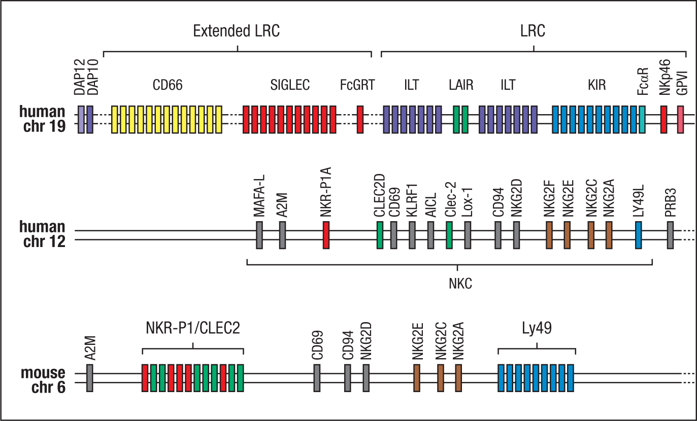
The receptors that regulate the activity of NK cells fall into several large families that contain a number of other cell-surface receptors in addition to NK receptors (Fig. 3.39). Members of the family of killer cell immunoglobulin-like receptors (KIRs) have differing numbers of immunoglobulin domains. Some, such as KIR-2D, have two immunoglobulin domains, whereas others, such as KIR-3D, have three. The KIR genes form part of a larger cluster of immunoglobulin-like receptor genes known as the leukocyte receptor complex (LRC). Another family, the killer cell lectin-like receptors (KLRs) are C-type lectin–like proteins whose genes reside in a gene cluster called the NK receptor complex (NKC). Mice lack KIR genes and instead predominantly express Ly49 receptors, encoded in the NKC on mouse chromosome 6, to control their NK-cell activity. These receptors can be activating or inhibitory and are highly polymorphic between different strains of mice. By contrast, humans lack functional Ly49 genes and rely on KIRs encoded in the LRC to control their NK-cell activity. An important feature of the NK-cell population is that any given NK cell expresses only a subset of the receptors in its potential repertoire, and so not all NK cells in an individual are identical.
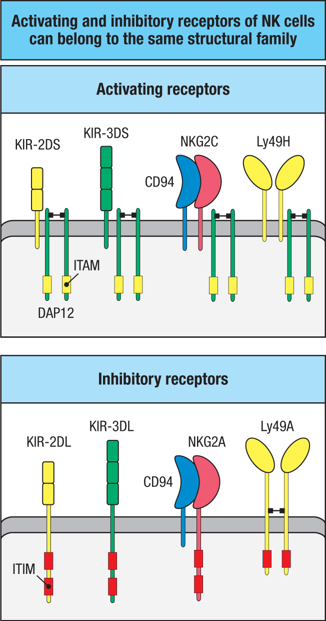
Activating and inhibitory receptors are present within the same structural family. Whether a KIR or Ly49 protein is activating or inhibitory depends on the presence or absence of particular signaling motifs in its cytoplasmic domain. Inhibitory KIRs and Ly49s have long cytoplasmic tails that contain an immunoreceptor tyrosine-based inhibition motif (ITIM). The consensus amino acid sequence for the ITIM is V/I/LxYxxL/V, where ‘x’ stands for any amino acid. For example, the cytoplasmic tails of the inhibitory receptors KIR-2DL and KIR-3DL each contain two ITIMs (Fig. 3.40). When ligands associate with an inhibitory KIR, the tyrosine in its ITIM becomes phosphorylated by the action of Src family protein tyrosine kinases. When phosphorylated, the ITIM can then bind the intracellular protein tyrosine phosphatases SHP-1 (Src homology region 2–containing protein tyrosine phosphatase-1) and SHP-2, which become localized near the cell membrane. These phosphatases inhibit signaling induced by other receptors (for example, the NK activating receptors) by removing phosphates from tyrosine residues on other intracellular signaling molecules.
Activating KIRs have short cytoplasmic tails and are designated, for example, as KIR-2DS and KIR-3DS (see Fig. 3.40). In mice, activating Ly49 proteins similarly have a short cytoplasmic tail that contains no signaling domain. Instead, these activating KIRs and Ly49 receptors have a charged residue in their transmembrane regions that associates with an accessory signaling protein called DAP12. DAP12 is a transmembrane protein that contains an immunoreceptor tyrosine-based activation motif (ITAM) with consensus sequence Yxx[L/I]x6–9Yxx[L/I]) in its cytoplasmic tail and forms a disulfide-linked homodimer in the membrane. When a ligand binds to an activating KIR or Ly49 receptor, the tyrosine residues in the ITAM of DAP12 become phosphorylated, turning on intracellular signaling pathways that activate the NK cell and lead to release of the cytotoxic granules. The phosphorylated ITAMs bind and activate intracellular tyrosine kinases such as Syk or ZAP-70, leading to further signaling events similar to those described for T cells in Chapter 7.
The KLR family also has both activating and inhibitory members. In mice, inhibitory Ly49 receptors have an ITIM in their cytoplasmic tail that recruits SHP-1. The latter’s importance is shown by the failure of Ly49 to inhibit NK activation upon binding to MHC class I in mice carrying the motheaten mutation, which inactivates SHP-1 protein. In humans and mice, NK cells express a heterodimer of two different C-type lectin–like receptors, CD94 and NKG2. This heterodimer interacts with nonpolymorphic MHC class I–like molecules, including HLA-E in humans and Qa1 in mice. HLA-E and Qa1 are unusual in that instead of binding peptides derived from pathogens, they bind fragments of the signal peptide derived from other MHC class I molecules during processing in the endoplasmic reticulum. This enables CD94:NKG2 to detect the presence of several different MHC class I variants, whose expression may be targeted by viruses, and kill cells in which overall MHC molecule expression is diminished. In humans there are four closely related NKG2 family proteins, NKG2A, C, E, and F (encoded by KLRC1–4), and a more distantly related protein, NKG2D (encoded by KLRK1). Of these, for example, NKG2A contains an ITIM and is inhibitory, whereas NKG2C has a charged transmembrane residue, associates with DAP12, and is activating (see Fig. 3.40). NKG2D is also activating but quite distinct from the other NKG2 receptors, and we will discuss it separately in the next section.
The overall response of NK cells to differences in MHC expression is further complicated by the extensive polymorphism of KIR genes, with different numbers of activating and inhibitory KIR genes being found in different people. The responsiveness of NK cells is thought to be tuned by their ability to recognize inhibitory ligands during their development. This process, referred to as licensing or education, is likely to underlie the effect of NK cells as a barrier to transplantation, because the NK cells educated in the environment of the recipient may fail to recognize the same inhibitory ligands on donor cells. A similar phenomenon may occur during pregnancy, because of differences between fetal and maternal MHC molecules (see Section 15-26). The advantage of such extensive KIR polymorphism is not yet clear, and some genetic epidemiologic studies even suggest an association between certain alleles of KIR genes and earlier onset (although not absolute frequency) of rheumatoid arthritis. The KIR gene cluster is not present in mice, but some species, including some primates, contain genes of both the KIR and KLR families. This might suggest that both gene clusters are relatively ancient and that for some reason, one or the other gene cluster was lost by mice and humans.
Signaling by the inhibitory NK receptors suppresses the killing activity and cytokine production of NK cells. This means NK cells will not kill healthy, genetically identical cells with normal expression of MHC class I molecules, such as the other cells of the body. Virus-infected cells, however, can become susceptible to being killed by NK cells by a variety of mechanisms. First, some viruses inhibit all protein synthesis in their host cells, so that synthesis of MHC class I proteins would be blocked in infected cells, even while their production in uninfected cells is being stimulated by the actions of type I interferons. The reduced level of MHC class I expression in infected cells would make them correspondingly less able to inhibit NK cells through their MHC-specific receptors, and they would become more susceptible to being killed. Second, many viruses can selectively prevent the export of MHC class I molecules to the cell surface or induce their degradation once there. This might allow the infected cell to evade recognition by cytotoxic T cells but would make it susceptible to being killed by NK cells. Virally infected cells can still be killed by NK cells even if the cells do not down-regulate MHC, provided that ligands for activating receptors are induced. However, some viruses target ligands for the activating receptors on NK cells, thwarting NK-cell recognition and killing of virus-infected cells.
3-27 NK cells express activating receptors that recognize ligands induced on infected cells or tumor cells.
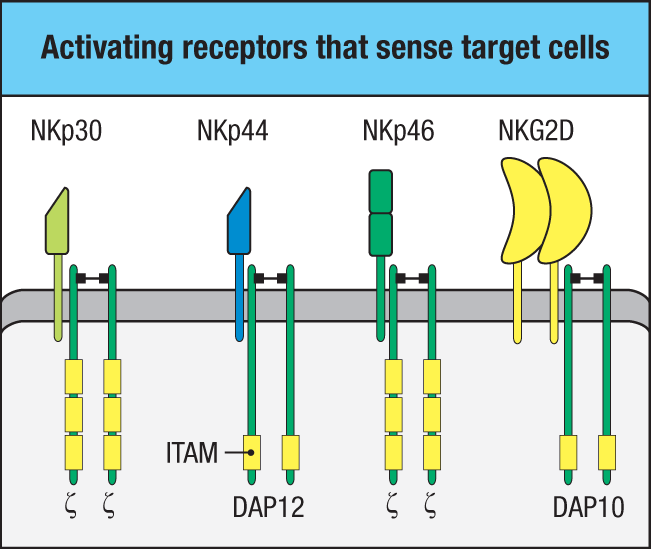
In addition to the KIRs and KLRs, which have a role in sensing the level of MHC class I proteins present on other cells, NK cells also express receptors that more directly sense the presence of infection or other perturbations in a cell. Activating receptors for the recognition of infected cells, tumor cells, and cells injured by physical or chemical damage include the natural cytotoxicity receptors (NCRs) NKp30, NKp44, and NKp46, which are immunoglobulin-like receptors, and the C-type lectin–like family members Ly49H and NKG2D (Fig. 3.41). Among NCRs, only NKp46 is conserved in humans and in mice, and it is the most selective marker of NK cells across mammalian species. The ligands recognized by the NCRs are still being defined, but some evidence suggests that they recognize viral proteins. Ly49H is an activating receptor that recognizes the viral protein m157, an MHC class I–like structure encoded by the murine cytomegalovirus. The ligand for NKp30 is a protein named B7-H6, a member of the B7 family of co-stimulatory proteins, further described in Chapters 7 and 9.
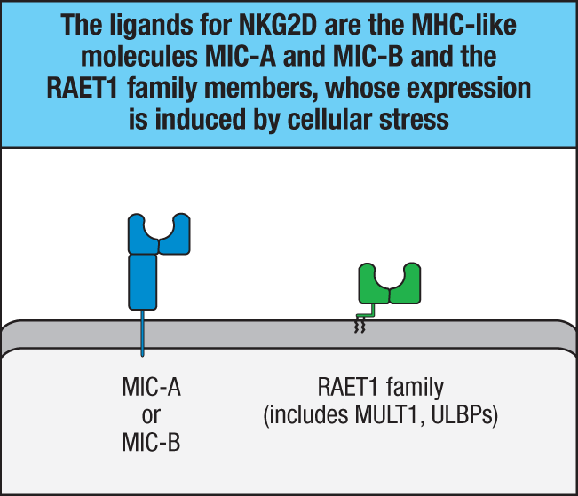
NKG2D has a specialized role in activating NK cells. NKG2A, NKG2C, NKG2E, and NKG2F form heterodimers with CD94 and bind the MHC class I molecule HLA-E. In contrast, two NKG2D molecules form a homodimer that binds to several MHC class I–like molecules that are induced by various types of cellular stress. These include the MIC molecules MIC-A and MIC-B and the RAET1 family of proteins, which are similar to the α1 and α2 domains of MHC class I molecules (Fig. 3.42). The RAET1 family has 10 members, three of which were initially characterized as ligands for the cytomegalovirus UL16 protein and are also called UL16-binding proteins, or ULBPs. Mice do not have equivalents of the MIC molecules; however, the ligands for mouse NKG2D have a very similar structure to that of the RAET1 proteins and are probably orthologs of them. In fact, these ligands were first identified in mice as the RAE1 (retinoic acid early inducible 1) protein family and also include related proteins H60 and MULT1 (see Fig. 6.26). We will return to these MHC-like molecules when we discuss the structure of the MHC molecule in Chapter 6.
The ligands for NKG2D are expressed in response to cellular or metabolic stress, and so are up-regulated on cells infected with intracellular bacteria and most viruses, as well as on incipient tumor cells that have become malignantly transformed. Thus, recognition by NKG2D acts as a generalized alarm signal to the immune system. In addition to expression by a subset of NK cells, NKG2D is expressed by various T cells, including all CD8 T cells and some γδ T cells and invariant NKT cells (described in Chapter 8). In these cells, recognition of NKG2D ligands provides a potent co-stimulatory signal that enhances their effector functions.
NKG2D differs from other activating receptors on NK cells in the signaling pathway it engages within the cell. The other activating receptors are associated intracellularly with signaling proteins such as the CD3ζ chain, the Fc receptor γ chain, and DAP12, which all contain ITAMs. In contrast, NKG2D binds a different adaptor protein, DAP10, which does not contain an ITAM sequence and instead activates the intracellular lipid kinase phosphatidylinositol 3-kinase (PI 3-kinase), initiating a different series of intracellular signaling events in the NK cell (see Section 7-4). Generally, PI 3-kinase is considered to enhance the survival of cells in which it is activated, thereby augmenting the cell’s overall effector activity. In NK cells, activation of PI 3-kinase is directly linked to the induction of cytotoxic activity. In mice, the workings of NKG2D are more complicated, because mouse NKG2D is produced in two alternatively spliced forms, one of which binds DAP12 and DAP10, whereas the other binds only DAP10. Mouse NKG2D can thus activate both signaling pathways, whereas human NKG2D seems to signal only through DAP10 to activate the PI 3-kinase pathway. Finally, NK cells express several receptors from the SLAM (signaling lymphocyte activation molecule) family, including 2B4, which recognizes the cell-surface molecule CD48 expressed by many cells including NK cells. Interactions between 2B4 and CD48 on nearby NK cells can release signals that promote survival and proliferation through SAP (SLAM-associated protein) and the Src kinase Fyn.
Summary.
Triggering of innate sensors on various cells—neutrophils, macrophages, and dendritic cells in particular—not only activates these cells’ individual effector functions but also stimulates the release of pro-inflammatory chemokines and cytokines that act together to recruit more phagocytic cells to the site of infection. Especially prominent is the early recruitment of neutrophils and monocytes. Furthermore, cytokines released by tissue phagocytic cells can induce more systemic effects, including fever and the production of acute-phase response proteins, such as mannose-binding lectin, C-reactive protein, fibrinogen, and pulmonary surfactant proteins, which add to a general state of augmented innate immunity. These cytokines also mobilize antigen-presenting cells that induce the adaptive immune response. The innate immune system has at its service several recently recognized subtypes of innate lymphoid cells to join the ranks of the long-recognized NK cells. ILCs exhibit specialized effector activity in response to different signals and act to amplify the strength of the innate response. The production of interferons in response to viral infections serves to inhibit viral replication and to activate NK cells. These in turn can distinguish healthy cells from those that are infected by virus or that are transformed or stressed in some way on the basis of expression of MHC class I molecules and MHC-related molecules that are ligands for some NK receptors. As we will see later in the book, cytokines, chemokines, phagocytic cells, and NK cells are all effector mechanisms that are also employed in the adaptive immune response, which uses variable receptors to target specific pathogen antigens.
Summary to Chapter 3.
Innate immunity uses a variety of effector mechanisms to detect infection and eliminate pathogens or to hold them in check until an adaptive immune response develops. These effector mechanisms are all regulated by germline-encoded receptors on many types of cells that can detect molecules of microbial origin or that sense pathogen-induced changes to host cells. The induced responses of the innate immune system are based on several distinct components. After the initial barriers—the body’s epithelia and the soluble antimicrobial molecules described in Chapter 2—have been breached, the most important innate defenses rely on tissue macrophages and other tissue-resident sensor cells, such as dendritic cells and ILCs. Macrophages provide a double service: they mediate rapid cellular defense at the borders of infection through phagocytosis and antimicrobial actions, and they also use their various innate sensors to activate the process of inflammation, which involves recruiting additional cells to sites of infection. Innate sensors activate signaling pathways that lead to the production of pro-inflammatory and antiviral cytokines, which in turn stimulate innate effector responses while also helping to initiate an adaptive immune response. The uncovering of the pathogen-sensing mechanisms described in this chapter is still extremely active. It is providing new insights into human autoinflammatory conditions such as lupus, Crohn’s disease, and gout. Indeed, the induction of powerful effector mechanisms by innate immune recognition based on germline-encoded receptors clearly has some dangers. It is a double-edged sword, as is illustrated by the effects of the cytokine TNF-α—beneficial when released locally, but disastrous when produced systemically. This illustrates the evolutionary knife edge along which all innate mechanisms of host defense travel. The innate immune system can be viewed as a defense system that mainly frustrates the establishment of a focus of infection; however, even when it proves limited in fulfilling this function, it has already set in motion the initiation of the adaptive immune response, which forms an essential part of human defenses against infection.
Glossary
- autocrine
- Describes a cytokine or other biologically active molecule acting on the cell that produces it.
- paracrine
- Describes a cytokine or other biologically active molecule acting on cells near to those that produce it.
- endocrine
- Describes the action of a biologically active molecule such as a hormone or cytokine that is secreted by one tissue into the blood and acts on a distant tissue. Cf. autocrine, paracrine.
- interleukin (IL)
- A generic name for cytokines produced by leukocytes. The more general term cytokine is used in this book, but the term interleukin is used in the naming of specific cytokines such as IL-2. Some key interleukins are listed in the glossary under their abbreviated names; for example, IL-1β and IL-2. Cytokines are listed in eAppendix III.
- IL-1 family
- One of four major families of cytokines, this family contains 11 cytokines that are structurally similar to IL-1α and are largely pro-inflammatory in function.
- hematopoietin superfamily
- Large family of structurally related cytokines that includes growth factors and many interleukins with roles in both adaptive and innate immunity.
- class I cytokine receptors
- A group of receptors for the hematopoietin superfamily of cytokines. These include receptors using the common β chain for IL-2, IL-4, IL-7, IL-15, and IL-21, and a common β chain for GM-CSF, IL-3, and IL-5.
- X-linked severe combined immune deficiency (X-linked SCID, or XSCID)
- An immune deficiency disease in which T-cell development fails at an early intrathymic stage, and no production of mature T cells or T cell–dependent antibody occurs. It is due to a defect in a gene that encodes the γc chain shared by the receptors for several different cytokines.
- class II cytokine receptors
- A group of heterodimeric receptors for a family of cytokines that includes interferon (IFN)-α, IFN-β, IFN-γ, and IL-10.
- TNF family
- Cytokine family, the prototype of which is tumor necrosis factor-α (TNF-α, or TNF). It contains both secreted (e.g., TNF-α and lymphotoxin) and membrane-bound (e.g., CD40 ligand) members.
- TNF receptors
- Family of cytokine receptors that includes some that lead to apoptosis of the cell on which they are expressed (e.g., Fas and TNFR-I), whereas others lead to activation.
- severe combined immune deficiency (SCID)
- Type of immune deficiency (due to various causes) in which both B-cell (antibody) and T-cell responses are lacking; it is fatal if not treated.
- signal transducers and activators of transcription (STATs)
- A family of seven transcription factors activated by many cytokine and growth factor receptors. See also Janus kinase (JAK) family.
- tyrosine phosphatases
- Enzymes that remove phosphate groups from phosphorylated tyrosine residues on proteins. See also CD45.
- CD45
- A transmembrane tyrosine phosphatase found on all leukocytes. It is expressed in different isoforms on different cell types, including the different subtypes of T cells. Also called leukocyte common antigen, it is a generic marker for hematopoietically derived cells, with the exception of erythrocytes.
- suppressor of cytokine signaling (SOCS)
- Regulatory protein that interacts with Janus kinases (JAKs) to inhibit signaling by activated receptors.
- protein inhibitors of activated STAT (PIAS)
- A small family of proteins that inhibit STAT family transcription factors.
- chemotaxis
- Cellular movement occurring in response to chemical signals in the environment.
- CC chemokines
- One of the two main classes of chemokines, distinguished by two adjacent cysteines (C) near the amino terminus. They have names CCL1, CCL2, etc. See eAppendix IV for a list of individual chemokines.
- CXC chemokines
- One of the two main classes of chemokines, distinguished by a Cys-X-Cys (CXC) motif near the amino terminus. They have names CXCL1, CXCL2, etc. See eAppendix IV for a list of individual chemokines.
- leukocyte functional antigens (LFAs)
- Cell-adhesion molecules on leukocytes that were initially defined using monoclonal antibodies. LFA-1 is a β2 integrin; LFA-2 (now usually called CD2) is a member of the immunoglobulin superfamily, as is LFA-3 (now called CD58). LFA-1 is particularly important in T-cell adhesion to endothelial cells and antigen-presenting cells.
- selectins
- Family of cell-adhesion molecules on leukocytes and endothelial cells that bind to sugar moieties on specific glycoproteins with mucin-like features.
- intercellular adhesion molecules (ICAMs)
- ICAM-1, ICAM-2, ICAM-3. Cell-adhesion molecules of the immunoglobulin superfamily that bind to the leukocyte integrin CD11a:CD18 (LFA-1). They are crucial in the binding of lymphocytes and other leukocytes to antigen-presenting cells and endothelial cells.
- integrins
- Heterodimeric cell-surface proteins involved in cell–cell and cell–matrix interactions. They are important in adhesive interactions between lymphocytes and antigen-presenting cells and in lymphocyte and leukocyte adherence to blood vessel walls and migration into tissues.
- immunoglobulin-like proteins
- Proteins containing one or more immunoglobulin-like domains, which are protein domains structurally similar to those of immunoglobulins.
- LFA-1 (CD11a:CD18, or αLβ2)
- Cell-adhesion molecules on leukocytes that were initially defined using monoclonal antibodies. LFA-1 is a β2 integrin; LFA-2 (now usually called CD2) is a member of the immunoglobulin superfamily, as is LFA-3 (now called CD58). LFA-1 is particularly important in T-cell adhesion to endothelial cells and antigen-presenting cells.
- CR3 (CD11b:CD18)
- Complement receptor 3. A β2 integrin that acts both as an adhesion molecule and as a complement receptor. CR3 on phagocytes binds iC3b, a breakdown product of C3b on pathogen surfaces, and stimulates phagocytosis.
- CR3 (CD11b:CD18)
- Complement receptor 3. A β2 integrin that acts both as an adhesion molecule and as a complement receptor. CR3 on phagocytes binds iC3b, a breakdown product of C3b on pathogen surfaces, and stimulates phagocytosis.
- talin
- An intracellular protein involved in the linkage of activated integrins, such as LFA-1, to the cytoskeleton to allow changes in cellular motility and migration, such as in the diapedesis of neutrophils across the vascular endothelium.
- leukocyte adhesion deficiencies (LADs)
- A class of immunodeficiency diseases in which the ability of leukocytes to enter sites infected by extracellular pathogens is affected, impairing elimination of infection. There are several different causes, including a deficiency of the common β chain of the leukocyte integrins.
- Weibel–Palade bodies
- Granules within endothelial cells that contain P-selectin.
- P-selectin
- Family of cell-adhesion molecules on leukocytes and endothelial cells that bind to sugar moieties on specific glycoproteins with mucin-like features.
- E-selectin
- Family of cell-adhesion molecules on leukocytes and endothelial cells that bind to sugar moieties on specific glycoproteins with mucin-like features.
- leukocyte adhesion deficiency type 2 (LAD-2)
- Disease caused by defects in the production of sulfated sialyl-LewisX that prevent neutrophils from interacting with P-selectin and E-selectin, eliminating their ability to migrate properly to sites of infection.
- CR4 (CD11c:CD18)
- A β2 integrin that acts both as an adhesion molecule and as a complement receptor. CR4 on phagocytes binds iC3b, a breakdown product of C3b on pathogen surfaces, and stimulates phagocytosis.
- CR4 (CD11c:CD18)
- A β2 integrin that acts both as an adhesion molecule and as a complement receptor. CR4 on phagocytes binds iC3b, a breakdown product of C3b on pathogen surfaces, and stimulates phagocytosis.
- BDCA-2 (blood dendritic cell antigen 2)
- A C-type lectin expressed selectively as a receptor on the surface of human plasmacytoid dendritic cells.
- BST2 (bone marrow stromal antigen 2)
- Bone marrow stromal antigen 2, also called tetherin and CD317, is a type II integral membrane protein that functions as a viral restriction element trapping virions in the plasma membrane and inhibiting viral budding and which serves as a marker of plasmacytoid dendritic cells (pDCs), but is also expressed by B cells.
- shear-resistant rolling
- The capacity of neutrophils to remain attached to the vascular endothelium under high rates of flow—or shear—enabled by specialized plasma membrane extensions called slings.
- slings
- The capacity of neutrophils to remain attached to the vascular endothelium under high rates of flow—or shear—enabled by specialized plasma membrane extensions called slings.
- PECAM
- A cell-adhesion molecule found both on lymphocytes and at endothelial cell junctions. CD31–CD31 interactions are thought to enable leukocytes to leave blood vessels and enter tissues.
- CD31
- A cell-adhesion molecule found both on lymphocytes and at endothelial cell junctions. CD31–CD31 interactions are thought to enable leukocytes to leave blood vessels and enter tissues.
- diapedesis
- The movement of blood cells, particularly leukocytes, from the blood across blood vessel walls into tissues.
- neutropenia
- Abnormally low levels of neutrophils in the blood.
- disseminated intravascular coagulation (DIC)
- Blood clotting occurring simultaneously in small vessels throughout the body in response to disseminated TNF-α, which leads to the massive consumption of clotting proteins, so that the individual’s blood cannot clot appropriately. Seen in septic shock.
- endogenous pyrogens
- Cytokines that can induce a rise in body temperature.
- exogenous pyrogen
- Any substance originating outside the body that can induce fever, such as the bacterial lipopolysaccharide LPS. Cf. endogenous pyrogens.
- acute-phase response
- A change in the proteins present in the blood that occurs during the early phases of an infection. It includes the production of acute-phase proteins, many of which are produced in the liver.
- acute-phase proteins
- Proteins with innate immune function whose production is increased in the presence of an infection (the acute-phase response). They circulate in the blood and participate in early phases of host defense against infection. An example is mannose-binding lectin.
- C-reactive protein
- An acute-phase protein that binds to phosphocholine, a constituent of the surface C-polysaccharide of the bacterium Streptococcus pneumoniae and of many other bacteria, thus opsonizing them for uptake by phagocytes.
- pentraxin
- A family of acute-phase proteins formed of five identical subunits, to which C-reactive protein and serum amyloid protein belong.
- surfactant proteins A and D (SP-A and SP-D)
- Acute-phase proteins that help protect the epithelial surfaces of the lung against infection.
- leukocytosis
- The presence of increased numbers of leukocytes in the blood. It is commonly seen in acute infection.
- type II interferon
- The antiviral interferon IFN-γ.
- interferon-producing cells (IPCs)
- A distinct lineage of dendritic cells that secrete large amounts of interferon on activation by pathogens and their products via receptors such as Toll-like receptors. Cf. conventional dendritic cells.
- natural interferon-producing cells
- A distinct lineage of dendritic cells that secrete large amounts of interferon on activation by pathogens and their products via receptors such as Toll-like receptors. Cf. conventional dendritic cells.
- IRF9
- A member of the IRF family of transcription factors that interacts with activated STAT1 and STAT2 to form the complex called ISGF3, which induces transcription of many ISGs.
- ISGF3
- A member of the IRF family of transcription factors that interacts with activated STAT1 and STAT2 to form the complex called ISGF3, which induces transcription of many ISGs.
- interferon-stimulated genes (ISGs)
- A category of genes induced by interferons, which include many whose products promote innate defense against pathogens, such as oligoadenylate synthetase, PKR, and the Mx, IFIT, and IFITM proteins.
- oligoadenylate synthetase
- Enzyme produced in response to stimulation of cells by interferon. It synthesizes unusual nucleotide polymers, which in turn activate a ribonuclease that degrades viral RNA.
- PKR
- Serine/threonine kinase activated by IFN-α and IFN-β. It phosphorylates the eukaryotic protein synthesis initiation factor eIF-2, inhibiting translation and thus contributing to the inhibition of viral replication.
- Mx (myxoma resistant) proteins
- Interferon-inducible proteins required for cellular resistance to influenza virus replication.
- IFIT (IFN-induced protein with tetratricoid repeats)
- A small family of host proteins induced by interferons that regulate protein translation during infection in part by interactions with eIF3.
- eukaryotic initiation factor 3 (eIF3)
- Multi-subunit complex that acts in formation of the 43S pre-initiation complex. It can bind IFN-induced protein with tetratricoid repeats (IFIT) proteins, which thereby suppress translation of viral proteins.
- interferon-induced transmembrane protein (IFITM)
- A small family of host transmembrane proteins induced by interferons that function in the cell’s vesicular compartment to restrain various steps in viral replication.
- natural killer (NK) cell
- A type of ILC that is important in innate immunity to viruses and other intracellular pathogens and in antibody-dependent cell-mediated cytotoxicity (ADCC). NK cells express activating and inhibitory receptors but not the antigen-specific receptors of T or B cells.
- innate lymphoid cells (ILCs)
- These are a class of innate immune cells having overlapping characteristics with T cells but lacking an antigen receptor. They arise in several groups, ILC1, ILC2, ILC3, and NK cells, which exhibit properties roughly similar to TH1, TH2, TH17, and CD8 T cells.
- common lymphoid progenitor (CLP)
- Stem cell that can give rise to all the types of lymphocytes but is biased toward producing B cells.
- group 1 ILCs (ILC1s)
- The subtype of innate lymphoid cells (ILCs) characterized by IFN-γ production.
- Nfil3
- A transcription factor important during the development of several types of immune cells including certain types of NK cells.
- Tbet
- A transcription factor active in many immune cell types but most typically associated with ILC1 and TH1 function.
- group 2 ILCs (ILC2s)
- A subset of innate lymphoid cells characterized by production of IL-4, IL-5, and IL-13.
- thymic stromal lymphopoietin (TSLP)
- Thymic stroma–derived lymphopoietin. A cytokine thought to be involved in promoting B-cell development in the embryonic liver. It is also produced by mucosal epithelial cells in response to helminthic infections and promotes type 2 immune responses through its actions on macrophages, ILC2s, and TH2 cells.
- group 3 ILCs (ILC3s)
- A subset of innate lymphoid cells characterized by production of IL-17 and IL-22.
- caspase 8
- An initiator caspase activated by various receptors that activates the process of apoptosis.
- granulocyte–macrophage colony-stimulating factor (GM-CSF)
- A cytokine involved in the growth and differentiation of cells of the myeloid lineage, including dendritic cells, monocytes and tissue macrophages, and granulocytes.
- activating receptors
- On NK cells, receptors whose stimulation results in activation of the cell’s cytotoxic activity.
- inhibitory receptors
- On NK cells, receptors whose stimulation results in suppression of the cell’s cytotoxic activity.
- dysregulated self
- Refers to changes that take place in infected or malignant cells that alter expression of various surface receptors that can be detected by the innate immune system.
- stress-induced self
- Refers to changes that take place in infected or malignant cells that alter expression of various surface receptors that can be detected by the innate immune system.
- missing self
- Refers to the loss of cell-surface molecules that engage with inhibitory receptors on NK cells, resulting in NK-cell activation.
- MHC class I molecules
- Polymorphic cell-surface proteins encoded in the MHC locus and expressed on most cells. They present antigenic peptides generated in the cytosol to CD8 T cells and also bind the co-receptor CD8.
- MHC class II molecules
- Polymorphic cell-surface proteins encoded in the MHC locus and expressed primarily on specialized antigen-presenting cells. They present antigenic peptides derived from internalized extracellular pathogens to CD4 T cells and also bind the co-receptor CD4.
- killer cell immunoglobulin-like receptors (KIRs)
- Large family of receptors present on NK cells, through which the cells’ cytotoxic activity is controlled. The family contains both activating and inhibitory receptors.
- leukocyte receptor complex (LRC)
- A large cluster of immunoglobulin-like receptor genes that includes the killer cell immunoglobulin-like receptor (KIR) genes.
- killer cell lectin-like receptors (KLRs)
- Large family of receptors present on NK cells, through which the cells’ cytotoxic activity is controlled. The family contains both activating and inhibitory receptors.
- NK receptor complex (NKC)
- A cluster of genes that encode a family of receptors on NK cells.
- Ly49 receptors
- A family of C-type lectins expressed by mouse, but not human, NK cells. These can be either activating or inhibitory in function.
- immunoreceptor tyrosine-based inhibition motif (ITIM)
- Sequence motifs in the signaling chains of inhibitory receptors that are sites of tyrosine phosphorylation, leading to inhibitory signaling, such as through recruitment of phosphatases that remove phosphate groups added by tyrosine kinases.
- Src family protein tyrosine kinases
- Receptor-associated protein tyrosine kinases characterized by Src homology protein domains (SH1, SH2, and SH3). The SH1 domain contains the kinase, the SH2 domain can bind phosphotyrosine residues, and the SH3 domain can interact with proline-rich regions in other proteins. In T cells and B cells they are involved in relaying signals from the antigen receptor.
- motheaten
- A mutation in the SHP-1 protein phosphatase that impairs the function of some inhibitory receptors, such as Ly49, resulting in overactivation of various cells, including NK cells. Mice with this mutation have a ‘motheaten’ appearance because of chronic inflammation.
- CD94
- A C-type lectin that is a subunit of the killer cell lectin-like receptors (KLRs) of NK cells.
- NKG2
- Family of C-type lectins that supply one of the subunits of the killer cell lectin-like receptors (KLRs) on NK cells.
- signal peptide
- The short N-terminal peptide sequence responsible for directing newly synthesized proteins into the secretory pathway.
- natural cytotoxicity receptors (NCRs)
- Activating receptors on NK cells that recognize infected cells and stimulate cell killing by the NK cell.
- Ly49H
- A family of C-type lectins expressed by mouse, but not human, NK cells. These can be either activating or inhibitory in function.
- NKG2D
- Activating C-type lectin receptor on NK cells, cytotoxic T cells, and γδ T cells that recognizes the stress-response proteins MIC-A and MIC-B.
- RAET1
- A family of 10 MHC class Ib proteins that are ligands for NKG2D. Also called RAET1 proteins.
- cytomegalovirus UL16 protein
- A nonessential glycoprotein of cytomegalovirus that is recognized by innate receptors expressed by NK cells.
- RAE1 (retinoic acid early inducible 1) protein family
- Several murine MHC class Ib proteins; these are orthologs of human RAET1 family proteins, including H60 and MULT1, and are ligands for murine NKG2D.
- phosphatidylinositol 3-kinase (PI 3-kinase)
- Enzyme involved in intracellular signaling pathways. It phosphorylates the membrane lipid phosphatidylinositol 4,5-bisphosphate (PIP2) to form phosphatidylinositol 3,4,5-trisphosphate (PIP3), which can recruit signaling proteins containing pleckstrin homology (PH) domains to the membrane.
- SLAM (signaling lymphocyte activation molecule)
- A family of related cell-surface receptors that mediate adhesion between lymphocytes and which includes SLAM, 2B4, CD84, Ly108, Ly9, and CRACC.
- 2B4
- A receptor belonging to the signaling lymphocyte activation molecule (SLAM) family expressed by NK cells, which binds to CD48, another SLAM receptor. These signal through SAP and Fyn to promote survival and proliferation.
- CD48
- A receptor belonging to the signaling lymphocyte activation molecule (SLAM) family expressed by NK cells, which binds to CD48, another SLAM receptor. These signal through SAP and Fyn to promote survival and proliferation.
- SAP (SLAM-associated protein)
- An intracellular adaptor protein involved in signaling by SLAM (signaling lymphocyte activation molecule). Inactivating mutations in this gene cause X-linked lymphoproliferative (XLP) syndrome.
- common β chain (βc)
- A transmembrane polypeptide (CD131) that is a common subunit for receptors of the cytokines IL-3, IL-5, and GM-CSF.
- common γ chain (γc)
- A transmembrane polypeptide chain (CD132) that is common to a subgroup of cytokine receptors.
- Janus kinase (JAK) family, Janus kinases (JAKs)
- Enzymes of the JAK–STAT intracellular signaling pathways that link many cytokine receptors with gene transcription in the nucleus. The kinases phosphorylate STAT proteins in the cytosol, which then move to the nucleus and activate a variety of genes.
- SH2 (Src homology 2) domain
- Receptor-associated protein tyrosine kinases characterized by Src homology protein domains (SH1, SH2, and SH3). The SH1 domain contains the kinase, the SH2 domain can bind phosphotyrosine residues, and the SH3 domain can interact with proline-rich regions in other proteins. In T cells and B cells they are involved in relaying signals from the antigen receptor.
- LFA-1 (CD11a:CD18, or αLβ2)
- Cell-adhesion molecules on leukocytes that were initially defined using monoclonal antibodies. LFA-1 is a β2 integrin; LFA-2 (now usually called CD2) is a member of the immunoglobulin superfamily, as is LFA-3 (now called CD58). LFA-1 is particularly important in T-cell adhesion to endothelial cells and antigen-presenting cells.
- sulfated sialyl-LewisX
- A sulfated tetrasaccharide carbohydrate structure attached to many cell surface proteins, it binds the P-selectin and E-selectin molecules on the surface of cells, such as neutrophils, that mediate interactions with the endothelium.
- TACE (TNF-α–converting enzyme)
- A protease responsible for cleavage of the membrane-associated form of the cytokine TNF-α into its soluble form, which can enter the systemic circulation.
- IFN-γ
- A cytokine of the interferon structural family produced by effector CD4 TH1 cells, CD8 T cells, and NK cells. Its primary function is the activation of macrophages, and it acts through a different receptor from that of the type I interferons.
- IFN-λ
- Also called type III interferons, this family includes IL-28A, IL-28B, and IL-29, which bind a common receptor expressed by a limited set of epithelial tissues.
- IFN-λ receptor
- Heterodimeric receptor composed of a unique IL-28Rα subunit and the β subunit of the IL-10 receptor that recognizes IL-28A, IL-28B, and IL-29.
- interferon-α receptor (IFNAR)
- This receptor recognizes IFN-α and IFN-β to activate STAT1 and STAT2 and induce expression of many ISGs.
- STAT1
- A member of the signal transducer and activator of transcription (STAT) family of transcription factors activated by type I and type II interferon signaling.
- STAT2
- A member of the signal transducer and activator of transcription (STAT) family of transcription factors activated by type II interferon signaling and which heterodimerizes with STAT1 and associates with IRF9 to form a complex called ISGF3.
- IFIT1, IFIT2
- IFN-induced protein with tetratricoid repeats (IFIT) 1 and 2 are proteins encoded by interferon-stimulated genes (ISGs) that regulate the translation of normally capped mRNAs and lead to reduced cellular proliferation. IFIT1 can also suppress translation of some viral RNAs that lack 2′-O-methylation (cap-0), such as Sindbis virus. In this way, IFIT1 and IFIT2 serve as restriction factors in certain viral infections.
- 2′-O-methyltransferase (MTase)
- An enzyme that transfers a methyl group to the 2′ hydroxyl of the first and second ribose groups in mRNA. Viruses that acquire MTase can produce cap-1 and cap-2 on their transcripts and thereby evade restriction by IFIT1.
- IFITM1, IFITM3
- Interferon-induced transmembrane protein (IFITM) 1 and 3 are proteins encoded by interferon-stimulated genes (ISGs) that interfere with membrane fusion between viral membranes and membranes of the lysosome (IFITM1) or endosome (IFITM3), and so serve as restriction factors in various viral infections.
- IFITM1, IFITM3
- Interferon-induced transmembrane protein (IFITM) 1 and 3 are proteins encoded by interferon-stimulated genes (ISGs) that interfere with membrane fusion between viral membranes and membranes of the lysosome (IFITM1) or endosome (IFITM3), and so serve as restriction factors in various viral infections.
- DR4, DR5
- Members of the TNFR superfamily expressed by many cell types and which can be activated by TRAIL to induce apoptosis.
- antibody-dependent cell-mediated cytotoxicity (ADCC)
- The killing of antibody-coated target cells by cells with Fc receptors that recognize the constant region of the bound antibody. Most ADCC is mediated by NK cells that have the Fc receptor FcγRIII on their surface.
- SHP-1, SHP-2
- Intracellular tyrosine phosphatases that are recruited to phosphorylated ITIM domains of various inhibitory receptors and which can remove phosphate residues from other signaling molecules, with effect of reducing signaling.
- SHP-1, SHP-2
- Intracellular tyrosine phosphatases that are recruited to phosphorylated ITIM domains of various inhibitory receptors and which can remove phosphate residues from other signaling molecules, with effect of reducing signaling.
- DAP10, DAP12
- Signaling chains containing ITAMs that are associated with the tails of some activating receptors on NK cells.
- DAP10, DAP12
- Signaling chains containing ITAMs that are associated with the tails of some activating receptors on NK cells.
- MIC-A, MIC-B
- MHC class Ib proteins that are induced by stress, infection, or transformation in many cell types and are recognized by NKG2D.
- UL16-binding proteins (ULBPs)
- A family of 10 MHC class Ib proteins that are ligands for NKG2D. Also called RAET1 proteins.