The role of adaptive immunity in regulating the intestinal mucosal immune system at homeostasis.
As is evident from the foregoing discussion, three principal tasks of the intestinal mucosa are nutrient and water absorption, maintenance of the commensal microbiota, and defense against pathogens. The balance between nutrient uptake and accommodation to the microbiota, which are constant needs, and defense of the host, a less frequent need, represents a daunting challenge. The adaptive immune system plays a central role in navigating this balance, informed by signals from the epithelium and the array of innate immune cells that are integrated into the intestinal mucosa. Perhaps in no other tissue in the body is the necessity for the maintenance of immune tolerance more imperative: the diversity of foreign antigens present in food and the commensal bacteria is immense but must be tolerated.
A simple solution would be complete partitioning of these antigens from adaptive immune cells. Indeed, the innate defenses are sufficiently robust that much of the host response to the microbiota progresses without involvement of CD4 T cell–dependent responses. Further, a portion of the secretory IgA (SIgA) that contributes to the partitioning of commensal organisms away from the epithelium is generated in isolated lymphoid follicles (ILFs) without help from T cells. However, complete exclusion of foreign antigens within food and the microbiota is not possible, and in fact, the system has evolved to actively sample the luminal antigens through the specialized mucosal lymphoid tissues, such as Peyer’s patches and ILFs. Accordingly, the mucosal immune system has acquired strategies to recognize and respond to innocuous antigens in a way that minimizes inflammation, unless absolutely necessary.
As the foreign antigens derived from food and the commensal microbiota are extracellular and therefore primarily presented by MHC II, it is the CD4 T-cell response that is central to the induction of homeostatic adaptive immunity in the intestines. As we will see, in the intestines, induction of T-cell immunity strongly favors the development of Treg cells at homeostasis, reflecting unique properties of the antigen-presenting cells (APCs) in the gut and the route by which antigen is introduced—including the delivery of innocuous antigens to intestinal APCs via SIgA. Here, we will discuss mechanisms active in the intestinal mucosa that promote noninflammatory recognition of antigens by macrophages and dendritic cells, followed by a consideration of the induction of Treg cells and SIgA-producing B cells.
12-7 The mucosal immune system must establish and maintain tolerance to harmless foreign antigens.
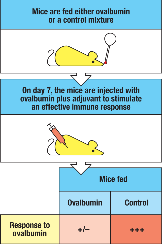
Antigens within food and commensal bacteria normally do not induce an inflammatory immune response, despite the lack of central (thymic) tolerance to them (Fig. 12.27). The mucosal immune system’s environment is inherently tolerogenic—a barrier to the development of nonliving vaccines, which need to overcome local regulatory mechanisms. Food proteins are not digested completely in the intestine; significant amounts are absorbed into the body in an immunologically relevant form. The default response to oral administration of a protein antigen is the development of a phenomenon known as oral tolerance. This is a form of peripheral tolerance that renders the systemic and mucosal immune systems relatively unresponsive to subsequent exposures to the same antigen. It can be demonstrated experimentally in mice by feeding them a foreign protein such as ovalbumin (Fig. 12.28). When the animals are then challenged with the antigen in adjuvant via a nonmucosal route, such as injection into the skin, the normal immune response one would expect is blunted. This suppression of systemic immune responses is long lasting and is antigen specific; responses to other antigens are not affected. A similar suppression of subsequent immune responses is observed after the administration of proteins into the respiratory tract, giving rise to the general concept of mucosal tolerance, because the usual response to such antigens is delivered via a mucosal surface. Systemic T-cell responses can also be inhibited by feeding humans protein antigens that they have not encountered previously, although it has proven an ineffective strategy for reversing reactivity to antigens to which individuals are already sensitized, such as food allergens. Thus, although mucosal tolerance can be used to avoid inflammatory disease in experimental animal models of type 1 diabetes mellitus, arthritis, and encephalomyelitis, clinical trials in humans have been less successful and have given way to other therapies, as we will discuss in greater detail in Chapters 14 and 16.
|
Protective immunity |
Mucosal tolerance |
|
|
Antigen |
Invasive bacteria, viruses, toxins |
Food proteins; commensal bacteria |
|
Primary Ig production |
Intestinal IgA and IgG Specific Ab present in serum |
Some local IgA Low or no Ab in serum |
|
Primary T-cell response |
Local and systemic effector and memory T cells |
pTreg cell induction; no local effector T-cell response |
|
Response to antigen reexposure |
Enhanced (memory) response |
Low or no response or systemic response |
Fig. 12.27 Immune priming and tolerance are different outcomes of intestinal exposure to antigen. The intestinal immune system generates protective immunity against antigens that are presented during infections by pathogenic organisms. IgA antibodies are produced within the GALT, serum IgG and IgA are made in draining lymph nodes, and the appropriate effector T cells are activated in the intestine and elsewhere. When the antigen is encountered again, there is effective memory, ensuring rapid protection. Antigens from food proteins induce tolerance locally and systemically, with little or no IgA antibody production. T cells are not activated, and subsequent responses to challenge are suppressed. In the case of commensal bacteria, there is local secretory IgA (SIgA) production, but typically no systemic antibody responses, and peripheral regulatory T cells (pTreg cells) rather than effector T cells are activated.
Various mechanisms are likely to account for oral tolerance to protein antigens, although the preferential generation of peripheral regulatory T (pTreg) cells appears to be dominant. Antigens delivered to the GALT or draining lymph nodes generally induce antigen-specific FoxP3+ Treg cells at homeostasis. As we will learn, migratory dendritic cells of the intestinal mucosa are conditioned by the local cytokine environment to produce two factors that induce Treg cell differentiation from naive CD4 T-cell precursors—retinoic acid and TGF-β—without producing pro-inflammatory signals, such as IL-1, IL-6, IL-23, or IL-12, which would otherwise stimulate the differentiation of effector CD4 T cells, such as TH17 or TH1 cells. These same pTreg cell–inducing factors also induce the expression of homing molecules that direct the migration of pTreg cells back to the intestinal mucosa, where they act to repress subsequent exposure to their cognate antigens. Although it is known that these same factors are also essential for the suppression of systemic immune responses to the same antigens, the mechanisms responsible for this link between the mucosal and peripheral immune systems are not yet understood. One plausible mechanism is that pTreg cells that have been primed in the intestines encounter antigen peripherally as they circulate from the intestines via draining lymph into the bloodstream. Alternatively, there is evidence that orally administered antigens can be found in lymph and the bloodstream, enabling access to resident dendritic cells in peripheral lymph nodes, although this remains to be proven. Notably, unlike soluble antigens, microbial antigens generally induce tolerance that is confined to the intestine; the systemic immune system normally remains ignorant of them, which may reflect their containment by the GALT and draining lymph nodes. Importantly, at times oral tolerance induction can fail, as is believed to occur in celiac disease (discussed in detail in Section 14-16) and peanut allergies (discussed in Sections 14-10 and 14-12).
 Case Study: Celiac Disease
12-8 Macrophages and dendritic cells have complementary roles in the maintenance of immune tolerance in the intestines.
Both inductive and effector sites within the intestinal mucosa contain abundant macrophages and dendritic cells (DCs). Indeed, the lamina propria of the healthy intestines contains the largest population of macrophages and conventional dendritic cells (cDCs) in the body, with the highest number residing in the small intestine. A major historical confounder in understanding the origins and function of macrophages and DCs in the intestines has been the crossover in expression of several markers between the two lineages in this tissue. Thus, the classical macrophage marker, F4/80, is expressed by some cDCs in the intestines, and many intestinal macrophages express the DC marker CD11c (Fig. 12.29). However, recent studies indicate that while both intestinal macrophages and DCs express CD11c and class II MHC, unlike most DCs in this site, intestinal macrophages generally lack expression of αE integrin (CD103). Instead, intestinal macrophages have been found to express FcγRI (CD64; see Fig. 10.38) and, in the mouse, CX3CR1, the receptor for the chemokine CX3CL1 (fractalkine); most intestinal DCs do not. On the basis of these markers, it is clear that intestinal macrophages are dependent on colony stimulating factor 1 (CSF1; also known as M-CSF) for their development and survival; in contrast, intestinal DCs are dependent on FLT3 ligand (FLT3L). Accordingly, CD64+CD11c+MHC II+ cells are tissue-resident and display characteristic macrophage morphology, with abundant cytoplasm and intracytoplasmic vacuoles, while CD64–CD11c+MHC II+ cells are migratory and localize to the GALT and mesenteric lymph nodes in a CCR7-dependent manner to participate in T-cell priming.
|
Characteristics of major mononuclear phagocytes in the intestines |
|||
|
Intestinal mononuclear phagocyte |
Tissue-resident macrophage |
Conventional dendritic cell 1 (cDC1) |
Conventional dendritic cell 2 (cDC2) |
|
Common surface markers |
MHC class II CD11c CCR2 |
MHC class II CD11c CCR2 |
MHC class II CD11c CCR2 |
|
Unique surface markers |
CX3CR1 FcγRI (CD64) αvβ8 |
XCR1 |
SIRPα |
|
Key inducing cytokines |
CSF1 (M-CSF) |
FLT3L |
FLT3L |
|
TLRs |
TLR-4lo, various |
TLR-3+, various |
TLR-3−, various |
|
Cytokines produced |
TGF-β (activation) (IL-10) |
IL-12 |
IL-6, IL-23, TGF-β |
|
Prevalence |
Abundant |
Frequent |
Abundant |
|
Major functions |
“Silent” bacterial uptake and destruction Antigen transfer to migratory DCs Efferocytosis Transepithelial antigen sampling |
Antigen presentation to naive CD8 T cells |
Antigen presentation to naive CD4 T cells |
Fig. 12.29 Characteristics of major mononuclear phagocytes in the intestines.
In contrast to many other tissues, where macrophages seed early in life and are maintained by local turnover, intestinal macrophages continually differentiate within the intestinal lamina propria from circulating Ly6Chi monocytes that are recruited there by homeostatic chemokines recognized by CCR2 (for example, CCL2, CCL8). The transition from incoming monocytes to mature macrophages takes several days, and although the mature cells typically have a tissue life span of days to weeks, some studies have shown that a minority of macrophages can persist for several months. In the intestines, macrophages are much more prevalent than cDCs and represent ~75% of all mononuclear phagocytes. They are positioned immediately under the epithelium and are highly phagocytic and microbicidal, and thus ideally suited to ingest and degrade any microbes or other particulates that penetrate the epithelial barrier. They also clear dying epithelial cells, which are found in large numbers in the intestine—an inevitable consequence of the high turnover of most IECs.
As mentioned earlier, intestinal macrophages, unlike macrophages in other parts of the body, are conditioned by the intestinal microenvironment to be non-inflammatory; they do not produce significant quantities of inflammatory cytokines (for example, TNF-α, IL-1β, IL-6) or reactive oxygen or nitrogen species in response to phagocytosis or exposure to stimuli such as bacteria or TLR ligands. They also lack receptors for IgA, complement, and the signaling components of the IgG receptor, and they have limited antigen-presentation capacity. Because of their anti-inflammatory properties, these cells are sometimes referred to as inflammation-anergic macrophages. Unlike other tissue macrophages, these cells appear to be terminally differentiated and do not give rise to M1 or M2 macrophages (see Section 11-6). Like other immune cells in the gut, intestinal macrophages are conditioned by exposure to the commensal microbiota, as evidenced by decreased numbers and lower turnover of these cells in germ-free mice and mice treated with broad-spectrum antibiotics.
The terminal differentiation of intestinal macrophages from incoming monocytes is regulated by TGF-β signaling. TGF-β is abundant in the intestinal lamina propria, where it is produced in its latent form by IECs, stromal cells, Treg cells, mast cells, and macrophages themselves, and binds to the extracellular matrix. Once in the mucosa, signaling via TGF-β receptor 1 (TGF-βRI) and TGF-β receptor 2 (TGF-βRII) on incoming monocytes is essential for their differentiation into intestinal macrophages. The up-regulation of genes that are characteristic of the homeostatic profile of intestinal macrophages, such as CX3CR1 and CD11c, and the down-regulation of others, such as Ly6C, is mediated by the TGF-β–TGF-βR axis. Also up-regulated by TGF-β signaling is αvβ5 integrin, which is used by intestinal macrophages to ingest apoptotic cells in a process referred to as efferocytosis (Latin for ‘to bury’). This integrin also activates latent TGF-β, and the process of efferocytosis induces TGF-β expression by intestinal macrophages, providing a feed-forward mechanism by which macrophage uptake of apoptotic cells contributes to tissue homeostasis. In this regard, apoptotic up-regulation of the ligand for CX3CR1 (that is, CX3CL1) by apoptotic epithelial cells serves to attract tissue macrophages to ensure their prompt removal, contributing also to their positioning immediately beneath the epithelium. CX3CR1 and αvβ5 integrin also contribute to the extension of transepithelial dendrites (TEDs), cellular processes that extend across the epithelium to sample the luminal contents without perturbing tight junctions and epithelial integrity, as will be discussed further in Section 12-9.
While TGF-β signaling is required for induction of the inflammation-anergic phenotype of intestinal macrophages, IL-10 signaling also contributes by down-regulating components of TLR signaling, including MyD88, TRIF, and TRAF6, as well as CD14, a component of the LPS recognition complex. Thus, despite expressing TLR-3 and TLRs 5–9, intestinal macrophages have impaired abilities to activate MyD88-dependent and -independent NFΚB signaling downstream of TLR ligation. IL-10 signaling also up-regulates inhibitors of NFΚB signaling. Although intestinal macrophages can express IL-10, they appear to be a minor source, as macrophage-specific deletion of IL-10 does not disrupt intestinal homeostasis. Instead, CD4 T cells, particularly FoxP3+ Treg cells and FoxP3– T regulatory 1 (TR1) cells, appear to be the major source of IL-10 that restrains intestinal macrophages from assuming a pro-inflammatory function (see Section 12-10). Thus, T cell–specific deletion of IL-10 and macrophage-specific deletion of the IL-10 receptor both lead to severe intestinal inflammation. Dietary and microbiota-derived metabolites, such as the short-chain fatty acid butyrate, can also repress the expression of pro-inflammatory cytokines.
In the inflamed intestines, whether the inflammation is caused by infection or immune-mediated disease (for example, inflammatory bowel disease), the accelerated influx of monocytes from the circulation results in a large number of phagocytic cells that express high levels of CD14, CD11c, and MHC II (CD14hiCD11hiMHC IIhi cells) and couple TLR signaling to an inflammatory response. These cells can produce abundant pro-inflammatory cytokines, such as TNF-α, IL-1β, IL-6, and IL-23, as well as chemokines that recruit both innate and adaptive immune cells. Monocytes recruited by inflammatory signals also demonstrate enhanced antigen processing and presentation capabilities, and they deploy the typical array of functional Fc and complement receptors. Notably, the CX3CR1hi resident macrophages that persist in an inflammatory setting retain their anti-inflammatory signature, consistent with their fixed, terminal differentiation.
Dendritic cells that populate the intestinal mucosa at homeostasis share many features with those in other tissues. At homeostasis, these cells actively sample antigens transported across the epithelium, and they spend a relatively short time in the intestine before migrating into T-cell zones of the GALT or draining lymph nodes. As elsewhere, migration of dendritic cells to lymph nodes depends on the chemokine receptor CCR7 (see Fig. 9.7). It is estimated that 5–10% of the mucosal DC population emigrates to mesenteric lymph nodes every day, allowing constant delivery of antigens from the mucosa to lymph nodes. However, intestinal DCs differ from peripheral DCs in two fundamental ways: first, in the GALT, the lines between migratory and lymphoid tissue–resident DCs are somewhat blurred because of the close proximity of the antigen-sampling regions beneath the follicle-associated epithelium (FAE) to T-cell zones and the lack of afferent lymphatics; and second, at homeostasis, intestinal DCs are conditioned by the TGF-β–rich microenvironment to induce primarily noninflammatory adaptive immune responses dominated by the production of peripheral Treg cells and T follicular helper (TFH) cells that favor B-cell class switching to secretory IgA. These functions are critical to the induction of antigen-specific immune tolerance of food and the commensal microbiota, and they reinforce the anti-inflammatory tone of the intestines in a complementary, reinforcing feedback loop with intestinal macrophages. We will discuss further the role of DCs in maintaining homeostasis in Section 12-10.
12-9 The intestines have multiple routes for uptake and delivery of antigen to antigen-presenting cells.
A key challenge to the intestinal immune system is the task of surveying the immense intestinal surface area for the diversity of soluble, particulate, and microbial luminal antigens to which it must respond—all while maintaining an intact barrier. The intestinal mucosa must monitor what is ‘out there’ so that it can regulate its external and internal environments while remaining vigilant for potential threats. Accordingly, multiple mechanisms for the uptake of luminal antigens have evolved. These follow two general routes: across the specialized follicle-associated epithelium (FAE) that overlies Peyer’s patches and ILFs; and across the non-FAE intestinal epithelium that overlies the lamina propria (Fig. 12.30). In both locations, antigens can be transported directly across the epithelial cells themselves (transepithelial transport, or transcytosis), between epithelial cells through regulated passages within the tight junctions (paracellular transport), or via transepithelial processes of macrophages and DCs that extend across the epithelium between IECs (phagocyte-mediated transport). Finally, antigens can be delivered across the epithelium in association with dying epithelial cells that are taken up by phagocytes. Although many luminal antigens are sampled as ‘free’ molecules, microbial antigens in particular are often taken up bound to secretory IgA (SIgA). Whereas antigens taken up across the non-FAE epithelium are transported to regional lymph nodes via afferent lymphatics, as is the case for other tissues, those taken up across the FAE epithelium are typically transported directly to the underlying GALT, which lacks afferent lymphatics but shares with other lymphoid tissues a similar network of reticular cells and conduits through which antigens are delivered for recognition by naive T cells (see Section 9-4).
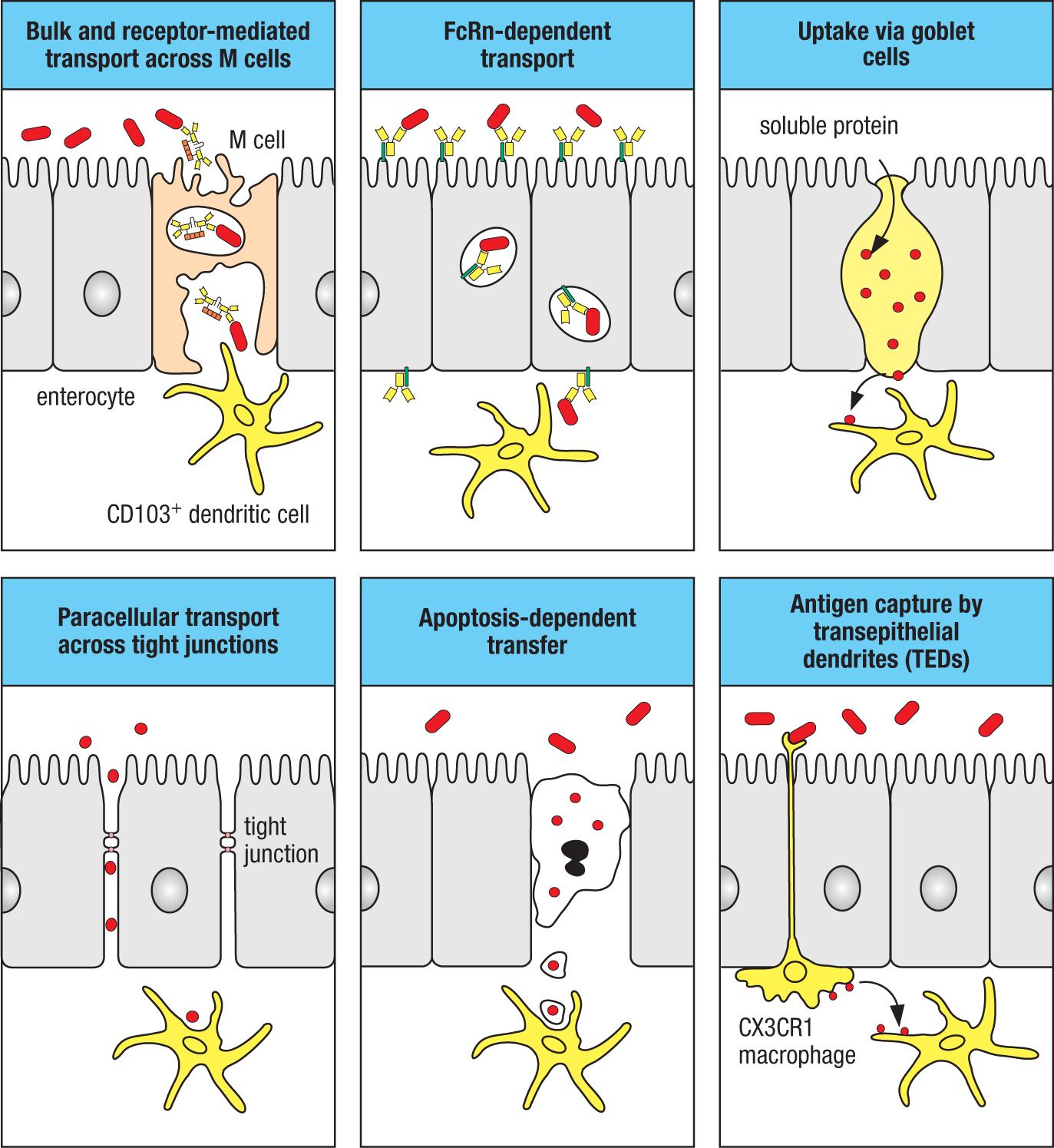
As noted previously, specialization of the FAE includes the absence of a glycocalyx and the presence of M (microfold) cells. M cells continually sample luminal antigens that range from macromolecules and small particulates to viruses and whole bacteria particles. This material is transported through the interior of the cell in membrane-bound vesicles to the basolateral cell membrane, where it is released. The basal cell membrane of an M cell makes close contacts with processes of dendritic cells to facilitate local antigen presentation within Peyer’s patches and ILFs (see Fig. 12.30; see also Fig. 12.9). These dendritic cells are recruited to the subepithelial dome, or even into the FAE, in response to chemokines that are released constitutively by FAE epithelial cells; CCL20 and CCL9 produced by FAE cells bind to their receptors (CCR6 and CCR1, respectively) on dendritic cells. Although many antigens are taken up by macropinocytosis and micropinocytosis, intact microbes in particular can be taken up by receptor-mediated endocytosis. For a number of bacteria this may involve specific recognition of the bacterial FimH protein found in type 1 pili by a glycoprotein (GP2) on the M cell or receptors that recognize SIgA bound to the bacteria. Because antigens delivered via M cells promote SIgA responses in the GALT, the receptor-mediated uptake of SIgA-coated microbes provides a positive feedback loop for SIgA production. In addition to receptor-mediated uptake of SIgA-bound antigens by the M cell, non-M cells of both FAE and non-FAE express the neonatal Fc receptor (FcRn), which binds and transcytoses IgG bidirectionally across the epithelium. Although primarily a pathway to import maternal IgG from breast milk to the newborn for passive transfer of antibodies (see Section 10-17), this mechanism can also mediate the uptake of IgG-bound antigens, as well as transport IgG from the lamina propria into the lumen, typically in the context of infections (see Section 12-20).
Although the FAE is enriched for M cells, rare M cells can also be found outside of the FAE, particularly within the epithelium of villi of the small intestine. Here they transcytose antigens to DCs in the villous lamina propria, which migrate to mesenteric lymph nodes to present antigen. Although continual sampling by M cells represents an important pathway for sensing of luminal antigens, a number of pathogens have evolved strategies to target M cells and gain access to the subepithelial space, even though they then find themselves in the heart of the intestinal adaptive immune system. These include strains of Salmonella enterica that are the causative agents of typhoid fever or bacterial food poisoning (see Section 12-20); Shigella species that cause dysentery; and Yersinia pestis, which causes plague. Poliovirus, reoviruses, some retroviruses (such as HIV), and prions (such as the causal agent of scrapie) follow the same entry route.
Another pathway for the uptake of particulate antigens is via transepithelial dendrites (TEDs) of CX3CR1+ intestinal macrophages (see Fig. 12.30; see also Section 12-8). The subepithelial recruitment of intestinal macrophages positions them to eliminate any bacteria that cross the epithelial barrier. In addition, they can extend TEDs containing tight-junction proteins across the epithelial barrier without perturbing epithelial integrity, allowing them to capture bacteria and other antigens directly in the intestinal lumen. As these macrophages are nonmigratory, antigens taken up in this manner must be transferred to local conventional dendritic cell 2 (cDC2) cells to be ferried to mesenteric lymph nodes for presentation. The transfer of antigens from macrophages to DCs may be mediated by gap junctions, which allow direct connections between the two cells. In addition to the uptake of antigens from CX3CR1+ macrophages, intestinal DCs may also enter the epithelium for direct uptake of luminal antigens. Although each of these activities has been identified at homeostasis, they are particularly evident during infection and appear to provide a key mechanism to sample pathogens prior to their compromise of the epithelial barrier. Finally, antigen derived from apoptotic epithelial cells may be processed by cross-presenting dendritic cells (see Section 6-5) for induction of immune responses against enteric (that is, intestinal) viruses, such as rotaviruses, which cause diarrheal disease because of their specialized ability to infect enterocytes.
While the pathways described above play important roles in the acquisition of particulate antigens, including microbes, other pathways appear to dominate in the uptake of soluble antigens at homeostasis; the relatively limited surface area of follicle-associated epithelium and the small number of M cells restricts the flux of antigens that can be sampled via this route, and direct TED-mediated uptake at homeostasis is limiting. Recently, it was found that in addition to their role in secreting mucins, goblet cells have an important role in the transport of soluble antigens across the epithelial barrier. This occurs via goblet cell–associated passages (GAPs) that conduct low-molecular-mass soluble antigens (<70 kDa) to underlying CD103+ cDC2 cells in the lamina propria (see Fig. 12.29). Precisely how antigen is transferred to DCs is unclear, but it appears to be linked to the process of goblet-cell secretion; when goblet-cell mucin secretion is stimulated, there is increased antigen uptake by goblet cells. DCs that have taken up antigen by this mechanism are found to contain goblet-cell proteins, indicating that goblet-cell components are transferred to DCs along with the luminal antigens. Importantly, in mice in which goblet cells were deleted, luminal antigen delivery to lamina-propria dendritic cells was undetectable, indicating that GAPs are a major pathway for soluble-antigen delivery at homeostasis and likely play a central role in oral-tolerance induction.
Another route for the uptake of soluble antigens is via paracellular transport, also referred to as paracellular ‘leak.’ As discussed in Section 12-4, because of the network of tight junctions between adjacent IECs, the intestinal epithelium is a selectively permeable barrier, allowing water, solutes, small molecules, and ions through, but preventing the transit of larger proteins (see Fig. 12.17). However, the permeability of tight junctions can be regulated by the differential expression of certain claudin proteins (for example, claudin-2), allowing macromolecules up to 10 kDa to traverse the epithelium. In the small intestine, paracellular transport appears to vary between villi at homeostasis, suggesting that it is actively regulated locally. Small antigens delivered via paracellular transport are not taken up efficiently by intestinal DCs. Instead, they are transported via lacteals and lymphatics to mesenteric lymph nodes, where they are delivered to resident DCs via reticular conduits (see Section 9-4). In inflammatory intestinal disease, such Crohn’s disease or ulcerative colitis, increased permeability and disruption of tight junctions results in increased paracellular leak, possibly contributing to a breakdown in tolerance to antigens of the commensal microbiota.
12-10 Intestinal dendritic cells favor the induction of antigen-specific Treg cells that are critical for the maintenance of mucosal immune homeostasis.
Peripheral FoxP3+ Treg cells are central players in intestinal immune tolerance to food and the commensal microbiota and are preferentially induced in the gut at homeostasis. Although thymic-derived regulatory T cells (tTreg cells) circulate to GALT and mesenteric lymph nodes, just as they do to peripheral lymphoid tissues, the dominant Treg cells in the intestinal tissues are pTreg cells, which express a distinct TCR repertoire that reflects their development in response to nonself antigens taken up from the intestines, not to self antigens expressed in the thymus. These two populations can be distinguished by expression of the transcription factor helios and the surface receptor neuropilin-1 (NRP1), both of which are preferentially expressed by FoxP3+ tTreg cells, but not by pTreg cells; hence the majority of Treg cells in the intestines are typically FoxP3+helios–NRP1– pTreg cells. Global deficits in FoxP3+ Treg cells, such as those seen in people or mice with mutations in the FOXP3 gene, are associated with systemic autoimmunity that includes inflammatory bowel disease (IBD) and food allergies (see Section 15-4). In mice deficient for a conserved intronic enhancer element in the Foxp3 gene called CNS1 (conserved nucleotide sequence 1), which controls the development of pTreg cells, but not tTreg cells, there is intestine-specific breakdown in tolerance that results in inflammation and an imbalance in the microbiota in the colon. Thus, intestinal immune homeostasis is primarily controlled by pTreg cells responsive to antigens introduced via the gut.
As elsewhere, conventional dendritic cells (cDCs) are important in initiating and shaping immune responses in mucosal tissues. They are deployed both in intestinal lymphoid tissues (that is, GALT) and throughout the lamina propria. Both of the major subtypes of conventional dendritic cells (cDC1 and cDC2) are present in the intestine (see Fig. 12.29; see also Section 6-5 and Section 9-6). Given their dominant role in presentation of extracellular antigens, including those derived from food and commensal microbes, cDC2 cells are critical to the induction of Treg cells in the gut. As mentioned earlier (see Section 12-8), the anti-inflammatory, pro-regulatory behavior of mucosal dendritic cells in the healthy gut is promoted by factors that are constitutively produced in the mucosal microenvironment. These include thymic stromal lymphopoietin (TSLP) produced by IECs; short-chain fatty acids (SCFAs), such as butyrate, which are produced by anaerobic commensal bacteria; prostaglandin E2 (PGE2) produced by stromal cells; neuropeptides, such as vasointestinal peptide (VIP), produced by local neurons; IL-10 produced by intestinal macrophages and FoxP3+ Treg cells; and TGF-β and retinoic acid (RA) produced by IECs and stromal cells (Fig. 12.31). The collective action of these factors induces the expression by intestinal DCs of RA and TGF-β, both of which are critical to the induction of FoxP3+ pTreg cells (see Section 9-15 and Fig. 9.37).
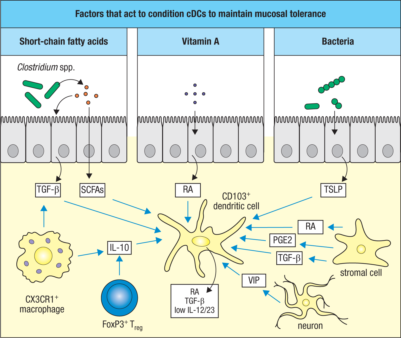
Because vitamin A (retinol), the substrate from which RA is generated, cannot be synthesized and must be absorbed by the intestine from the diet, pTreg cell induction is dependent on a dietary factor. Retinol stored in the liver and delivered to the small intestine via bile provides an additional source for the local generation of retinoic acid in the intestines. Production of RA from vitamin A requires the sequential actions of enzymes expressed by intestinal DCs: alcohol dehydrogenases (ADHs) and retinal dehydrogenases (RALDHs). RA is a ligand for a nuclear receptor, retinoic acid receptor (RAR), activation of which promotes the development of pTreg cells while strongly repressing the differentiation of TH17 cells (see Fig. 9.37). RA also induces the expression of gut-specific homing receptors, including α4β7 and CCR9 (see Section 12-11). The induction of regulatory T cells in intestinal tissues is also dependent on TGF-β, which is produced by migratory and lymphoid tissue–resident cDC2 cells that take up antigens in the lamina propria and GALT, respectively. TGF-β also induces expression of the integrin αEβ7 (CD103) on developing Treg cells. The combined effects of RA and TGF-β therefore, are to induce the differentiation of FoxP3+ pTreg cells and to direct their trafficking to the intestines.
After trafficking to the intestines, pTreg cells can acquire additional functional features that are important in maintaining immune homeostasis. It is thought that this transition involves further differentiative signals received when Treg cells enter effector sites in the intestines, such as the lamina propria. Referred to as effector regulatory T (eTreg) cells, these cells are characterized by their expression of the anti-inflammatory cytokines IL-10 and TGF-β, both of which contribute to the tolerogenic nature of homeostatic intestinal cDCs in a positive feedback loop and are indispensable for restraining inflammatory responses to the commensal microbiota in the large intestine. Studies that examined the expression of FoxP3 and IL-10 in tissues throughout the body using transgenic reporter mice found that the highest frequency of IL-10–expressing cells in the body was in the intestines (Fig. 12.32), particularly in the large intestine, where it is primarily expressed by FoxP3+helios–NRP1– pTreg cells at homeostasis. The functional importance of IL-10 expression by eTreg cells has been demonstrated in studies in which selective deletion of the gene Il10 in FoxP3+ T cells resulted in spontaneous inflammation of the large intestine, or colitis. Thus, IL-10 produced by eTreg cells is important for immune homeostasis in the large intestine.
|
Treg cells are highly enriched in the intestinal mucosa |
||||
|
CD4+FoxP3− |
CD4+FoxP3+ |
|||
|
IL-10− |
IL-10+ |
IL-10− |
IL-10+ |
|
|
Mesenteric LNs |
80–90% |
∼1% |
5–10% |
∼1% |
|
Small intestine |
70–80% |
5–10% |
5–10% |
5–10% |
|
Large intestine |
60–70% |
3–5% |
10–15% |
15–20% |
|
Large intestine, GF |
80–90% |
∼1% |
5–10% |
∼1% |
Fig. 12.32 Treg cell subsets are major populations in the large and small intestines. Flow cytometry was used to analyze Treg cell subsets in murine intestines and mesenteric lymph nodes, defined by concordant or differential expression of IL-10 and FoxP3 within the total fraction of CD4 T cells. The analysis of large intestinal cells compared those isolated from conventionally housed and germ-free (GF) mice. LNs, lymph nodes.
Elegant experiments in the 1990s led to the now generally accepted idea that T cells with the potential to respond to commensal microbiota are present in normal animals but are usually kept in check through active repression by Treg cells (Fig. 12.33). In these studies, the transfer of naive CD4 T cells into immunodeficient mice led to spontaneous IBD, or colitis, unless a separate population that included Treg cells was co-transferred. The cell types that drove this disease were later shown to be microbiota-reactive TH17 and TH1 cells that developed from the transferred naive CD4 T cells. That the inflammatory response was directed against the intestinal microbiota was demonstrated by the absence of disease in germ-free mice. An essential requirement for IL-10 expression by Treg cells in preventing disease was found, as the transfer of Treg cells deficient in IL-10 failed to control the development of colitis. Similar studies have shown that Treg cells deficient for TGF-β are impaired in their prevention of colitis. Accordingly, mutations in the genes IL10RA, IL10RB, TGFBR1, or TGFBR2 in humans are associated with severe early-onset inflammatory bowel disease, such as Crohn’s disease, presumably due, at least in part, to defects in the function of eTreg cells reactive to commensal microbial antigens.
|
Cells transferred |
Microbiota |
Disease |
Normal |
Colitis |
|
|
Unfractionated CD4+ T cells |
+ |
No |
 |
 |
|
|
CD4+CD45RBhi T cells (naive) |
+ |
Colitis |
|||
|
CD4+CD45RBhi T cells + CD4+CD45RBlo T cells (includes Treg cells) |
+ |
No |
|||
|
CD4+CD45RBhi T cells + IL-10–deficient CD4+CD45RBlo T cells (includes Treg cells) |
+ |
Colitis |
|||
|
CD4+CD45RBhi T cells |
– |
No |
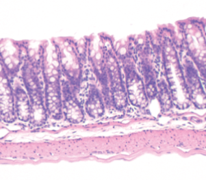
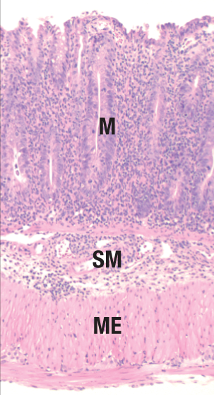
Fig. 12.33 T cells with the potential to produce inflammation in response to commensal bacteria are present in normal animals, but are controlled by regulatory T cells. Transfer of total (unfractionated) CD4+ T cells from a normal mouse into an immunodeficient mouse, such as one lacking the gene Rag1 (Rag1–/–), will lead to reconstitution of the CD4 T-cell compartment. However, if naive CD4 T cells (CD4+CD45RBhi) are isolated from the total CD4 T-cell population and transferred, the recipient mice develop severe inflammation of the colon. This is prevented by co-transferring CD4+CD45RBlo T cells that were removed during the isolation of the naive (CD45RBhi) fraction of the total CD4+ T-cell population. The CD4+CD45RBlo fraction comprises nearly all of the Treg cells within the CD4 T-cell population. If the CD4+CD45RBlo fraction is isolated from IL-10–deficient mice, the protective effect of these cells is lost. The intestinal inflammation caused by naive CD4+ T cells requires the presence of the microbiota; it does not occur in germ-free mice or in mice treated with antibiotics. These experiments demonstrate that some CD4+ T cells in normal animals are capable of provoking inflammatory responses against the intestinal microbiota, but they are normally held in check by regulatory T cells. Shown in the two right panels are examples of the histopathology of the normal mouse colon (left) and the colitic colon (right), the latter demonstrating a much thickened mucosa (M) due to elongation of the crypts and dense infiltrates of inflammatory cells, as well as the expansion of the submucosa (SM) and thickening of the muscularis externa (ME).
12-11 Lymphocytes primed within the mucosal immune system are directed to return to the mucosal tissue by tissue-specific adhesion molecules and chemokine receptors.
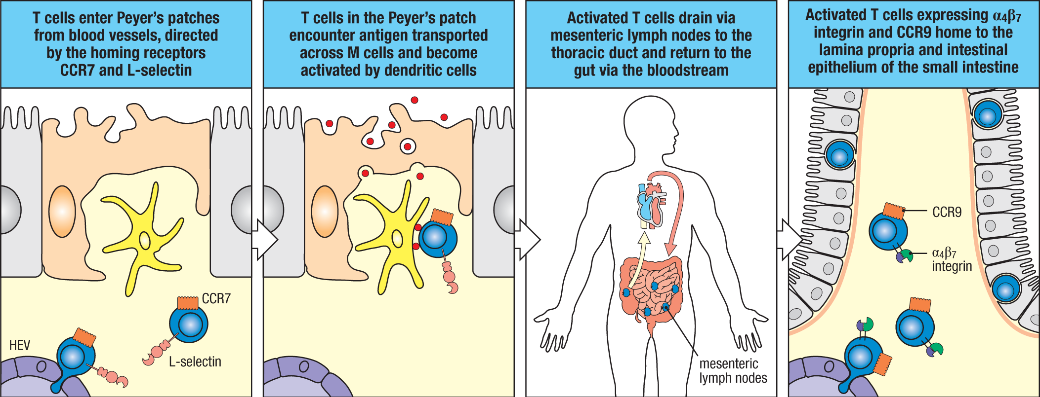
The destination of naive T cells and B cells circulating in the bloodstream is not predetermined, and like those in other peripheral lymphoid tissues, the cells destined to be primed in the GALT or mesenteric lymph nodes enter through high endothelial venules (HEVs) (see Fig. 9.4). As in the systemic immune system, this process is controlled largely by the chemokines CCL21 and CCL19, which are released from stromal cells in these sites and bind the receptor CCR7 on naive lymphocytes. In the Peyer’s patches and ILFs, this process is assisted by the binding of the mucosal vascular addressin MAdCAM-1 on HEVs to the L-selectin expressed on naive T cells. CXCR5 responding to CXCL13 produced in B-cell areas is also important for recruitment of naive B cells to Peyer’s patches and ILFs. As in other peripheral lymphoid tissues, if the naive lymphocytes do not see their antigen, they do not down-regulate sphingosine 1-phosphate receptor 1 (S1PR1), and they return to the bloodstream via the lymphatics. If they encounter antigen in the GALT or regional lymph nodes, they are imprinted by intestinal DCs to return to the intestinal lamina propria.
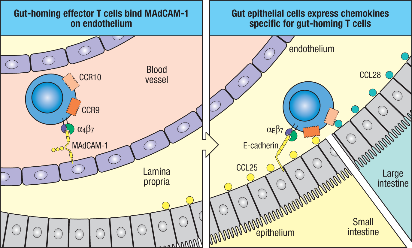
Although some T and B lymphocytes initially activated in Peyer’s patches or ILFs may migrate directly to adjacent parts of the lamina propria, most leave via the lymphatics, pass through mesenteric lymph nodes, and eventually end up in the thoracic duct. From there they circulate in the bloodstream (Fig. 12.34) and selectively reenter the intestinal lamina propria via small blood vessels. Gut-specific homing by antigen-stimulated T and B cells is determined in large part by the expression of the integrin α4β7, which binds to MAdCAM-1 expressed on microvessels of intestinal tissues (Fig. 12.35). Homing of these lymphocytes to the small intestinal or large intestinal mucosa is further regulated by differential expression of the chemokine receptors, including CCR9 or CCR10, which respectively bind the homeostatic chemokine CCL25 (small intestine) or CCL28 (large intestine) produced by the local epithelium. The chemokine receptor–like orphan G-coupled protein receptor 15 (GCPR15) also appears to be important for trafficking of effector T cells to the large intestine, although its ligand(s) is not yet known. Notably, the epithelium of the lactating breast also expresses CCL28. In this way, SIgA-producing B lymphoblasts primed to commensal microbial antigens in the large intestine are recruited to the breast where they produce maternal SIgA, which is passively transferred to the nursing neonate.
12-12 Secretory IgA is the dominant class of antibody associated with the mucosal immune system at homeostasis.
The dominant class of antibody in the mucosal immune system is immunoglobulin A (IgA), which is produced locally by plasma cells in the lamina propria, including that of the intestines. Hundreds of thousands of IgA-producing plasma cells are present in the normal human intestine, and they produce 3–4 g of IgA each day, by far the most of any immunoglobulin class produced in the gut. This continual production of large quantities of IgA occurs in the absence of pathogenic invasion and is driven almost entirely by recognition of the commensal microbiota. The nature of IgA differs between the two main compartments in which it is found—the blood and mucosal secretions. IgA in the blood is mainly in the form of a monomer (monomeric IgA, or MIgA) that is produced in the bone marrow by plasma cells that home there following their initial development in secondary lymphoid tissues. In mucosal lymphoid tissues, IgA is produced almost exclusively as a polymer, typically a dimer in which the two immunoglobulin monomers are linked by a J chain (see Section 10-16).
Naive B-cell precursors of IgA-secreting plasma cells in the intestinal lamina propria are activated in Peyer’s patches, ILFs, and mesenteric lymph nodes. Class switching of activated B cells to IgA is controlled by the cytokine TGF-β, whether generated via T cell–dependent or T cell–independent mechanisms. The development of T cell–independent IgA class switching is discussed later (see Section 12-13). In lymphoid tissues with developed germinal centers (for example, Peyer’s patches), IgA class switching is entirely T-cell dependent and regulated by T follicular helper (TFH) cells by the same mechanisms described in Chapter 10. Notably, a substantial fraction of the TFH cells that support IgA class switching in the intestines appears to derive from pTreg cells. This makes regulatory sense, as these cells are producers and activators of TGF-β, and by inducing the production of SIgA to antigens they recognize, they provide a self-reinforcing positive feedback loop, due to the fact that delivery of antigens in complex with SIgA favors the generation of pTreg cells. In this regard, it has been shown in mice that depletion of Treg cells leads to a decline in IgA+ plasma cells and intestinal IgA over a few days, indicating that intestinal plasma cells are relatively short-lived and that Treg cells are critical for their maintenance and/or their replacement.
Unlike mice, which produce a single isotype of monomeric and dimeric IgA, humans produce two IgA isotypes, IgA1 and IgA2. The ratio of IgA1 to IgA2 varies markedly depending on the tissue, being about 10:1 in the blood and upper respiratory tract, about 3:2 in the small intestine, and 2:3 in the colon. Some common pathogens of the respiratory mucosa (such as Haemophilus influenzae) and the genital mucosa (such as Neisseria gonorrhoeae) produce proteolytic enzymes that can cleave IgA1, whereas IgA2 is cleavage-resistant. The higher proportion of plasma cells secreting IgA2 in the large intestine might result because the high density of commensal microorganisms at this site drives the production of cytokines that cause its selective class switching. The IgA isotype produced in mice is most like human IgA2.
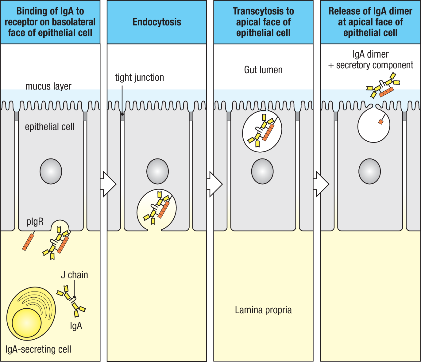
IgA-expressing B plasmablasts produced in the GALT or regional lymph nodes express the mucosal homing integrin α4β7 and the chemokine receptors CCR9 or CCR10, which cause them to localize to the small or large intestinal lamina propria. Once there, these cells undergo final differentiation into plasma cells that synthesize IgA dimers and secrete them into the subepithelial space (Fig. 12.36). To reach antigens in the gut lumen, the dimeric IgA must be transported across the epithelium by the polymeric immunoglobulin receptor (pIgR) (see Section 10-16). pIgR is expressed constitutively on the basolateral surfaces of immature epithelial cells located at the base of intestinal crypts where it binds dimeric IgA. pIgR transports the dimeric IgA to the luminal surface of the epithelium, where it is released by proteolytic cleavage of the extracellular domain of the receptor. Part of the cleaved pIgR remains associated with IgA and is known as secretory component (SC). The resulting antibody is protected from proteolytic cleavage and is referred to as secretory IgA (SIgA) (see Section 10-16). The same pathway is used to transport pentameric IgM across the intestinal epithelium—so-called secretory IgM (SIgM)—which appears to play an important role in the colonization of the intestines by the commensal microbiota early in life.
In some animals there is a second route of IgA secretion into the intestine—the hepatobiliary route. Dimeric IgA that has not bound pIgR is taken up into venules in the lamina propria, which drain intestinal blood to the liver via the portal vein (see Fig. 12.7). In the liver, the portal vein delivers the dimeric IgA to sinusoids that are lined by a fenestrated endothelium containing ‘holes’ that allow the antibodies direct access to underlying hepatocytes, which express pIgR on their surface. IgA is taken up into the hepatocytes and transported by transcytosis into an adjacent bile duct. In this way, secretory IgA can be delivered back to the duodenum via the common bile duct. This hepatobiliary route allows dimeric IgA to eliminate antigens that have invaded the lamina propria and have been bound there by IgA. Although highly efficient in rats and other rodents, this route does not seem to be of great significance in humans and other primates, in whom hepatocytes do not express pIgR.
IgA secreted into the gut lumen binds to the layer of mucus coating the epithelial surface via carbohydrate determinants in secretory component. There it prevents invasion by pathogenic organisms and also has a crucial role in maintaining the homeostatic balance between the host and the commensal microbiota. IgA does this in a number of ways (Fig. 12.37). First, it inhibits microbial adherence to the epithelium, its ability to bind bacteria being assisted by the unusually wide and flexible angle between the Fab pieces of the IgA molecule, particularly the IgA1 isotype, allowing very efficient bivalent binding to large antigens such as bacteria. Secretory IgA also aggregates bacteria such that they are less able to penetrate the network of mucins in the mucus. Secretory IgA can also neutralize microbial toxins or enzymes.
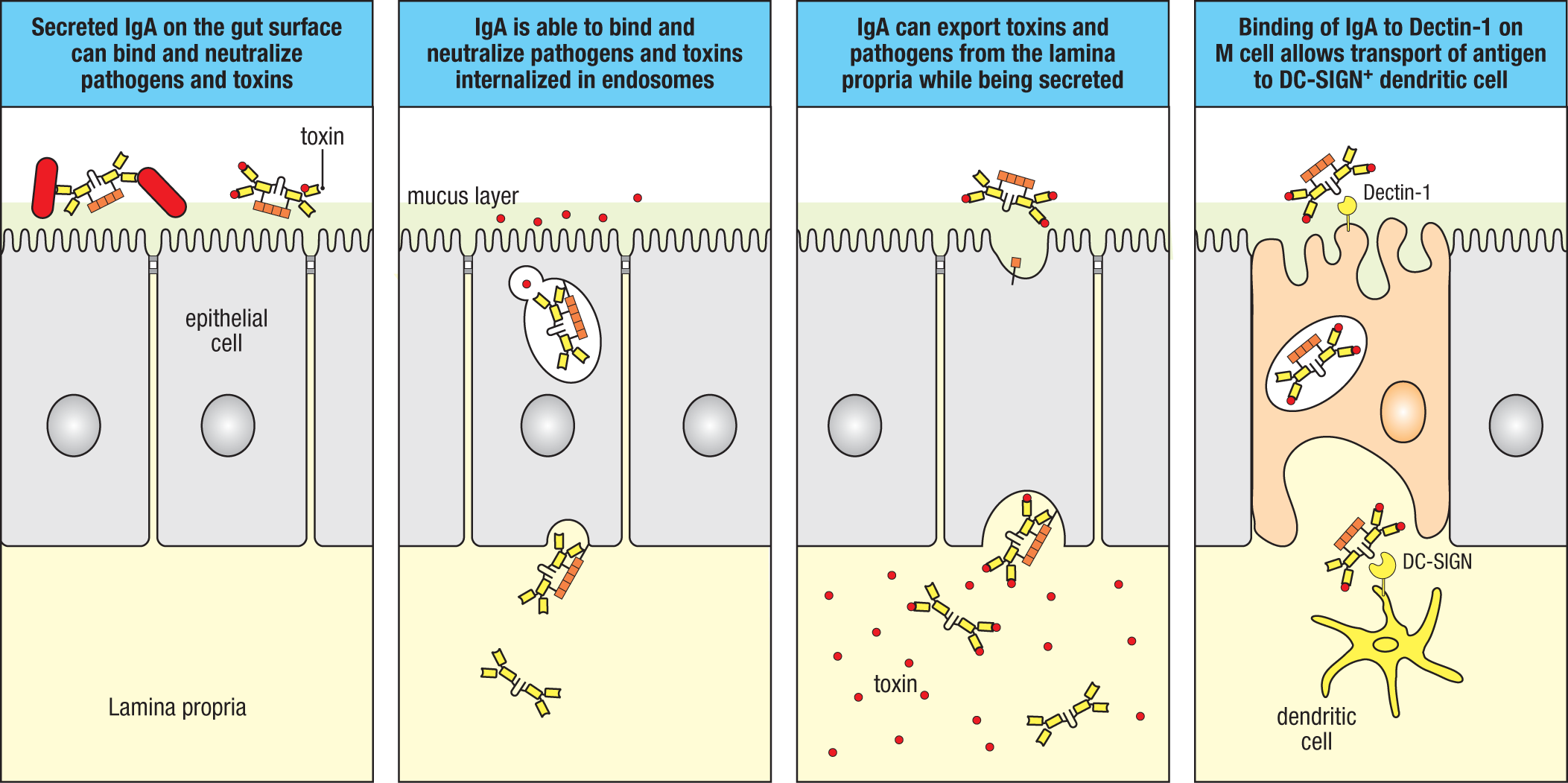
In addition to its activities in the lumen, IgA can neutralize bacterial lipopolysaccharide and viruses it encounters within endosomes inside epithelial cells, as well as across the epithelial barrier in the lamina propria after bacteria and viruses have penetrated there. The resulting IgA:antigen complexes are then reexported into the gut lumen. From there they are excreted from the body. Complexes containing dimeric IgA formed in the lamina propria can also be excreted via the hepatobiliary route described earlier. In addition to enabling the elimination of antigens, the formation of IgA:antigen complexes can enhance the uptake of luminal antigen by M cells and local dendritic cells, via binding of carbohydrate residues on IgA to lectin receptors such as Dectin-1 and DC-SIGN, respectively. In addition to these antigen-specific effects, secretory IgA can restrict the entry of bacteria in a nonspecific manner, because the high carbohydrate content of the Fc part of the IgA heavy chain allows it to act as a decoy for receptors that bacteria use to bind carbohydrates on the epithelial surface. Importantly, SIgA has little capacity to activate the classical pathway of complement or to act as an opsonin, and so does not induce inflammation. Uptake of IgA:antigen complexes by dendritic cells also induces these cells to produce anti-inflammatory IL-10. Together these properties mean that IgA can limit the penetration of microbes into the mucosa without risking inflammatory damage to these fragile tissues, something that would be potentially harmful in the intestine. For the same reasons, secretory IgA fosters the beneficial symbiosis between the host and its commensal microbiota.
12-13 T cell–independent processes can contribute to IgA production in some species.
A significant proportion of intestinal SIgA is derived from T cell–independent B-cell activation and class switching. This is clearly true in mice, and probably similar in humans, although experimental support for the latter is more limited. T cell–independent B-cell activation depends on activation of the innate immune system by the products of commensal microbes and results from the direct interaction of B cells with intestinal cDCs and follicular dendritic cells in isolated lymphoid follicles. Most of this SIgA production seems to involve lymphocytes of the B-1 subset (see Section 8-10), which arise from precursor B cells in the peritoneal cavity and migrate to the intestinal wall in response to microbial constituents such as lipopolysaccharide (LPS). Once in the mucosa, TGF-β–dependent class switching to IgA occurs under the influence of local factors, including IL-6, retinoic acid, and BAFF and APRIL (see Fig. 10.6), which bind to TACI on B cells, substituting for signals otherwise supplied by CD4 helper T cells (see Section 10-1). Intestinal epithelial cells produce BAFF and APRIL, while local macrophages, DCs, and eosinophils may contribute by producing APRIL, IL-6, and TGF-β.
The IgA antibodies produced in these T cell–independent responses are generally of more limited diversity and lower affinity, with little evidence of somatic hypermutation. They are nevertheless an important source of ‘natural’ antibodies directed at commensal bacteria and may play a particularly important role in modulating the developing intestinal microbiota in neonates prior to establishment of more mature T- and B-cell responses. With progressive maturation of the immune system during the first years of life, there is a shift to a greater fraction of SIgA contributed by T-dependent B-cell activation and class switching; hence, a greater proportion of SIgA has undergone somatic hypermutation with increasing age. T-independent SIgA production in the intestines may offer a glimpse into the evolutionary history of specific antibody responses in the mucosa, and it appears to serve as a ‘backup’ for the generation of SIgA when T cell–dependent IgA production is compromised, as it is in AIDS or in individuals with an inherited deficiency of CD40.
12-14 IgA deficiency is relatively common in humans but may be compensated for by secretory IgM.
Selective deficiency of IgA production is one of the most common inherited, or primary, immune deficiencies in humans, occurring in about 1 in 500 to 700 Caucasians. (It is somewhat rarer in other ethnic groups.) The most frequent genetic mutation that has been identified in this condition is in the TACI receptor for BAFF. A slightly higher incidence of respiratory infections, atopy (a tendency for allergic reactions to harmless environmental antigens), and autoimmune disease has been reported in older people with IgA deficiency. However, most individuals with IgA deficiency are not overly susceptible to infections. The dispensability of IgA probably reflects the ability of IgM to replace IgA as the predominant antibody in secretions, and increased numbers of IgM-producing plasma cells are indeed found in the intestinal mucosa of IgA-deficient people. Because IgM is a J chain–linked polymer, IgM produced in the gut mucosa is bound efficiently by the pIgR and is transported across epithelial cells into the gut lumen as secretory IgM. The importance of this backup mechanism has been shown in knockout mice. Animals lacking IgA alone have a normal phenotype, but those lacking the pIgR are susceptible to mucosal infections. They also show increased penetration of commensal bacteria into tissues and a consequent systemic immune response to these bacteria. Genetic absence of the pIgR has never been reported in humans, suggesting that such a defect is lethal.
12-15 Large numbers of antigen-experienced T cells are present in the intestinal lamina propria even in the absence of disease.
Most of the T cells in the healthy lamina propria have been activated by dendritic cells and express markers of effector or memory T cells. Just as for Treg cells and B cells, the largest deployment of effector T cells at homeostasis is in the intestines. The T-cell population of the lamina propria has a ratio of CD4 to CD8 T cells of 3:1 or more, similar to that in systemic lymphoid tissues. In industrialized countries, where intestinal helminth infections are less common, the major effector T-cell subsets resident in the gut express cytokines characteristic of TH17 and TH1 cells. And like Treg cells and B cells, the development of these effector subsets is largely dependent on the dominant component of the microbiota—bacteria. TH17 cells in particular, in their role as responders to extracellular antigens, are particularly abundant, due in part to their developmental ties to pTreg cells with which they share a requirement for the abundant intestinal cytokine, TGF-β (see Section 9-14). Their large number likely reflects the constant state of immune recognition of the microbiota and other environmental antigens that takes place in the intestine. At least some TH17 cells in the lamina propria appear to be resident memory (TRM) cells, although the proportion varies depending on the antigenic exposure history. Cytokines produced by intestinal TH17 cells are important components of intestinal homeostasis and defense. IL-17 is needed for full expression of the polymeric immunoglobulin receptor involved in secretion of IgA into the lumen, while IL-22 stimulates intestinal epithelial cells to produce antimicrobial peptides that help maintain epithelial barrier integrity. Effector CD8 T cells are also present in the normal lamina propria and are capable of both cytokine production and cytotoxic activity when a protective immune response to a pathogen is required.
Whether at steady state, in response to infection, or in the context of chronic inflammation induced by inflammatory bowel disease (IBD), the intestine contains, in addition to effectors that express IL-17, cells that express IFN-γ, or both IL-17 and IFN-γ. Although a fraction of these T cells is generated in response to the microbiota and are therefore presumed to be specific for antigens of the commensal flora, their developmental origins and functions remain incompletely defined. Notably, however, in contrast to TH1 and TH2 cells, TH17 cells retain the capacity for divergent cytokine expression profiles and function after their commitment to the TH17 pathway and can give rise to IL-17+IFN-γ + effectors, as well as IL-17–IFN-γ+ effectors that resemble classical TH1 cells, so-called TH17/TH1 cells (see Section 11-14 and Fig. 11.21). This has been demonstrated for both human and mouse TH17 cells and has raised the possibility that many of the IFN-γ–producing ‘TH1’ cells in the intestine arise from the TH17 pathway. While the relative contributions of TH17 and TH17/TH1 cells to host defense or inflammatory disease remain to be elucidated, IFN-γ–producing cells are required for disease pathogenesis in at least some mouse models of IBD. Although these TH17/TH1 cells share many features of classical TH1 cells, gene-expression analyses indicate important differences that are likely to have implications for immune disease mediated by these cells. This suggests generic categorization of IFN-γ–producing CD4 T cells as ‘TH1’ cells is an oversimplification, and so further studies aimed at dissecting the contributions of branches of the TH17 pathway to immune protection or pathogenesis will be important.
In any other situation, the presence of such large numbers of differentiated effector T cells would suggest the presence of a pathogen and likely would lead to inflammation. The fact that it does not so do in the healthy lamina propria is because the generation of TH1, TH17, and cytotoxic T cells is balanced by the presence of substantial numbers of IL-10–producing T cells. In the large intestine, these are almost entirely FoxP3+ Treg cells, whereas in the small intestine, FoxP3+ Treg cells and FoxP3– CD4 T cells (TR1) produce IL-10 and are likely involved in preventing unrestrained effector T cell–driven inflammation, as occurs in IBD.
12-16 Priming of lymphocytes in one mucosal tissue may induce protective immunity at other mucosal surfaces.
Not all parts of the mucosal immune system use the same tissue-specific chemokines, allowing localized compartmentalization of lymphocyte recirculation within the system. Thus, effector T and B cells primed in lymphoid organs draining the small intestine (mesenteric lymph nodes and Peyer’s patches) are most likely to return to the small intestine; similarly, those primed in the respiratory tract migrate most efficiently back to the respiratory mucosa. This programming of tissue homing is useful in returning antigen-specific effector cells to the mucosal organ in which they will be most effective in fighting an infection or controlling immune responses against foreign proteins and commensals. Nevertheless, some lymphocytes that have been primed in the GALT, for example, can also circulate as effector cells to other mucosal tissues such as the respiratory tract, urogenital tract, and lactating breast. This overlap between mucosal recirculation routes gave rise to the idea of a common mucosal immune system, which is distinct from other parts of the immune system. Although this is now understood to be an oversimplification, it does have important implications for vaccine development, because it may enable immunization by one mucosal route to be used to protect against infection at another mucosal surface. An important example of this is the induction of SIgA antibody production in the lactating breast by natural infection or vaccination of mucosal surfaces such as the intestine. As we have seen, this is because the vasculature of the lactating breast expresses MAdCAM-1 and chemokines recognized by CCR10 (see Section 12-11). This “cross-talk” is a crucial means of generating protective immunity that can be transmitted to nursing infants by passive transfer of the antibodies in maternal milk. A further example has been shown in experimental animals, in which nasal immunization has a special ability to prime immune responses in the urogenital tract against HIV. The mechanisms behind this are currently unknown.
Summary.
A fundamental challenge of the intestinal immune system is the maintenance of tolerance in the face of considerable nonpathogenic antigenic challenges, both microbial and nonmicrobial. Multiple mechanisms contribute to this, but central to immune homeostasis in the intestines is the delivery of antigens under conditions that favor the differentiation of pTreg cells and SIgA-producing plasma cells—adaptive immune cells that both reinforce non-inflammatory responses to their inductive antigens. This is achieved by the creation of a microenvironment rich in anti-inflammatory cytokines, principal among which are TGF-β, which is produced by IECs and stromal cells, and IL-10, which is produced primarily by pTreg cells themselves. Among other local factors important to intestinal immune homeostasis is retinoic acid (RA), which is produced from vitamin A by IECs and stromal cells, as well as cDCs that are conditioned in the intestines. TGF-β has potent actions on macrophages and cDCs, but also has direct effects on developing T cells and B cells to favor pTreg cell differentiation and IgA class-switching, respectively, as does RA. IL-10 potently suppresses the production of pro-inflammatory cytokines by intestinal macrophages, playing a nonredundant role in maintaining the anti-inflammatory tone of the intestinal mucosa. The critical role of pTreg cells and IL-10 has been demonstrated in murine studies wherein deletion of either results in spontaneous intestinal inflammation. SIgA promotes intestinal homeostasis as a non-complement-fixing antibody isotype that promotes pro-tolerogenic delivery of antigens to which it binds.
Many of the same factors that contribute to tolerogenic adaptive immunity in the intestines also promote homing of lymphocytes to effector sites in the intestinal mucosa. Thus, TGF-β and RA both contribute to the expression of integrins (for example, α4β7) and chemokine receptors (for example, CCR9 and CCR10) that direct lymphocytes to the intestines, ensuring that pTreg cells and IgA plasmablasts are directed to the site where they can reinforce tolerance to the antigens that induced their differentiation. Importantly, expression of the mucosal addressin (MadCAM-1) and chemokines that can attract gut-primed B cells by the lactating breast provides an important mechanism for delivery of maternal SIgA reactive to the mother’s microbiota to her newborn, providing a mechanism to transmit immune tolerance to her offspring.
Glossary
- oral tolerance
- The suppression of specific systemic immune responses to an antigen by the prior administration of the same antigen by the oral (enteric) route.
- peripheral tolerance
- Tolerance acquired by mature lymphocytes in the peripheral tissues, as opposed to central tolerance, which is acquired by immature lymphocytes during their development.
- mucosal tolerance
- The suppression of specific systemic immune responses to an antigen by the previous administration of the same antigen by a mucosal route.
- CX3CR1
- Chemokine receptor expressed by monocytes, macrophages, NK cells, and activated T cells that binds CX3CL1 (fractalkine).
- inflammation-anergic macrophages
- Macrophages in the homeostatic, noninflamed gastrointestinal mucosa that are hyporesponsive to inflammatory stimuli because of down-regulation of the LPS co-receptor CD14, IgA and IgG Fc receptors, and NFκB signaling molecules downstream of tonic TGF-β signaling.
- efferocytosis
- The process of removal of apoptotic cells by phagocytes (from the Latin effere, meaning ‘to take to the grave’).
- transepithelial dendrites (TEDs)
- Cellular processes of intestinal macrophages and dendritic cells that extend across the epithelial barrier to sample antigens in the intestinal lumen.
- neonatal Fc receptor (FcRn)
- A receptor that transports IgG from mother to fetus across the placenta and across other epithelia such as the epithelium of the gut.
- goblet cell–associated passages (GAPs)
- Route by which soluble antigens are transported across goblet cells in the intestinal tract. May be a principal route for the induction of oral tolerance.
- inflammatory bowel disease (IBD)
- General name for a set of inflammatory conditions in the gut, such as Crohn’s disease and ulcerative colitis, that have an immunological component.
- CCL21
- Chemokine made by dendritic cells and stromal cells in T-cell zones of lymph nodes that binds CCR7 and functions to attract naive T cells.
- CCL19
- Chemokine made by dendritic cells and stromal cells in T-cell zones of lymph nodes that binds CCR7 and functions to attract naive T cells.
- CCR7
- Chemokine receptor expressed by all naive T and B cells, and some memory T and B cells, such as central memory T cells, and which binds CCL19 and CCL21 made by dendritic cells and stromal cells in lymphoid tissues.
- MAdCAM-1
- Mucosal addressin cell-adhesion molecule-1. A mucosal addressin that is recognized by the lymphocyte surface proteins L-selectin and VLA-4, enabling the specific homing of lymphocytes to mucosal tissues.
- L-selectin (CD62L)
- Adhesion molecule of the selectin family found on lymphocytes. L-selectin binds to CD34 and GlyCAM-1 on high endothelial venules to initiate the migration of naive lymphocytes into lymphoid tissue.
- CXCR5
- A chemokine receptor expressed by circulating B cells and activated T cells that binds the chemokine CXCL13 and directs cell migration into the follicle.
- CXCL13
- Chemokine produced in the follicle and the light zone of the germinal center that binds CXCR5 expressed on circulating B cells and centrocytes.
- sphingosine 1-phosphate receptor 1 (S1PR1)
- A G protein–coupled receptor activated by sphingosine 1-phosphate, a lipid mediator in the blood that regulates several physiological processes, including the trafficking of naive lymphocytes from tissues into the blood.
- J chain
- Small polypeptide chain made by B cells that attaches to polymeric immunoglobulins IgM and IgA by disulfide bonds and is essential for formation of the binding site for the polymeric immunoglobulin receptor.
- polymeric immunoglobulin receptor (pIgR)
- The receptor for polymeric immunoglobulins IgA and IgM on basolateral surfaces of mucosal and glandular epithelial cells that transports IgA (or IgM) into secretions.
- secretory component (SC)
- Fragment of the polymeric immunoglobulin receptor that remains after cleavage and is attached to secreted IgA after transport across epithelial cells.
- hepatobiliary route
- Route whereby mucosally produced dimeric IgA enters the portal veins in the lamina propria, is transported to the liver, and reaches the bile duct by transcytosis. This pathway is not of great significance in humans.
- common mucosal immune system
- The mucosal immune system as a whole, the name reflecting the fact that lymphocytes that have been primed in one part of the mucosal system can recirculate as effector cells to other parts of the mucosal system.
- FcγRI (CD64)
- Fc receptor highly expressed by monocytes and macrophages that has the highest affinity of the Fc receptors for IgG.
- effector regulatory T (eTreg) cells
- Treg cells that have received additional differentiative signals that cause them to migrate into peripheral tissues where they can regulate effector T-cell response. A signature cytokine of eTreg cells is IL-10.
- high endothelial cells, high endothelial venules (HEVs)
- Specialized small venous blood vessels in lymphoid tissues. Lymphocytes migrate from the blood into lymphoid tissues by attaching to the high endothelial cells in the walls of the venules and squeezing between them.
- T follicular helper (TFH)
- An effector T cell found in lymphoid follicles that provides help to B cells for antibody production and class switching.