 12.1 Antigenic Drift
Immunity at other barrier tissues.
Like the intestines, other barrier tissues present a considerable surface area for interaction with the external environment. Indeed, infections of the respiratory tract and skin are among the most common, and respiratory infections cause more deaths than do any other type of infection (see Fig. 12.4). While the innate and adaptive immune cells that participate in barrier immunity in these tissues are similar, there are anatomic differences in the organs and the epithelia that invest them that deserve special consideration. Moreover, the unique physiological functions of these tissues present special challenges for the immune system. Here, we briefly examine features of the respiratory tract and skin that bear on these challenges.
12-22 Anatomy and mucosal immunity of the airways.
Although there are many similarities in mucosal immunity of the gastrointestinal and respiratory tracts, there are also important differences that reflect their unique anatomy and physiological functions. The primary function of the respiratory tract is gas exchange—the intake of oxygen and release of CO2. Because life is not supported by even a brief interruption in this function, the lungs, where gas exchange occurs, are continually exposed to 5–10 liters of air per minute at rest. The respiratory tract is divided into upper and lower segments, separated by the vocal cords of the larynx (Fig. 12.43). Like the intestines, the upper respiratory tract—including the nose and nasal sinuses, pharynx, and larynx above the vocal cords—is populated by a commensal microbiota that develops after birth. Unlike the intestines, which harbor a preponderance of anaerobes, particularly in the large intestine, a greater contribution by facultative anaerobes is found in the upper respiratory tract. The lower respiratory tract includes the conducting airways—the larynx below the vocal cords, trachea, and branching bronchi and bronchioles—through which air is conducted in inspiration and expiration, as well as the respiratory airspaces, including alveoli, in which gas exchange between the inspired air and blood occurs. Collectively, all the pulmonary tissues distal to the two major bronchi compose the lungs. Importantly, the lower respiratory tract is normally sterile; there is no resident microbiota below the vocal cords.
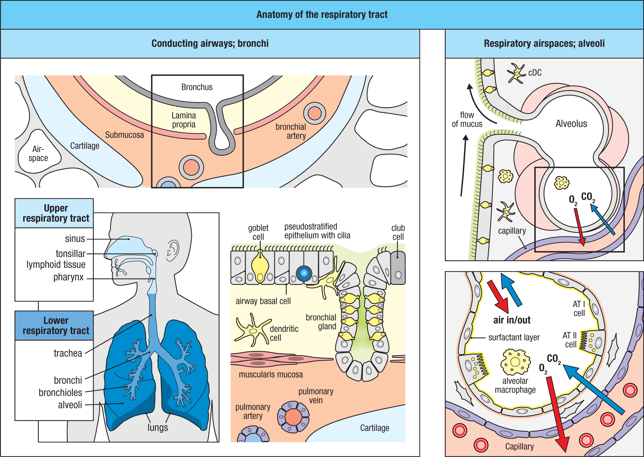
The larger conducting airways are lined by a pseudostratified columnar epithelium, which becomes progressively thinner and transitions to a simple columnar epithelium in the smaller conducting airways (terminal bronchioles; see Fig. 12.2). The dominant cell types in these airways are ciliated columnar cells, goblet cells, club cells, and airway basal cells. In addition, there are small numbers of respiratory tuft cells (also called brush cells) and neuroendocrine cells. Goblet cells, tuft cells, and neuroendocrine cells serve similar functions in the respiratory tract as in the GI tract. The basal cells are stem cells that differentiate to replenish all epithelial cell types in the larger airways (that is, above the bronchioles). Goblet cells become less numerous as the airways become smaller and are lost in the terminal bronchioles (the smallest airways). Club cells (formerly called Clara cells) are secretory cells that appear in the bronchioles, increasing in number as the goblet cells decrease in number. They produce a serous product that lubricates the bronchiole as the mucus layer terminates approaching the respiratory airspaces. They also produce antimicrobial factors (for example, lysozyme) and serve as stem cells for repopulation of the epithelium of the distal airways.
As in the intestines, the wall of conducting airways beneath the epithelium includes the lamina propria, muscularis mucosa, and submucosa (see Fig. 12.43). In contrast to the GI tract, where the outer wall is composed of muscle layers that produce the peristaltic waves that propel the luminal contents downstream, the outer wall of the conducting airways is supported by cartilage, which keeps them open. In a further distinction from the GI tract, the respiratory tract is blind-ended; that is, inspired air and any particulates or microbes that it contains must be eliminated by transport back up through the airways or locally by phagocytic cells. A central mechanism for clearance of the airways is the mucociliary escalator, a term that refers to the constant propulsion of mucus produced by goblet cells (and, in the larger conducting airways, bronchial glands) up and out of the respiratory tract by the constant unidirectional beating of cilia of the ciliated columnar cells. This constant clearance mechanism is enhanced by coughing and sneezing, which can propel air, particulates, and inhaled microbes from the airways at speeds up to 100 miles per hour.
The alveoli are lined by two epithelial cell types: type I and type II alveolar cells (AT I and AT II cells) (see Fig. 12.43). Most of the surface area of the alveoli is lined by the AT I cells, which are simple squamous cells that facilitate gas exchange with surrounding alveolar capillaries. AT II cells are more cuboidal and produce a surfactant composed of a variety of phospholipids and glycoproteins that reduces the surface tension of the alveolus to facilitate reinflation after exhalation. Although more numerous than AT I cells, AT II cells cover far less surface area due to their smaller footprint on the alveolar basement membrane. Within alveoli are alveolar macrophages, which reside in the alveolar lumen, where they act to scavenge small particulates that are deposited there, including microbes. Like macrophages at other mucosal sites, alveolar macrophages are anti-inflammatory at homeostasis, as gas exchange is highly sensitive to thickening of the space between the alveolar lumen and surrounding capillaries (the alveolo-capillary membrane). This is due to the production of TGF-β by alveolar epithelial cells, which is activated and ‘presented’ to alveolar macrophages by αvβ6 integrin expressed on the surface of AT I cells. The turnover of alveolar macrophage populations is very slow at steady state, and renewal is largely dependent on local cell division. However, as for tissue-resident macrophages in the airways and the gut, renewal of alveolar macrophages during inflammatory processes occurs via incoming monocytes.
As in the GI tract, the respiratory airways are populated by a complement of innate and adaptive immune cells that are found within the lamina propria and the epithelium itself. These cells include conventional dendritic cells (cDCs), plasmacytoid DCs (pDCs), resident CX3CR1+ macrophages, ILCs, T cells, and antibody-secreting plasma cells. Notably, the antibody isotypes that predominate in the upper and lower respiratory tract differ: in the upper respiratory tract, SIgA dominates, whereas in the lower respiratory tract, IgG is dominant, transported across the epithelium by pIgR and FcRn, respectively. This difference likely reflects the presence of a commensal microbiota in the upper respiratory tract, but not in the lower tract. As in the intestines, most intraepithelial T cells, including both conventional and nonconventional T cells, express CD8, whereas CD4 T cells dominate in the lamina propria. Also, the mucus layer contains SIgA and antimicrobial peptides that restrain invasion by microbes. Moreover, airway macrophages and dendritic cells (DCs) are located immediately beneath and within the airway epithelium and can extend cellular projections (transepithelial dendrites, or TEDs) into the airway lumen as they do within the gut.
However, unlike the intestinal mucosa, the respiratory mucosa of adults does not typically contain lymphoid tissues. While isolated lymphoid follicles can be found within the walls of the larger airways in childhood (bronchus-associated lymphoid tissue, or BALT), these are lacking in adults. Thus, the induction of adaptive immune responses in the adult respiratory tract, as in nonmucosal tissues, is initially mediated by draining lymph nodes (hilar, parabronchial, and mediastinal lymph nodes). Effector T and B cells primed in these sites return to the lung effector sites, such as lamina propria and the respiratory epithelium. As in the intestines, at homeostasis the immunologic ‘tone’ of these inductive sites in the upper airways favors the generation of FoxP3+ Treg cells and SIgA-expressing plasma cells, due to similar mechanisms (retinoic acid and TGF-β) that trigger the generation of cDC2 cells that promote these tolerogenic mechanisms. However, in response to infections, BALT can be induced rapidly in the lower airways. After BALT is established, it generates B-cell areas containing germinal centers and T-cell areas around high endothelial venules that are organized similarly to other MALT—including an overlying FAE and M cells—and supports the induction of adaptive immune responses as in other mucosal tissues.
12-23 Respiratory immunity to inhaled pathogens; respiratory viruses.
The extensive surface area (~70 m2 in adult humans), high rate of continual air exchange with the environment (~10,000 liters per day), and absence of a competing microbiota in the lower airways make the respiratory tract a major portal for the entry of infectious agents. At all barrier sites, the types of microbial exposure shape the types of disease, and, in the respiratory tract, pathogens generally enter suspended in air as particulates. This favors infections by organisms that can be transmitted in small droplets or as free particles—particularly bacteria and viruses.
The efficiency of the airways in removing particulates is size-dependent. The larger the particle, the less likely it is to be deposited in the smaller airways and alveoli. Larger particles are blocked by hairs in the nostrils, and particles with an aerodynamic diameter greater than 10 μm (roughly the size of an activated T cell) are cleared by the upper airways, especially when inhaled through the nose. This is due to the extensive mucus-coated ‘baffles’ formed by the turbinate bones, which project into the nasal passages and nasal sinuses and provide a large surface mucosal surface that helps to trap particulates that flow over them. In the lower respiratory tract, particulates and microbes entrapped by mucus in the conducting airways are directed by cilia toward the back of the throat, where they can be expectorated or swallowed. Most particles that do pass to the conducting airways are efficiently removed from the air by the highly branching nature of the tracheobronchial tree; with more than 20 generations of branches, the likelihood is great that even smaller particulates (1–5 μm) will be trapped in the mucus blanket and removed. However, those microbes that express receptors that allow their attachment to the respiratory epithelial cells (RECs) can establish a foothold if they penetrate the mucus layer—especially those that are delivered deeper into the airways and thus have a longer distance to travel to be cleared by the mucociliary escalator. Finally, particles less than ~1 μm in size (roughly the size of a bacterium) can be delivered to the terminal respiratory airways and alveoli, which are devoid of a mucus coating. In this instance, the last line of defense is alveolar macrophages, which are efficient at disposing of inhaled bacteria, but less able to eliminate viruses.
Common respiratory pathogens include a variety of viruses (Fig. 12.44). While some of these cause relatively mild, self-limiting upper respiratory infections (for example, rhinovirus), others frequently cause more serious lower respiratory infections (for example, respiratory syncytial virus, parainfluenza). Here we consider influenza and coronavirus, two classes of viral pathogens that cause disease that can be mild or life-threatening, depending on the particular strain. Both are RNA viruses, meaning that the viral genome is encoded by RNA, not DNA, and therefore must be replicated by RNA polymerases, which have a greater error rate than DNA polymerase (see Section 13-25). As a consequence, the mutation rate for these viruses is greater than for DNA viruses. The alteration of viral proteins—and thus of viral antigens that are the targets of immune recognition—resulting from these proofreading mutations is referred to as antigenic drift (Fig. 12.45). Moreover, because the influenza RNA genome is segmented, and the virus’s two major surface proteins—hemagglutinin and neuraminidase—are encoded on different segments, new viral strains can be generated by reassortment of the linear genetic elements encoding these two proteins when more than one strain of influenza infects the same cell. Consequently, prior B- and T-cell immunity generated from exposure to one strain of the virus may not be protective against the newly generated strain. The rapid changes in viral antigens caused by these reassortments of viral gene segments is referred to as antigenic shift. Similar assortment of viral nucleoproteins may also affect shifts in host recognition by T cells. Accordingly, influenza is prone to develop pandemics because prior herd immunity may not be protective against these new strains.
12.2 Antigenic Shift
|
Major respiratory viral pathogens |
|||||
|
Viruses |
Genome |
Receptors |
Cellular tropism |
Disease |
|
|
Primary |
Secondary |
||||
|
Influenza virus |
(–) ssRNA |
Sialylated glycan |
Epithelial cells |
DCs, macrophages, fibroblasts |
Influenza epidemics and pandemics |
|
Respiratory syncytial virus (RSV) |
(–) ssRNA |
GAG |
Epithelial cells |
Macrophages, T cells |
Infant ‘colds’ |
|
Adenovirus |
dsDNA |
CAR |
Epithelial cells |
Fibroblasts |
Bronchitis, croup, viral pneumonia |
|
Rhinovirus |
(+) ssRNA |
ICAM-1 |
Epithelial cells |
Macrophages |
Upper respiratory infections, sinusitis |
|
Coronavirus |
(+) ssRNA |
APN, ACE2, DPP4 |
Epithelial cells |
Type I alveolar epithelial cells |
Common cold |
|
Coxsackie virus |
(+) ssRNA |
CARs |
Epithelial, vascular endothelial cells |
– |
Sore throat, upper respiratory infections |
|
Paramyxovirus (Parainfluenza) |
(–) ssRNA |
Gangliosides, sialylated glycoproteins |
Epithelial cells |
– |
Parainfluenza |
Fig. 12.44 Major respiratory viral pathogens. See text for details. Abbreviations: APN, aminopeptidase N; ACE2, angiotensin-converting enzyme 2; CAR, Coxsackie–adenovirus receptor; DPP4, dipeptidyl peptidase 4; GAG, glycosaminoglycan; ICAM-1, intracellular adhesion molecule-1.
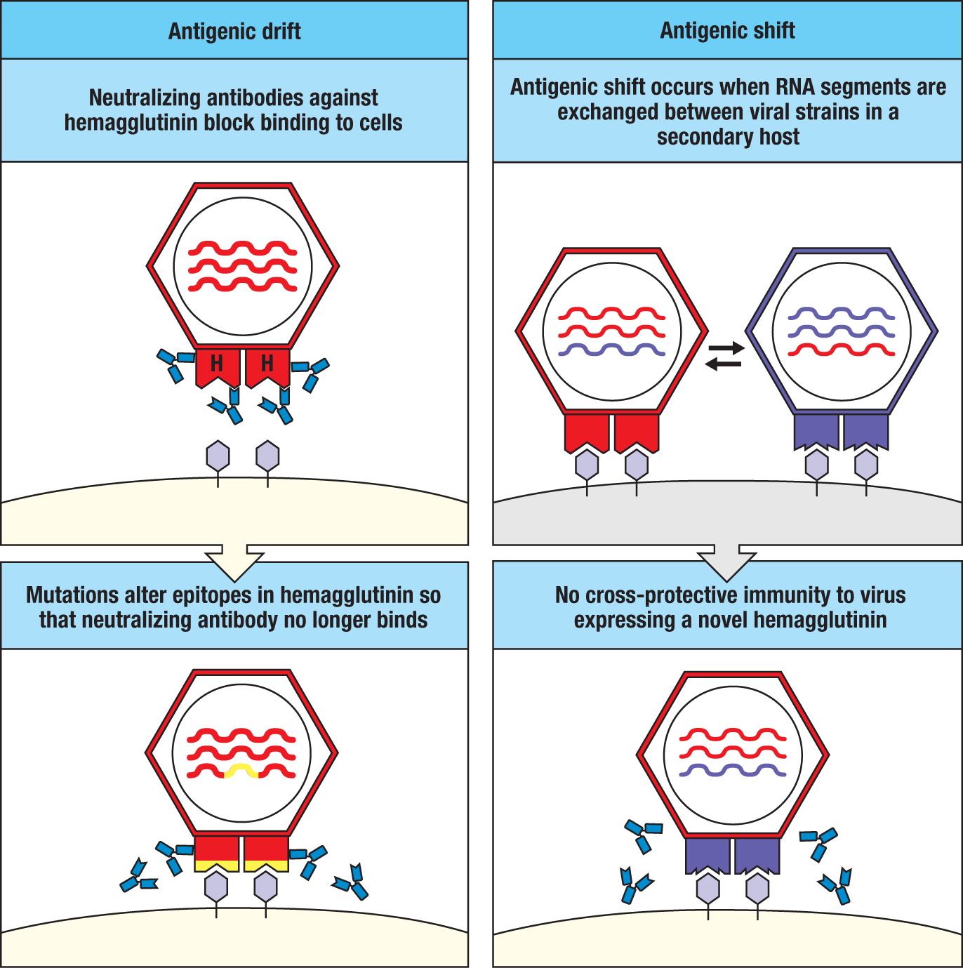
Individual strains of influenza are identified by type and the combination of hemagglutinin (H) and neuraminidase (N) variants. There are three major types of influenza that infect humans (A, B, C), two of which, types A and B, are responsible for most seasonal human disease. In general, type B strains cause less virulent infections; type A strains are the only viruses known to cause flu pandemics. At least 18 type A hemagglutinin and 11 neuraminidase subtypes have been identified to date, resulting in more than a hundred influenza A subtypes, which are identified by their combination of H and N usage [for example, influenza A (H3N2)]. Seasonal influenza viruses cause 3–5 million serious infections and up to a half million or more deaths worldwide each year. The outbreaks typically occur in the winter, when people are clustered indoors, facilitating spread from person to person. Influenza viruses also infect other species (for example, birds, pigs), which can be a source of further antigenic shifts due to zoonotic infections; that is, the transmission of a related infectious agent from other animals to humans. When this occurs, the existing immunity within humans can be ineffective, resulting in particularly lethal pandemics. A prime example is the swine flu pandemic of 1918, which was caused by an influenza A (H1NI) strain and caused an estimated 40 million deaths worldwide.
The attachment of influenza virus to the respiratory epithelium is mediated by the viral hemagglutinin surface molecule, which recognizes sialic acid residues on host-cell surface glycoproteins. Because of the broad expression of sialic acid on the cells of many tissues, the cellular tropism, or ability of the virus to infect specific host-cell types, is broad. Therefore, the route of delivery of influenza, typically in microdroplets inhaled via the airways, determines its localization to the respiratory tract more than its targeting to a particular cell type in the respiratory tract. And because the virus can infect cells outside the respiratory system, it can undergo systemic spread, accounting for the extrapulmonary symptoms that frequently accompany disease. The binding of hemagglutinin to host cells stimulates virion uptake into endocytic vesicles, in which the virus fuses with the endosomal membrane to gain entry to the host-cell cytoplasm, where viral replication is initiated. Viral neuraminidase does not contribute to the entry, replication, or budding of the virus, but appears to be important in the release of new virions from the infected cell surface and removal of sialic acid residues on viral surface proteins to prevent aggregation of virions. As for other viruses that invade via the respiratory tract, RECs are a primary target of both infection and propagation of influenza virus.
The detection of, and initial innate immune response against, influenza virus occurs in RECs, but also involves airway macrophages and DCs, including pDCs. Collectively, these innate immune sensor cells express a complete complement of pattern-recognition receptors (PRRs), including TLR-3, TLR-7, TLR-8, RIG-I, and NLRP3, each of which has been shown to contribute to the innate response to influenza. TLR-3 (dsRNA), TLR-7 (ssRNA), and, in humans, TLR-8 (ssRNA) detect viral RNA within endosomes, whereas RIG-I and NLRP3 detect virus within the cytosol of infected cells. This triggers the release of pro-inflammatory cytokines and type I and type III interferons (Fig. 12.46; and see Fig. 13.25). Engagement of receptors for these cytokines on epithelial cells and other cell types present in the lungs (for example, pDCs, γδ T cells, and NK cells) induces intracellular resistance mechanisms to viral replication and also amplifies the initial innate response and contributes to the release of chemokines that promote the recruitment of circulating inflammatory cells, such as pro-inflammatory monocytes and neutrophils. These signals also activate resident cells in the airways that can kill infected epithelial cells, such as NK cells and intraepithelial lymphocytes, as well as initiate the adaptive immune response. The uptake of dying RECs by cDC1 cells is important for the induction of an influenza-specific cytotoxic T lymphocyte (CTL) response, which is coordinated with a T helper type 1 response induced either by the same cDC1 cells or by cDC2 cells on which type I interferons have acted. This results in the recruitment of CTLs and TH1 cells to the respiratory mucosa where they act to eliminate infected cells and amplify the IFN response by releasing type II interferon (IFN-γ), which also enhances local antigen presentation of viral antigens. There is also production of IgG-producing plasmablasts that traffic to the respiratory mucosa where they mature and produce antibodies that target the elimination of free virus by phagocytes.
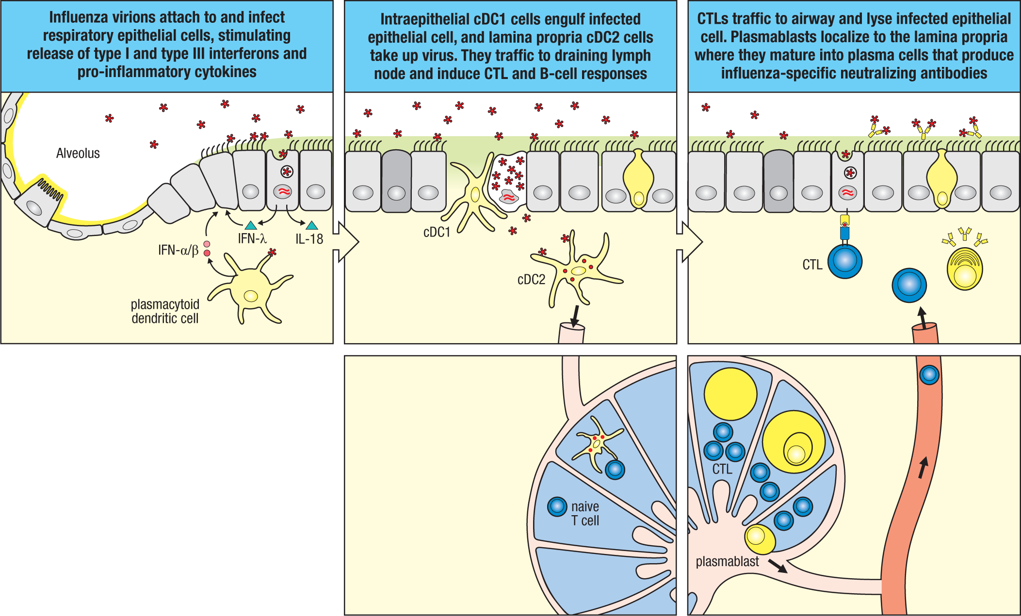
Antibody responses to the influenza virus surface glycoproteins, particularly those directed against hemagglutinin, also block viral attachment and are therefore neutralizing, conferring protection against cell-to-cell transmission of the influenza virus during active infection, as well as preventing viral reinfection after recovery from initial infection. Antigenic sites on the head domain of hemagglutinin that contribute to binding of the virus to the sialic acid residues on host cells are the dominant target of the neutralizing antibody response and are immunodominant compared to other viral B-cell epitopes; they block the interactions of hemagglutinin with host-cell sialic acid residues, thereby preventing viral attachment and entry into RECs. The importance of antibodies in protection against influenza infection was discovered in the 1930s, when it was found that the transfer of serum from infected individuals was protective. The relatively rapid mutation of the gene encoding hemagglutinin results in antigenic drift, which is a principal reason that influenza virus vaccines have to be updated yearly. This is also true for new pandemic viruses that result from antigenic shift, for which antigenically matched vaccines have to be produced. This is despite the fact that antibody responses against influenza virus are typically robust and long lasting.
In view of the importance of the antibody response to protection against influenza, it is evident that B-cell help mediated by TFH cells is central to a protective host response. Cytotoxic T cells (CTLs) are also required for optimal clearance of influenza virus. The CTL response targets antigenic components of the influenza viral core, or nucleocapsid, that include nucleoprotein (NP), membrane protein M1, and the viral polymerases, which, because of their conserved function to viral replication are less tolerant of antigenic drift. Thus, in contrast to the antibody response, the CTL response is more conserved across different influenza virus strains and subtypes. This may mitigate against the severity of disease caused by new viral strains in individuals who have been infected by influenza previously. Because current vaccines use killed virus, which provokes good antibody responses but not CTL responses, there is interest in developing better vaccines that also provide cross-protective immunity by eliciting robust CTL memory responses.
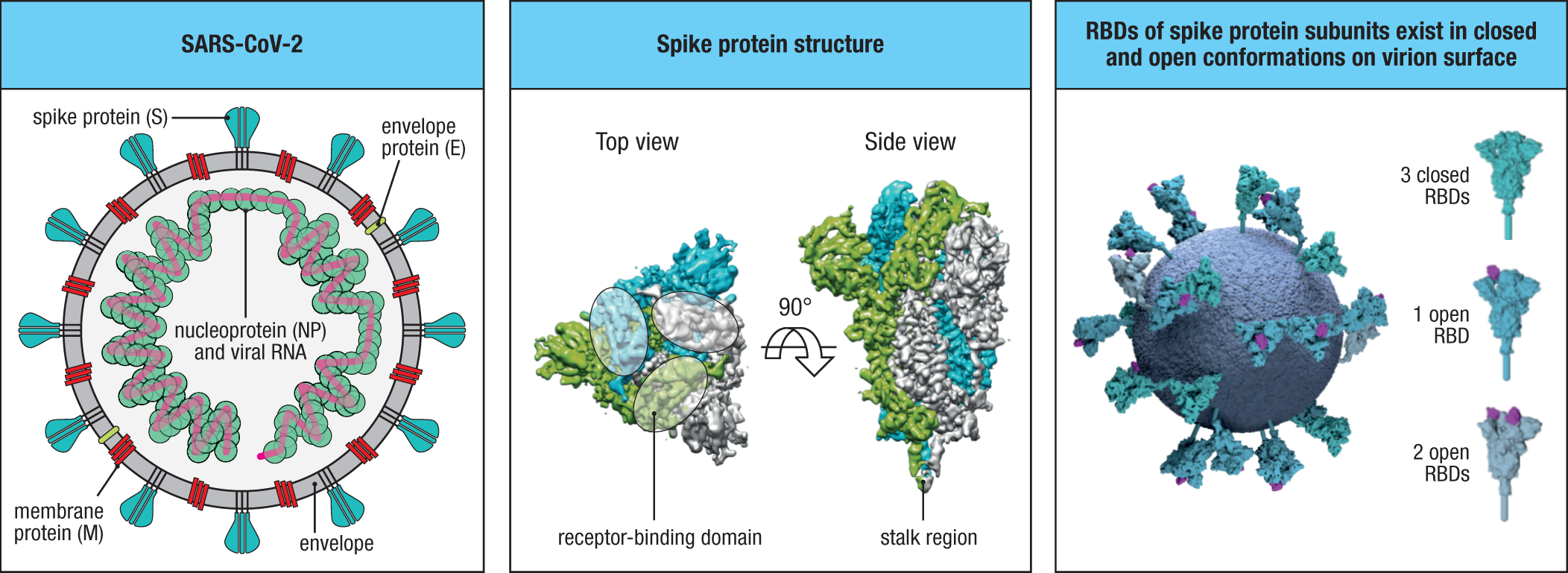
Another RNA virus family that can elicit mild or severe respiratory disease is the Coronaviridae, or coronaviruses, literally, ‘crown’ viruses, so named because of the appearance of their surface glycoproteins when viewed by electron microscopy (Fig. 12.47). Coronaviruses have the largest genomes of known RNA viruses (~25 to 32 kb; roughly twice the size of the influenza virus genome) and cause disease ranging from the common cold to life-threatening severe acute respiratory syndrome, or SARS. There are two major subfamilies of coronavirus, Torovirinae and Coronavirinae, the latter of which includes the Alphacoronavirus and Betacoronavirus genera that typically infect only mammals and are causes of human disease. Strains of both alphacoronaviruses and betacoronaviruses can cause the common cold. Less common strains of betacoronaviruses cause SARS, of which there have been three major outbreaks to date: SARS coronavirus (SARS-CoV; 2002–2004); Middle East respiratory syndrome coronavirus (MERS-CoV; 2012–2015); and most recently, SARS-CoV-2 (late 2019–present). The viral syndrome caused by SARS-CoV-2 has been referred to as coronavirus disease-2019, or COVID-19, reflecting its date of emergence.
Just as for influenza, the factors that determine the virulence of different strains of coronavirus are not well understood, although the emergence of zoonotic infections against which there is little preexisting immunity appears contributory. Although not proven, bats appear to be the likely source of each of the coronavirus strains that have caused the outbreaks responsible for SARS-CoV, MERS-CoV, and SARS-CoV-2. Whereas MERS-CoV originated in the Middle East and has remained largely endemic to the Arabian Peninsula where endemic infection of camels serves as a reservoir for transmission to humans, both SARS-CoV and SARS-CoV-2 originated in China and may have been amplified in, and been transmitted to, humans by intermediate hosts yet to be identified.
Although the scope of the initial SARS-CoV outbreak (~8000 cases and ~800 deaths worldwide) has now been dramatically eclipsed by SARS-CoV-2 (at least 175 million cases and over 3 million deaths worldwide to date), both of these pathogens, and all other strains of coronavirus, are primarily transmitted via respiratory spread. While MERS-CoV has diverged and uses the host ectoenzyme dipeptidyl peptidase 4 (DPP4) for virion attachment, SARS-CoV and SARS-CoV-2 use angiotensin-converting enzyme 2 (ACE2), an ectoenzyme that is preferentially expressed on the apical surface of airway epithelial cells. Attachment of these viruses to DPP4 or ACE2 is mediated by the coronavirus spike protein (S), a homotrimeric envelope protein that is the dominant protein expressed on the virus surface—the one responsible for the ‘crown’ motif for which the virus is named (see Fig. 12.47). The S protein has two fragments that are separated by a protease cleavage site: the S1 region contains the receptor-binding domain (RBD), and the S2 region mediates membrane fusion.
Binding of the S protein to the host cell stimulates its uptake into endosomes, wherein its cleavage by host proteases exposes the S2 fragment that causes viral membrane fusion with the endosomal membrane, triggering release of the viral genome into the host-cell cytosol. At least for SARS-CoV-2, the spike protein can bind ACE2 of multiple mammalian species, including bats, cats, civet cats, swine, ferrets, nonhuman primates, and possibly dogs, in addition to humans. Pangolins, which are scale-covered, ant-eating mammals that are a protected species that is traded illegally in Asia and elsewhere, have been implicated as an intermediate host in some studies. The distribution of the ACE2 viral receptor to epithelial cells of alveoli (in addition to those of the airways), like that of sialic acid residues targeted by influenza virus, may contribute to the severe respiratory syndromes that strains of each of these types of virus can cause by direct injury to the alveoli. In a further parallel with influenza virus, ACE2 is expressed by other epithelial cells of the body, including the epithelium of the intestines, which can be the primary site of infection if the virus is ingested rather than inhaled in respiratory droplets or a secondary site of involvement after spread from the lungs. ACE2 is also expressed by endothelial cells and may mediate infection of blood vessels.
In contrast to influenza, the coronavirus genome is not segmented, but rather is a single continuous positive-sense RNA strand (see Fig. 12.44), such that the virus may undergo antigenic drift but not antigenic shift. There is insufficient information at present as to the rate of antigenic drift in SARS-CoV-2. However, early evidence indicates that the virus has mutated several times during the current pandemic, producing several substrains that are more infectious than the parent strain. Indeed, it has been found that much of the antigenic drift observed in coronaviruses is focused on the RBD of the S1 fragment of the spike protein, which mediates attachment to the ACE2 host-cell receptor. Because the RBD of each monomer of the spike protein is an extended loop that projects from its surface and is not critical for its structural integrity, mutations in RBDs can occur rapidly without threat to virion integrity and function. In the case of SARS-CoV-2, the affinity of the RBD for ACE2 is substantially higher than that of SARS-CoV, suggesting a mechanism for its more efficient spread and rapid emergence as a pandemic. Thus, somewhat similar to influenza, the virus may be able to alter the antigenic targets of the host antibody response to evade blockade of virion uptake. In the case of SARS-CoV-2 this may be particularly selected for in bats, which represent ~20% of all mammalian species and live in dense populations that facilitate rapid selection of new strain variants that can sustain propagation within the host community.
Although there is limited information at present on details of the host immune response to SARS-CoV-2, there is reason to believe that the host response shares many features with the response to influenza, in which both neutralizing antibodies and CTLs play central roles (see Fig. 12.46). Neutralizing antibodies against SARS-CoV and MERS-CoV have been characterized from survivors of these infections, and some cross-protective antibodies against SARS-CoV-2 have been identified in individuals previously infected by SARS-CoV. Indeed, there is some indication that prior exposure to less pathogenic strains of betacoronaviruses that cause the common cold and also use ACE2 as a host receptor can afford at least partial protection against SARS-CoV-2. Although some reports have suggested that protective antibody responses to SARS-CoV-2 may, for unknown reasons, be short-lived, experience from SARS-CoV and MERS-CoV indicates that at least some neutralizing antibodies persist for many years, raising the possibility that the vaccines that have recently been implemented for SARS-CoV-2 may provide long-lived protection. Especially in view of the global health crisis caused by SARS-CoV-2, the potential for vaccines that induce neutralizing antibody responses against multiple variants of the RBD are an area of active investigation, although greater conservation of non-RBD components of the virus that are targeted by CTLs may also hold promise (see Chapter 16).
12-24 Cutaneous immunity.
Our focus so far has been on the immune responses at mucosal surfaces of the intestines and respiratory tract. Although not a mucosal tissue, the skin is nevertheless a highly important barrier. Unlike the intestinal and respiratory tracts, the skin develops from ectoderm rather than endoderm, and though smaller in total surface area than these mucosal tissues, it is the largest organ of the body by weight. Although the skin contributes to the generation of essential metabolites, including active vitamin D, and plays a major role in regulating body temperature and sensing the external environment, its main function is protective. Thus, in contrast to the GI and respiratory tracts, where physiological functions necessitate a thin epithelium that facilitates exchange with the external environment, the skin forms a tight but not impermeable seal that is ideal for providing protection against physical damage (for example, lacerations and radiative and chemical threats), microbial incursion, and water loss. However, despite structural differences between skin and other barrier tissues, many of the immune-cell populations present in skin are similar to those in other barrier tissues as are the host-protective strategies, although there are some unique features of skin immunity that we will highlight here.
The skin consists of the outer epidermis and the inner dermis (Fig. 12.48; and see Fig. 12.2), which are in many ways analogous in function to the epithelium and lamina propria, respectively, of mucosal tissues. However, unlike the single-cell epithelium of the intestines or the pseudostratified epithelium of the respiratory tract, the epithelium of the skin lacks goblet cells and, therefore, mucus. Similarly, the skin lacks standing inductive lymphoid tissues, such as the Peyer’s patches and ILFs of the intestinal tract, and given that SIgA is a specialized adaptation of mucosal tissues, it is absent in skin. Rather, the first line of defense of the skin is the continually proliferating keratinocytes—so named because of their high content of keratin, a highly cross-linked fibrous protein that is a major structural component of epidermal cells as well as hair and nails. Keratinocytes differentiate as they mature toward the skin surface to create an outer, densely keratinized and cross-linked matrix of anucleated, dead squamous cells, which are continually sloughed and replaced. This outermost layer, or stratum, of the epidermis is the stratum corneum, which is composed of dead keratinocytes sealed by extracellular lipids, which prevents fluid loss and acts as a relatively impenetrable barrier. The stratum corneum is replaced about every 10 days. The other layers of the skin include (from outside, in) the stratum granulosum and stratum spinosum, which are populated by immune cells, and the stratum basale, which is attached to the basement membrane and acts as a reservoir for renewal of the superficial layers of keratinocytes. Another epithelial cell type is the melanocyte, which produces pigment granules that are transferred to developing keratinocytes; melanocyte pigment absorbs UV irradiation to protect skin cells from solar damage. The underlying dermal tissue consists of fibroblasts within a connective matrix that supports hair follicles, some glands, and nerve endings and has a rich supply of lymphatic and blood vessels. This vascularization enables the extravasation of circulating immune cells mostly to the dermis but also to the epidermis.
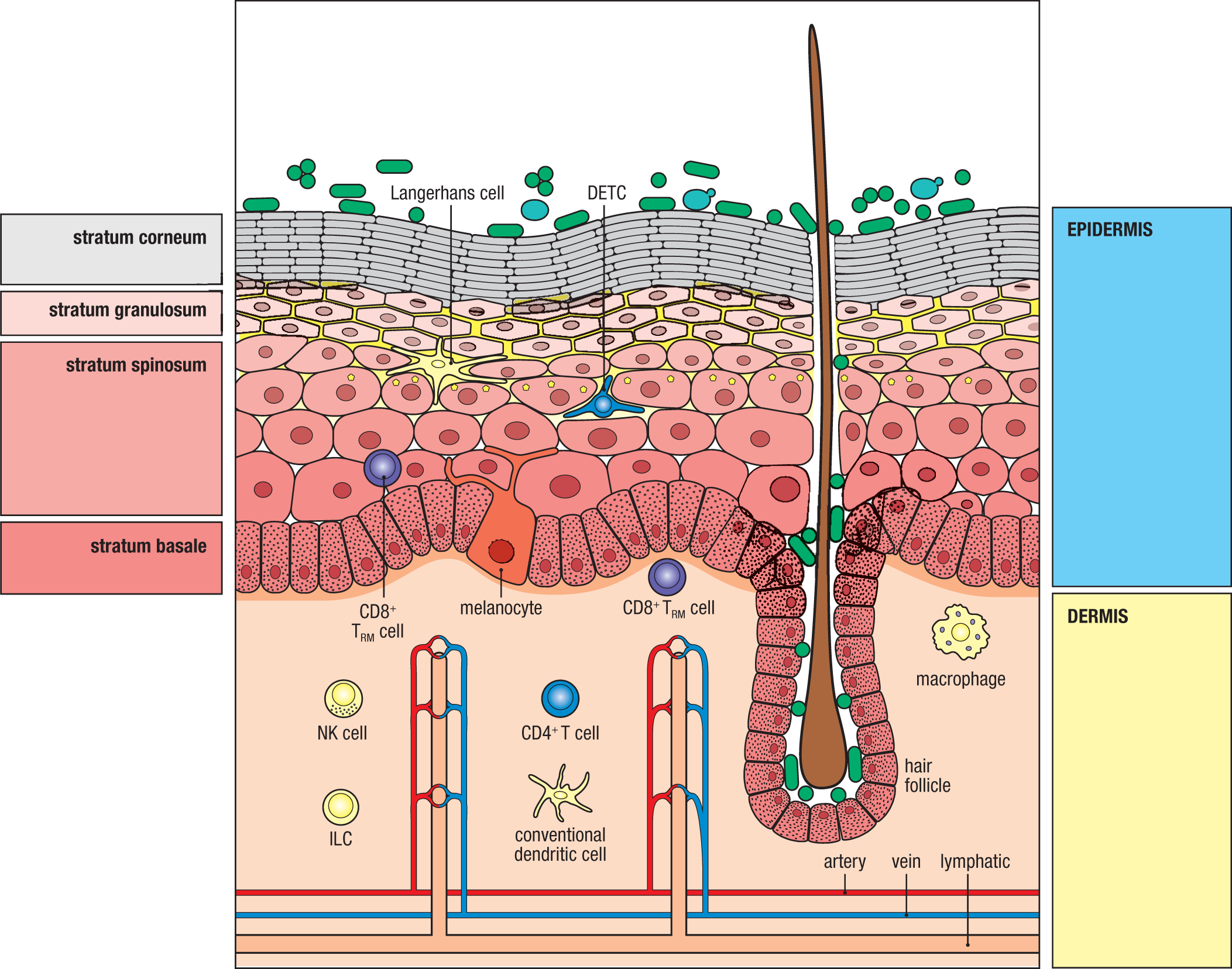
Keratinocytes account for ~90% of the cells in the epidermis, although this percentage varies from site to site, depending on skin thickness. Included among the remaining cells are epidermal immune cells, primarily Langerhans cells and T cells. In mice, intraepidermal T cells are dominated by a γδ T-cell subset termed dendritic epidermal T cells (DETCs), which are unconventional T cells with a restricted antigenic specificity (Vγ3+Vδ1+). They populate the skin from the thymus before birth (see Section 8-XX), and they remain there as long-lived resident T cells. DETC precursors up-regulate expression of CCR10 before exiting the thymus to home to the epidermis in response to CCL27 expressed by keratinocytes. Functionally, DETCs are thought to regulate epidermal repair during wound healing, and they may act as innate immune sensor cells. There is no direct equivalent of DETCs in human skin, although the epidermis of humans does contain γδ T cells that appear to be functionally comparable to DETCs in the mouse. Both species contain αβ T cells in the epidermis, which, akin to the shift from unconventional to conventional TRM cells in the intestinal epithelium with age (see Section 12-5), tend to replace the γδ T cells throughout life, depending on the history of antigenic exposure. This has led to speculation that skin γδ cells present at birth represent a primitive adaptive immune system that is replaced by αβ TRM cells contingent on an individual’s exposure to skin pathogens.
The dermis is enriched in collagen and elastic fibers, which provide the structural framework for vascular and lymphatic vessels that deliver circulating immune cells to and from the skin. Immune cells that reside in the dermis under steady-state conditions are similar to those in the lamina propria of mucosal tissues: cDCs, tissue-resident macrophages, ILCs, γδ T cells, αβ T cells, B cells, macrophages, and natural killer (NK) cells. There are also small numbers of innate effector cells, such as mast cells. Dermal γδ T cells are considerably more diverse than their epidermal counterparts, and although their activating ligands are incompletely characterized, they appear to be mainly restricted by host stress proteins, such as MICA and NKG2B. Dermal αβ T cells are largely memory cells that have homed to the skin after activation and differentiation in draining lymph nodes. Although details of the signals that imprint skin-trafficking features are not completely understood, some evidence suggests that vitamin D, the active form of which is generated locally in the skin in response to UVB radiation, may imprint T cells to traffic to skin, as in the programming of gut T cells by retinoic acid derived from vitamin A. In any case, up-regulation of E-selectin ligands, such as cutaneous lymphocyte antigen (CLA; see Section 11-14), and CXCR3, CCR4, CCR8, and CCR10 are contributory.
A unique feature of immunity induced in the skin is the contribution of Langerhans cells (LCs). While cDCs from dermis elicit the activation of CD4 and CD8 T cells in draining lymph nodes, cDCs are not typically positioned in the epidermis, where LCs constitute the largest single population of antigen-presenting cells in the entire skin. Like tissue-resident macrophages in mucosal tissues, LCs are derived from monocytic, not dendritic, cell precursors and are a long-lived, self-renewing population that populates the epidermis before birth. Langerhans cells are particularly enriched in the stratum spinosum of the epidermis. Like resident macrophages at mucosal sites, LCs constantly extend and retract dendrites between keratinocytes while maintaining barrier integrity by establishing tight junctions with adjacent keratinocytes. However, unlike mucosa-resident macrophages, LCs are migratory upon activation by inflammatory signals received from keratinocytes (for example, IL-1β and TNF-α) and traffic to draining lymph nodes where they can activate naive T cells. LCs may directly present antigen, but they can also transfer antigens transported from the epidermis to resident lymphoid DCs.
Although there is evidence supporting a role for LCs in the initiation of a variety of adaptive immune responses (for example, type 1 and type 3), LCs have limited ability to phagocytose bacteria and are particularly important in antiviral immunity in the skin. Nevertheless, cutaneous infection models with the type 3 pathogens Candida albicans or Staphylococcus aureus have demonstrated clear roles for LCs in priming protective CD4 T-cell responses. Moreover, activation of LCs in response to ionizing radiation has been shown to prime skin-tropic Treg cells. In humans, but not mice, LCs also express high levels of the invariant MHC-like molecule CD1a and appear particularly adapted to present lipid antigens to T cells. Thus, LCs appear especially adapted to activate immunity to topical sensitizers that do not reach the dermis, such as the poison ivy lipid urushiol.
Like other barrier tissues, the skin harbors a resident microbiota that is established soon after birth and plays an important role in shaping local immunity in the skin. Across the ~2 m2 of the skin surface of humans resides a diversity of bacteria, fungi, and viruses (see Fig. 12.5 and Fig. 12.48). Like the commensal microbiota in other sites, the skin microbiota provides colonization resistance to limit pathogen invasion and promote homeostasis. The latter includes the induction of populations of FoxP3+ Treg cells that maintain local tolerance. As in the GI and respiratory tracts, this reflects the local production and activation of TGF-β by keratinocytes. Thus, Treg cells specific for components of the skin microbiota arise early in neonatal life in response to skin colonization. Cutaneous Treg cells reside principally in the dermis, where they constitute one of the highest frequencies of Treg cells within the body. In addition to their role in accommodating to the skin microbiota, cutaneous Treg cells also appear to play an important role in non-immunological functions in skin. They have been shown to regulate cutaneous wound healing and the function of stem cells in hair follicles. Accordingly, soon after a skin wound occurs, skin Treg cells accumulate in large numbers at the affected site, where they limit the effector T-cell response and promote epidermal regeneration via production of ligands of the epidermal growth factor receptor pathway. A large fraction of Treg cells in skin is found near hair follicles, which serve as a major habitat for skin-resident microbes. Treg cell responses in that niche may contribute to the development of hair follicles soon after birth and may promote the resident commensal microbes to thwart occupation of the niche by pathogens.
Shifts in composition of the skin microbiota, or dysbiosis, have been documented in many skin inflammatory disorders and are likely cofactors in the etiology of many immune skin disorders. For example, atopic dermatitis, a chronic inflammatory disorder of the skin, has long been associated with colonization of the skin by Staphylococcus aureus. Similarly, enrichment in Streptococcus spp. was observed in skin lesions of psoriasis. Defined microorganisms or microbial communities could potentially contribute to the initiation or amplification of skin pathologies through several mechanisms. Local expansion of defined members of the skin microbiota with enhanced inflammatory potential could alter tissue homeostasis. For example, S. aureus isolates that release δ-toxin trigger local allergic responses as the δ-toxin induces degranulation of dermal mast cells, which in turn promotes both innate and adaptive type 2 immune responses. S. aureus α-toxin can also promote IL-17 production from human CD4 T cells, a property that could be explained by the ability of this toxin to induce IL-1β production by monocytes. The skin milieu and defined factors such as vitamin B12 can have a profound effect on the metabolism of the skin microbiota and thus the prevention or promotion of colonization by pathogens. Additional studies will be needed to better understand the role of shifts in the skin microbiota in promoting immune-mediated disease in skin.
12-25 The sensory nervous system communicates with the immune system to enhance defense against infections in the skin.
In this section, we will describe a recently recognized mechanism that enhances the immune barrier of the skin. This involves neuroimmune interactions between the peripheral nervous system and dendritic cells in the epidermis that act to activate immune defenses not only at the direct site of infection but also at neighboring skin regions that are innervated by these neurons (Fig. 12.49). This mechanism involves c-fiber sensory neurons, which are unmyelinated small-diameter neurons that carry sensory information, often in the form of pain, to indicate damage. Such neurons are called nociceptive neurons (from the Latin nocere, ‘to do harm’). Among these neurons is a subtype expressing the transient receptor potential cation channel subfamily V member 1 (TRPV1), which is a receptor for capsaicin, the molecule in chili peppers that induces its burning sensation. Further, some TRPV1+ neurons also express the sodium ion channel NaV1.8, and it is this subset that seems to be important in this neuroimmune pathway.
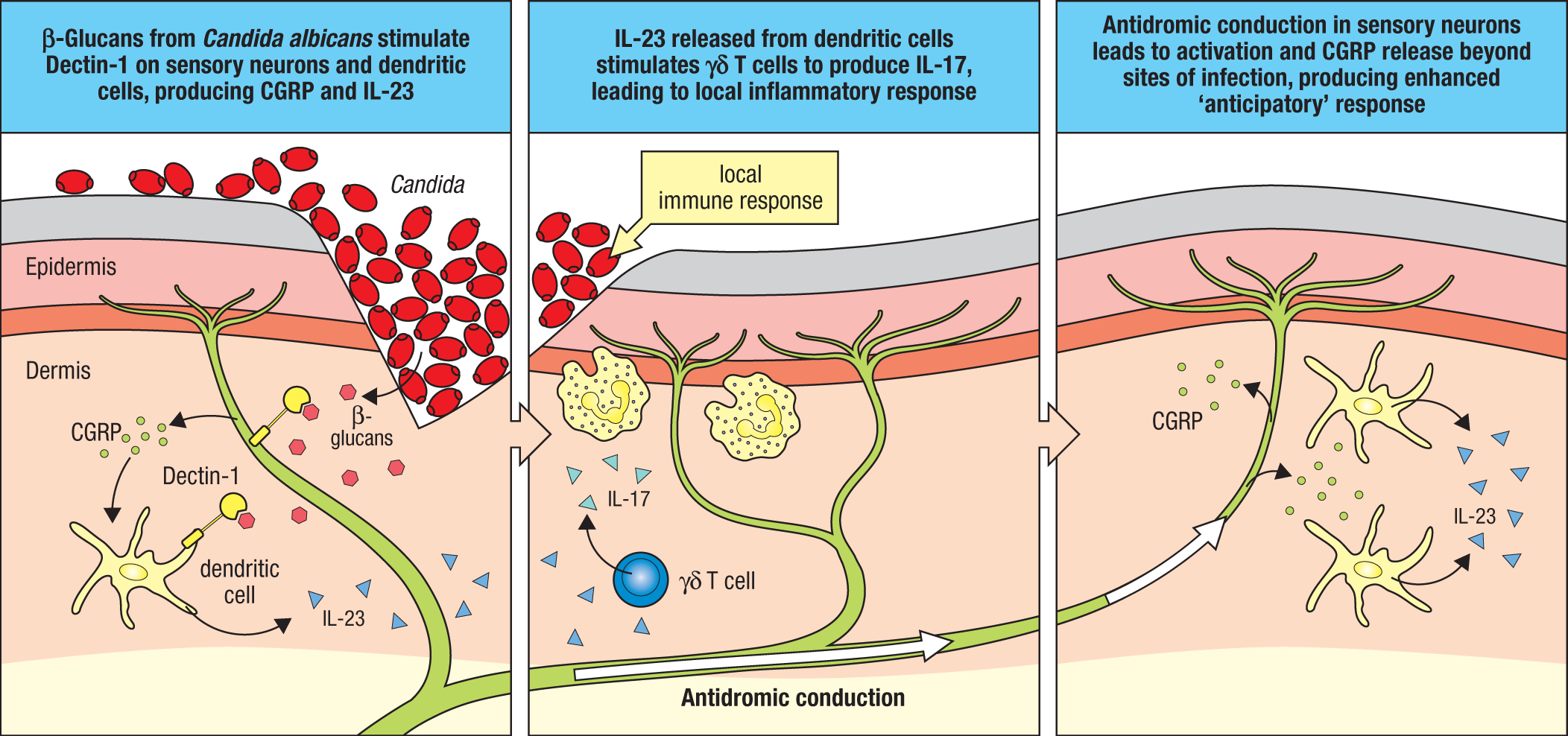
The role of the nervous system in cutaneous defense against pathogens was recognized in an experimental system of skin inflammation induced by cutaneous administration of imiquimod (IMQ), a small compound that activates TLR-7. Normally, IMQ administration to skin produces an inflammatory response that is dependent on the production of IL-23 by dermal dendritic cells. This IL-23 can activate the production of IL-17 from local T cells, predominantly dermal γδ T cells, and leads to measurable skin thickening in regions such as the ear. As we introduced in Section 9-14, extracellular bacteria and fungi, common microbes encountered by the skin, typically induce type 3 responses, in which IL-17 family cytokines help recruit neutrophils and other cells that are effective in eliminating these types of organisms. When sensory neurons expressing TRPV1 were eliminated prior to treatment with IMQ by the administration of resiniferatoxin (RTX), a toxin and extremely strong agonist of TRPV1, it was discovered that IMQ no longer induced inflammation in treated skin. However, the administration of IL-23 to skin treated with IMQ restored its ability to induce inflammation. Further, the majority of dermal dendritic cells were located close to TRPV1+ sensory fibers. These findings suggested that TRPV1 sensory neurons somehow regulated the ability of dermal dendritic cells to produce IL-23.
These findings were later extended to the setting of skin infection by the environmental fungus Candida albicans, which typically induces a type 3 response involving IL-23 and IL-17 production. Recognition of C. albicans is mediated, at least in part, by Dectin-1, a C-type lectin family member (CLEC7A) that recognizes polysaccharides known as β-glucans, which are derived from the cell walls of bacteria and fungi, and even plants. In addition to its expression by dendritic cells, Dectin-1 also appears to be expressed by dermal keratinocytes and TRPV1+ sensory neurons (see Fig. 12.49), suggesting that each of these cells might recognize β-glucans released by the cell walls of C. albicans. Indeed, it was found that infection by C. albicans activated neurons to release calcitonin gene–related peptide (CGRP), which acts to facilitate IL-23 production by dermal dendritic cells by an unknown mechanism. A second component of neuronal sensing of C. albicans seems to involve the purinoreceptor P2X3, which recognizes extracellular ATP. ATP can be released from keratinocytes activated by Dectin-1 signaling in response to β-glucans made by C. albicans. Additionally, some evidence suggests that some strains of Candida may directly release significant amounts of ATP. In either case, the combination of signaling by Dectin-1 and P2X3 leads to release of CGRP, promoting enhanced IL-23 from dermal dendritic cells, thus enhancing type 3 immunity in the vicinity of these cells.
Besides promoting IL-23 production by dendritic cells locally at the site of infection, the activation of TRPV1+ neurons by C. albicans also has an important secondary benefit. Because sensory neurons extend branches into an entire region of skin, the activation of one branch of a sensory neuron can lead to active nerve conduction through its entire sensory field, which can enhance immune activation to areas of skin outside the initial site of infection. This effect was discovered in clever experiments that used optogenetics in which a channelrhodopsin, a light receptor activated by blue light, was selectively expressed in TRPV1+ neurons of mice. Activation of neurons in a small region by shining a small focus of light onto a small patch of skin was observed to result in enhanced responses to C. albicans not only in the region activated by light, but also in adjacent regions of uninfected skin. This expansion of the area in which immunity is heightened occurs by conduction of nerve potential throughout the area served by the sensory neurons. This form of conduction is called antidromic nerve conduction, meaning that a nerve signal passes from the peripheral site of stimulation, first toward a neuronal cell body, but then into other local regions at the sites of axonal branching (see Fig. 12.49). In this way, the communication between sensory neurons and immune cells can provide an anticipatory response at sites not yet infected. Although this is still an active area of research, the anticipatory response is likely due to the release of CGRP by neurons induced by this antidromic conduction at distant sites independently of Dectin-1 signaling.
Summary.
Among barrier tissues, the respiratory tract is unique in that its major surface geography—the lower respiratory tract—is normally sterile; it is devoid of a commensal microbiota, reflecting the requirement for close contact between the airspaces and the blood to accomplish its major physiological function of gas exchange. Much of the protection from pathogens—and from commensals inhaled from the upper respiratory tract—lies in the unusual anatomy of the respiratory tract, which is highly efficient at trapping inhaled particulates of varying sizes and transporting them out of the airspaces via the mucociliary escalator. As in the gut, a tolerogenic tone is strongly favored in the respiratory tract to prevent the dire consequences attendant to overexuberant inflammation in the airways and airspaces, compromise of which can be rapidly fatal. These consequences, coupled with the relative ease of airborne spread of respiratory pathogens, accounts for the fact that respiratory infections are the greatest cause of death among all mucosal infections. This is painfully evident in the COVID-19 pandemic that has recently gripped the world and has emphatically exposed the need for better vaccine strategies that prevent, rather than treat, airborne infections.
The skin differs from other barrier sites in the keratinized epithelium of its epidermis, reflecting its exposure to the greatest physical injury among barrier tissues and its role in minimizing water loss. The epidermis also represents a formidable barrier to pathogen entry, unless it is physically damaged (for example, by cuts or burns) or penetrated by other organisms (for example, insects that transmit microbial pathogens). Unlike many mucosal tissues, such as the intestines, which harbor lymphoid tissues that are in direct contact with the epithelium and its local microbiota, the skin does not normally contain local lymphoid tissues; adaptive immunity in the skin is initiated in draining lymph nodes. Unique to the skin is its population by a specialized resident macrophage, the Langerhans cell, which appears to be important in delivering antigens acquired in the epidermis to lymph nodes. Intraepidermal lymphoid cells, including conventional TRM cells and unconventional γδ T cells, likely play important roles in immune defense, but the unconventional γδ T cells, including DETCs in mice, also have an important role in injury repair—representing an important immune function in supporting barrier function in a tissue so exposed to physical injury. The skin is also among the most important sensory organs of the body, literally the main interface with the outside world. Recent discoveries highlight the remarkable interplay of the neural and immune networks in skin that appear to enhance local immunity.
Summary to Chapter 12.
The barrier immune system is an extensive and diverse apparatus that has a crucial role in health, not just by protecting physiologically vital organs but also by helping to regulate the immunological tone to preserve immunological tolerance to nonthreatening antigens. Because of their extensive exposure to the external environment, the skin and mucosal surfaces of the body are at greatest risk of infection and possess unique, site-specific characteristics that distinguish their immune mechanisms from nonbarrier tissues. These include, but are not limited to: the immediate juxtaposition of mucosal epithelium and lymphoid tissue; immune regulation of mucus production that characterizes mucosal tissues; specialized antigen uptake mechanisms; a standing population of activated/memory lymphocytes and innate lymphoid cells (ILCs) even in the absence of infection; a diverse population of conventional and unconventional T cells resident within epithelia; the production of dimeric secretory IgA as the predominant antibody in mucosal tissues; and the down-regulation of immune responses to innocuous antigens such as food antigens and commensal microorganisms. Many of the most remarkable advances in immunobiology are being made in studies of barrier immunity, where there is a growing understanding both of the complexity of the role of innate and adaptive immune cells in supporting the mutualism between the host and the commensal microbiota and of the import of neuroimmune interactions that likely reflect the central role of barrier tissues in sensing, responding to, and protecting the host from the outside world.
Glossary
- club cells
- Columnar cells that are decorated with microvilli and are found in the small airways of the lungs, where they protect the bronchiolar epithelium through a variety of secretory molecules.
- mucociliary escalator
- The system of cilia-transported mucus by which the airways of the respiratory system trap and transport particles toward the pharynx and out of the lungs.
- alveolar macrophages
- Also referred to as ‘dust cells’; resident macrophages within the pulmonary alveoli that are responsible for clearing inspired microbes and other particles.
- antigenic drift
- The process by which influenza virus varies genetically in minor ways from year to year. Point mutations in viral genes cause small differences in the structure of the viral surface antigens.
- hemagglutinin
- Substances that can cause hemagglutination, such as human antibodies that recognize the ABO blood group antigens on red blood cells, or the influenza virus hemagglutinin (H), a glycoprotein that functions in viral fusion with endosome membranes.
- neuraminidase
- An influenza virus protein that cleaves sialic acid from host cells to allow viral detachment; a common antigenic determinant and a target of antiviral neuraminidase inhibitors.
- antigenic shift
- A radical change in the surface antigens of influenza virus, caused by reassortment of its segmented genome with that of another influenza virus, often from an animal.
- zoonotic
- Describes a disease of animals that can be transmitted to humans.
- cellular tropism
- The host-cell types that are infected by a particular pathogen.
- severe acute respiratory syndrome (SARS)
- A coronavirus-caused respiratory illness of zoonotic origin that was responsible for a global disease outbreak in 2003.
- severe acute respiratory syndrome (SARS)
- A coronavirus-caused respiratory illness of zoonotic origin that was responsible for a global disease outbreak in 2003.
- coronavirus disease-2019 (COVID-19)
- A respiratory illness caused by SARS-CoV-2, likely of zoonotic origin and responsible for the COVID-19 pandemic.
- angiotensin-converting enzyme 2 (ACE2)
- An ectoenzyme expressed on respiratory epithelial cells and other cells throughout the body that normally cleaves peptides, including the blood pressure regulator, angiotensin II. ACE2 is the host-cell entry receptor for SARS-CoV-2, bound by the receptor-binding domain (RBD) of the SARS-CoV-2 spike protein.
- Langerhans cells
- A unique immune lineage with features of dendritic cells and monocyte-derived macrophages that resides in the epidermis but can migrate upon activation to lymph nodes and present antigens to T cells.
- Dectin-1
- A phagocytic receptor on neutrophils and macrophages that recognizes β-(1,3)–linked glucans, which are common components of fungal cell walls.