1.2 Microbes Shape Human History

Today our knowledge of microbes is enormous, and it keeps growing. We know that microbes produce half the oxygen in our atmosphere, and that overall, microbial ecology is fundamental to our biosphere, mediating much of the human-caused change in global climate.

Yet throughout most of human history, we were unaware of how microbes shaped our culture. Yeasts and bacteria made foods such as bread and cheese (Fig. 1.7A), as well as alcoholic beverages (discussed in Chapter 16). “Rock-eating” bacteria, known as “lithotrophs,” leached copper and other metals from ores exposed by mining, enabling ancient human miners to obtain these metals. The lithotrophic oxidation of minerals for energy generates strong acids, which accelerate breakdown of the ore. Today, about 20% of the world’s copper, as well as some uranium and zinc, is produced by bacterial leaching. Unfortunately, microbial acidification also consumes the stone of ancient monuments (Fig. 1.7B)—a process intensified by airborne acidic pollution.

A
Two photographs provide examples of production and destruction by microbes. A photograph of the ripening step of the cheese production process.
More information

Two photographs provide examples of production and destruction by microbes. The first image is a photograph of the ripening step of the cheese production process. The second image is a photograph of a statue with decay.

A photograph of the ripening step of the cheese production process. Four individuals are seen organizing blocks of cheese in a long-term storage space with brick walls.

B
A close up photograph of decay on a human statue.
More information

A close up photograph of decay on a human statue. The surface of the statue is mottled with dark gray blotches. A small patch of bright orange growth is also visible.

FIGURE 1.7 Production and destruction by microbes. A. Roquefort cheeses ripening in France. B. Statue at the cathedral of Cologne, Germany, decaying from the action of lithotrophic microbes. The process is accelerated by acid rain. BETTMANN/GETTY IMAGESJOHNER IMAGES/GETTY IMAGES

How did people find out about microbes? Table 1.2 lists the discoveries throughout history that have brought us to our current level of knowledge. Microscopists in the seventeenth and eighteenth centuries formulated key concepts about microbes and their existence, including their means of reproduction and death. In the nineteenth century, the “golden age” of microbiology, scientists established the fundamental principles of disease pathology and microbial ecology that are still in use today. This period laid the foundation for modern biology, in which genetics and molecular biology provide powerful tools for scientists to manipulate microorganisms for medicine, research, and industry.

TABLE 1.2

Microbes and Human History

Date

Microbial discovery

Discoverer(s)

Microbes impact human culture without detection

10,000 BCE

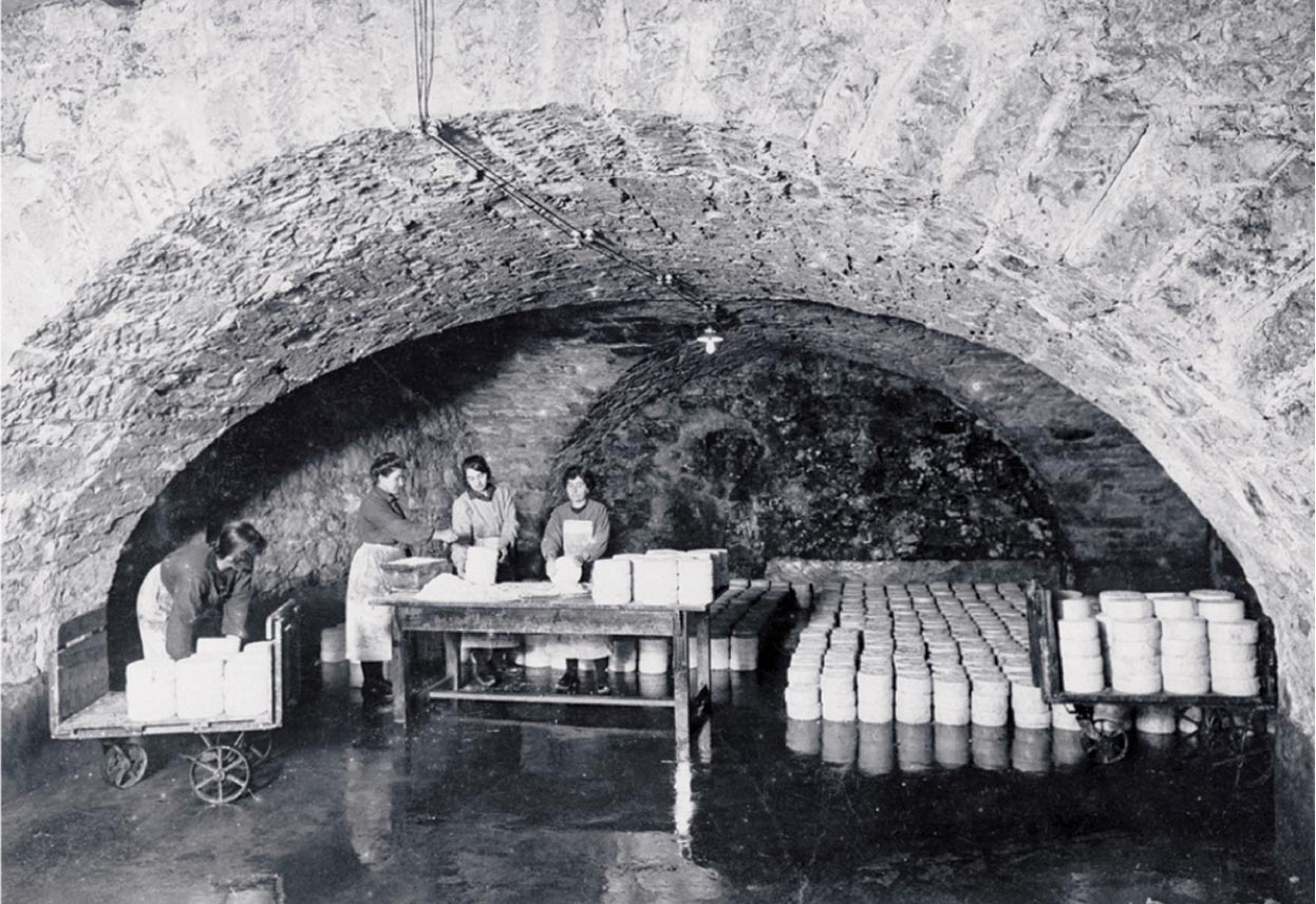
Food and drink are produced by microbial fermentation.

Egyptians, Chinese, and others

1500 BCE

Tuberculosis, polio, leprosy, and smallpox are evident in mummies and tomb art.

Egyptians

50 BCE

Copper is recovered from mine water acidified by sulfur-oxidizing bacteria.

Roman metal workers under Julius Caesar

1362 CE

Plague transmission is observed.

Ibn al-Khatib (Granada)

1546 CE

Syphilis and other diseases are seen to be contagious.

Girolamo Fracastoro (Padua)

Early microscopy and the origin of microbes
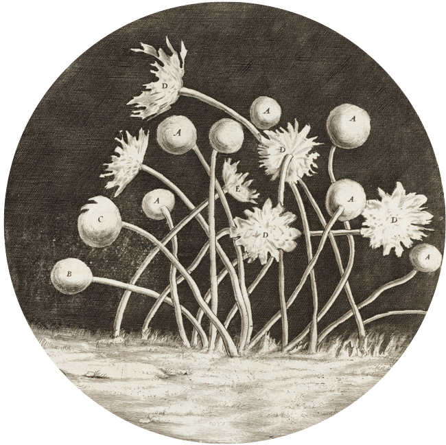
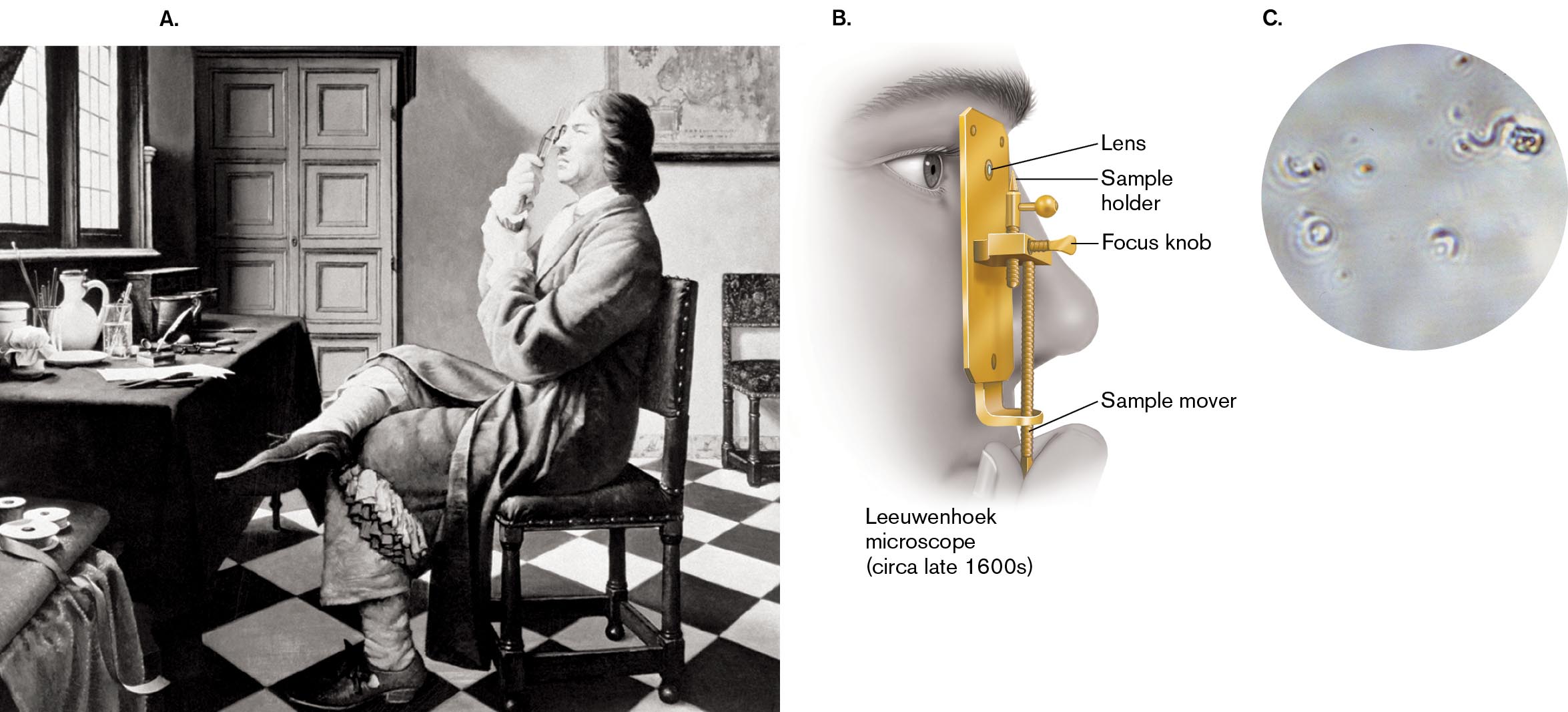
1676

Microbes are observed under a microscope.

Antonie van Leeuwenhoek (Netherlands)

1688

Spontaneous generation is disproved for maggots.

Francesco Redi (Italy)

1717

Smallpox is prevented by inoculation of pox material, a form of immunization.

Turkish women taught Lady Mary Montagu, who brought the practice to England
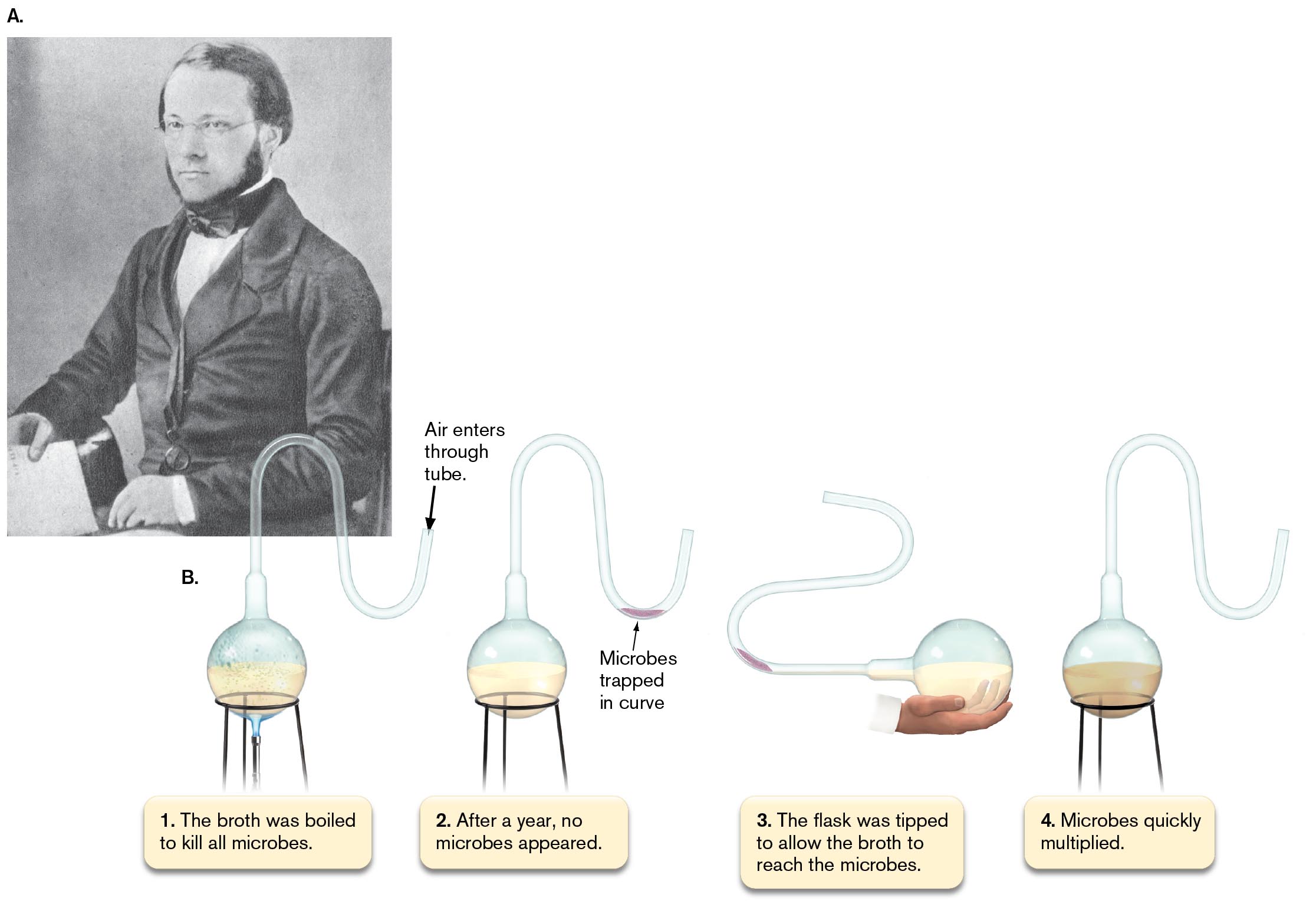
1765

Microbe growth in organic material is prevented by boiling in a sealed flask.

Lazzaro Spallanzani (Padua)

1798

Cowpox vaccination prevents smallpox.

Edward Jenner (England)

1835

Fungus causes disease in silkworms (first pathogen to be demonstrated in animals).

Agostino Bassi de Lodi (Italy)

1847

Chlorine as antiseptic wash for doctors’ hands decreases pathogens.

Ignaz Semmelweis (Hungary)

1881

Bacterial spores survive boiling but are killed by cyclic boiling and cooling.

John Tyndall (Ireland)

“Golden age” of microbiology: principles and methods established
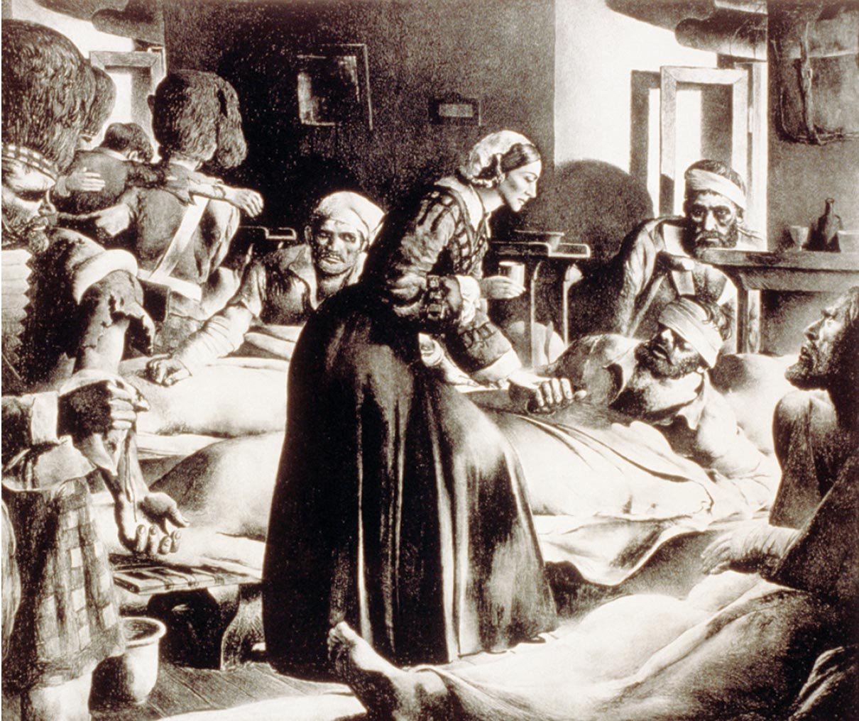
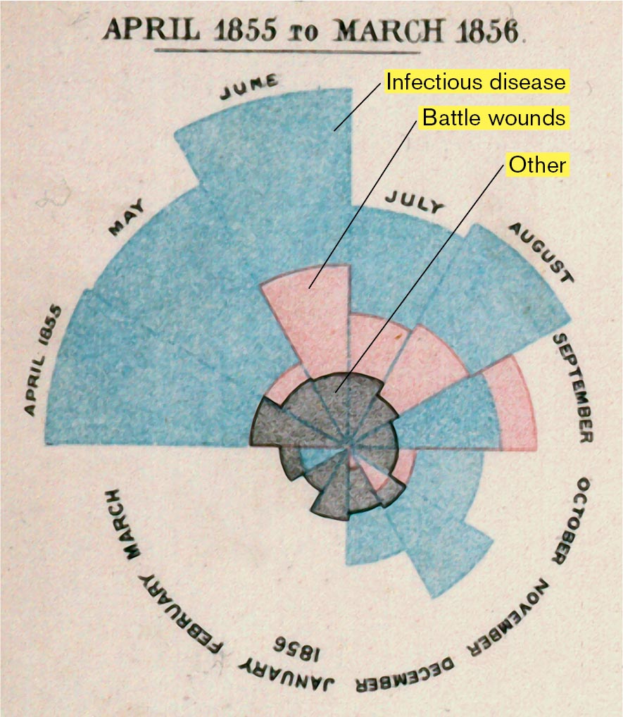
1855

Sanitation shows statistical correlation with mortality (Crimean War).

Florence Nightingale (England)

1857

Microbial fermentation produces lactic acid or alcohol.

Louis Pasteur (France)

1864

Microbes fail to appear spontaneously, even in the presence of oxygen.

Louis Pasteur (France)

1866

Microbes are defined as a class distinct from animals and plants.

Ernst Haeckel (Germany)

1867

Antisepsis during surgery prevents patient death.

Joseph Lister (England)

1881

First artificial vaccine is developed (against anthrax).

Louis Pasteur (France)

1882

First pure culture of colonies, Mycobacterium tuberculosis, is grown on solid medium.

Robert Koch (Germany)

1877–1884

Koch’s postulates are based on anthrax and tuberculosis.

Robert Koch (Germany)

1884

Gram stain is devised to distinguish bacteria from human cells.

Hans Christian Gram (Denmark)

1886

Intestinal bacteria include Escherichia coli, the future model organism.

Theodor Escherich (Austria)

1889

Bacteria oxidize iron and sulfur and fix CO2 (lithotrophy).

Sergei Winogradsky (Russia)

1889

Bacteria isolated from root nodules are proposed to fix nitrogen.

Martinus Beijerinck (Netherlands)

1892, 1899

The concept of a virus is proposed to explain tobacco mosaic disease.

Dmitri Ivanovsky (Russia) and Martinus Beijerinck (Netherlands)

Cell biology, biochemistry, and genetics

1908

Antibiotic chemicals are synthesized and identified (chemotherapy).

Paul Ehrlich (Germany)

1911

Viruses are found to be a cause of cancer in chickens.

Peyton Rous (USA)

1917

Bacteriophages are recognized as viruses that infect bacteria.

Frederick Twort (England) and Félix d’Herelle (France)

1924

The ultracentrifuge is invented and used to measure the size of proteins.

Theodor Svedberg (Sweden)

1928

Streptococcus pneumoniae bacteria are transformed by material from dead cells.

Frederick Griffith (England)

1929

Penicillin, the first widely successful antibiotic, is isolated from a fungus in 1941.

Alexander Fleming (Scotland), Howard Florey (Australia), and Ernst Chain (England)

1933

First African-American earns a PhD in microbiology, on the bacteriology of tuberculosis.

Ruth E. Moore (USA)

1933–1945

The transmission electron microscope is invented and used to observe cells.

Ernst Ruska and Max Knoll (Germany)

1937

The tricarboxylic acid cycle is discovered.

Hans Krebs (Germany)

1938

The microbial “kingdom” is subdivided into prokaryotes (Monera) and eukaryotes.

Herbert Copeland (USA)

1938

Bacillus thuringiensis spray is produced as the first bacterial insecticide.

Insecticide manufacturers (France)

1941

One gene encodes one enzyme in Neurospora.

George Beadle and Edward Tatum (USA)

1941

Poliovirus is produced in human tissue culture.

John Enders, Thomas Weller, and Frederick Robbins (USA)

1944

DNA is the genetic material that transforms S. pneumoniae.

Oswald Avery, Colin MacLeod, and Maclyn McCarty (USA)

1945

The bacteriophage replication mechanism is elucidated.

Salvador Luria (Italy) and Max Delbrück (Germany), working in the USA

1946

Bacteria transfer DNA by conjugation.

Edward Tatum and Joshua Lederberg (USA)

1946–1956

X-ray diffraction crystal structures are obtained for the first complex biological molecules: penicillin and vitamin B12.

Dorothy Hodgkin, John Bernal, and co-workers (England)

1950

Anaerobic culture technique is devised to study anaerobes of the bovine rumen.

Robert Hungate (USA)

1950

The E. coli K-12 genome carries a latent bacteriophage lambda.

Esther Lederberg (USA) and André Lwoff (France)

1951

Transposable elements in DNA are discovered in maize and later shown in bacteria.

Barbara McClintock (USA)

1952

DNA is injected into a cell by a bacteriophage.

Martha Chase and Alfred Hershey (USA)

Molecular biology and recombinant DNA

1953

Overall structure of DNA is identified by X-ray diffraction analysis as a double helix.

Rosalind Franklin and Maurice Wilkins (England)

1953

Double-helical DNA consists of antiparallel chains connected by the hydrogen bonding of AT and GC base pairs.

James Watson (USA) and Francis Crick (England)

1959

Expression of the messenger RNA for the E. coli lac operon is regulated by a repressor protein.

Arthur Pardee (England); François Jacob and Jacques Monod (France)

1960

Radioimmunoassay for detection of biomolecules is developed.

Rosalyn Yalow and Solomon Bernson (USA)

1961

The chemiosmotic theory, which states that biochemical energy is stored in a transmembrane proton gradient, is proposed and tested.

Peter Mitchell and Jennifer Moyle (England)

1966

The genetic code by which DNA information specifies protein sequences is deciphered.

Marshall Nirenberg, Har Gobind Khorana, and others (USA)

1967

Bacteria can grow at temperatures above 80°C in hot springs at Yellowstone National Park.

Thomas Brock (USA)

1968

Serial endosymbiosis is proposed to explain the evolution of mitochondria and chloroplasts.

Lynn Margulis (USA)

1969

Retroviruses contain reverse transcriptase, which copies RNA to make DNA.

Howard Temin, David Baltimore, and Renato Dulbecco (USA)

1972

Inner and outer membranes of Gram-negative bacteria (Salmonella) are separated by ultracentrifugation.

Mary Osborn (USA)

1973

A recombinant DNA molecule is made in vitro (in a test tube).

Stanley Cohen, Annie Chang, Robert Helling, and Herbert Boyer (USA)

1974

A rotary motor drives the bacterial flagellum.

Howard Berg, Michael Silverman, and Melvin Simon (USA)

1975

mRNA-rRNA base pairing initiates protein synthesis in E. coli.

Joan Steitz and Karen Jakes (USA); Lynn Dalgarno and John Shine (Australia)

1975

The dangers of recombinant DNA are assessed at the Asilomar Conference.

Paul Berg, Maxine Singer, and others (USA)

1975

Monoclonal antibodies are produced indefinitely in tissue culture by hybridomas, antibody-producing cells fused to cancer cells.

George Köhler (Germany) and Cesar Milstein (UK)

1977, 1980

A DNA sequencing method is invented and used to sequence the first genome of a virus.

Fred Sanger, Walter Gilbert, and Allan Maxam (England and USA)

1977

Archaea are identified as a third domain of life, the others being eukaryotes and bacteria.

Carl Woese (USA)

1978

The first protein catalog, based on 2D gels, is compiled for E. coli.

Fred Neidhardt, Peter O’Farrell, and colleagues (USA)

1978

Biofilms are a major form of existence of microbes.

William Costerton and others (Canada)

1979

Smallpox is declared eliminated—a global triumph of immunology and public health.

World Health Organization

Genomics, structural biology, and molecular ecology

1981

Invention of the polymerase chain reaction (PCR) makes available large quantities of DNA.

Kary Mullis (USA)

1981–1986

Self-splicing and self-replicating RNA is discovered in the protist Tetrahymena.

Thomas Cech, Sidney Altman, Jennifer Doudna, and Jack Szostak (USA)

1982

Archaea are discovered with optimal growth above 100°C.

Karl Stetter (Germany)

1982

Viable but noncultured bacteria contribute to ecology and pathology.

Rita Colwell and Norman Pace (USA)

1982

Prions, infectious agents consisting solely of protein, are characterized.

Stanley Prusiner (USA)

1983

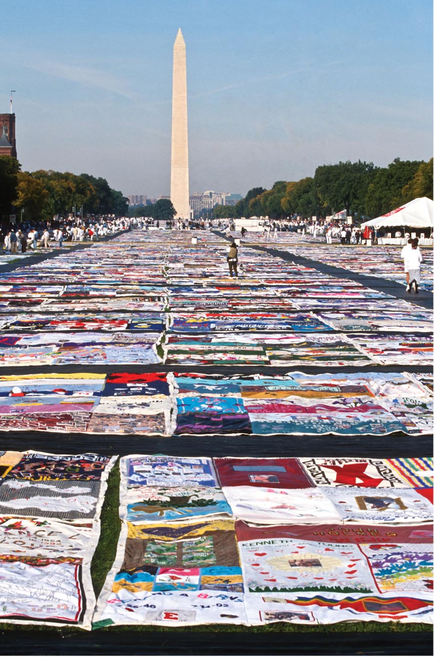
Human immunodeficiency virus (HIV) is discovered as the cause of AIDS.

Françoise Barré-Sinoussi and Luc Montagnier (France); Robert Gallo (USA)

1983

Genes are introduced into plants by use of Agrobacterium tumefaciens plasmid vectors.

Eugene Nester, Mary-Dell Chilton, and colleagues (USA)

1984

Acid-resistant Helicobacter pylori grow in the stomach, where they cause gastritis.

Barry Marshall and J. Robin Warren (Australia)

1987

Geobacter bacteria that can generate electricity are discovered.

Derek Lovley and colleagues (USA)

1988

Prochlorococcus is identified as Earth’s most abundant marine phototroph.

Sallie Chisholm and colleagues (USA)

1995

First genome is sequenced for a cellular organism, Haemophilus influenzae.

Craig Venter, Hamilton Smith, Claire Fraser, and others (USA)

2006

First metagenomes are sequenced, from Iron Mountain acid mine drainage and from the Sargasso Sea.

Jillian Banfield, Craig Venter, and others (USA)

2006

Gardasil vaccine prevents genital human papillomavirus (HPV), the most common sexually transmitted infection.

Patented by Georgetown University and other institutions (USA and Australia)

2012

CRISPR-Cas9 bacterial self-defense mechanism is used for programmable gene editing.

Jennifer Doudna (USA) and Emmanuelle Charpentier (France)

2013

A lentiviral vector, a genetically modified form of HIV, cures a person of cancer.

Michael Kalos, Stephan Grupp, Carl June, and colleagues (USA)

1988–2022

Escherichia coli long-term evolution experiment reaches 50,000 generations and continues.

Richard Lenski, Zachary Blount, and colleagues (USA)

2019

A coronavirus (SARS-CoV-2) is found to be the cause of the COVID-19 pandemic.

Li Wenliang (China)

2020

First mRNA vaccines are approved for human use, to prevent SARS-CoV-2 infection.

Pfizer-BioNTech, Moderna, and National Institutes of Health (USA and Germany)

Microbial Disease Devastates Human Populations

Microbial infectious diseases such as the bubonic plague and tuberculosis have profound effects on human history (Fig. 1.8). The Black Death (bubonic plague) wiped out a third of Europe’s population in the fourteenth century. Bubonic plague is caused by Yersinia pestis, a bacterium spread by fleas of rats and humans. Ironically, the plague-induced population decline enabled the social transformation that led to the Renaissance, a period of unprecedented cultural advancement. In the nineteenth century, the bacterium Mycobacterium tuberculosis stalked overcrowded cities, and tuberculosis was so common that the pallid appearance of tubercular patients became a symbol of tragic youth in European arts, such as Puccini’s opera La Bohème. Today, strains of tuberculosis that resist all known antibiotics stalk human communities throughout the world. A leading infectious cause of death, M. tuberculosis infects one-third of the world’s population.

A bar graph displays death estimates for infectious disease pandemics throughout history.
More information

A bar graph displays death estimates for infectious disease pandemics throughout history. The first bar represents the Plague of Athens, from 429 to 426 B C E. 75,000 to 100,000 deaths are estimated. The second bar represents the Plague of Justinian, from 521 to 542. 15 to 100 million deaths are estimated. The third bar represents The Black Death, from 1346 to 1353. 75 to 200 million deaths are estimated. The fourth bar represents The Third Plague, from 1855 to 1960. 12 to 15 million deaths are estimated. It is noted that The Plague of Justinian, The Black Death, and The Third Plague are parts of The Bubonic Plagues. The fifth bar represents H 1 N 1, from 1918 to 1920. 17 to 100 million deaths and 500 million infections are estimated. The sixth bar represents H I V or AIDS, from 1981 to the present day. 35 million deaths and 75 million infections are estimated. The seventh bar represents H 1 N 1 Influenza, from 2009 to 2010. 284,000 deaths and 0.7 to 1.4 billion infections are estimated. The eighth bar represents COVID 19, from 2019 to the present. 6 million deaths and 500 million infections are estimated.

FIGURE 1.8 Infectious disease pandemics throughout human history.

In the twentieth century, global pandemics were caused by viruses (see Chapter 11). Influenza virus in 1918 caused more deaths than World War I, so swiftly that coffins were stacked in city streets. The AIDS pandemic spread more slowly, over decades, but the deaths of so many young people profoundly shaped culture and economies in the 1980s and ’90s. The AIDS Memorial Quilt consists of 48,000 panels, each stitched in memory of an individual who died of AIDS (Fig. 1.9). The quilt can be viewed interactively online at aidsmemorial.org.

A number of quilts are spread out on the ground amongst a crowd in front of the Washington Monument.
More information

A number of quilts are spread out on the ground amongst a crowd in front of the Washington Monument.

FIGURE 1.9 The AIDS Memorial Quilt. Panels representing individuals who died of AIDS were displayed before the Washington Monument in 1992. A traveling exhibition continues, and the entire collection of panels is presented online.RICHARD ELLIS/ALAMY STOCK PHOTO

The coronavirus SARS-CoV-2, which caused the COVID-19 pandemic, infected more than 500 million people and caused more than 6 million deaths between late 2019 and mid-2022. It caused social and economic devastation for the United States and many countries around the globe. This book presents SARS-CoV-2 in several contexts: devising an mRNA vaccine (Chapter 1); the coronavirus replication cycle (Chapter 6); the production of the Pfizer-BioNTech mRNA vaccine (Chapter 16); and COVID-19 epidemiology (Chapter 28).

Medical Statistics and Health Disparities

Historians traditionally emphasize the role of warfare in shaping human destiny and the brilliance of leaders or the advantage of new technology in determining which civilizations rise or fall. Yet throughout history, more soldiers have died of microbial infections than of wounds in battle. Assessment of the effects of disease on large populations required medical statistics—a important invention for public health.

The significance of disease in warfare was first recognized by the British nurse and statistician Florence Nightingale (1820–1910; Fig. 1.10A). Better known as the founder of professional nursing, Nightingale also founded the science of medical statistics. She used methods invented by French statisticians to demonstrate the high mortality rate due to disease among British soldiers during the Crimean War. To show the deaths of soldiers due to various causes, she devised the “polar area chart” (Fig. 1.10B). In this chart, blue wedges represent deaths due to infectious disease, red wedges represent deaths due to wounds, and black wedges represent all other causes of death. Infectious disease accounts for more than half of all mortality.

A
An illustration of Florence Nightingale tending to a patient and a diagram of medical statistics from 1856 are shown. An illustration of Florence Nightingale tending to a wounded patient in a busy hospital.
More information

An illustration of Florence Nightingale tending to a patient and a diagram of medical statistics from 1856 are shown.

An illustration of Florence Nightingale tending to a wounded patient in a busy hospital.

B
A pie chart of mortality data from April 1855 to March 1856.
More information

A pie chart of mortality data from April 1855 to March 1856. Between April 1855 and May 1855, the total mortality rates stayed relatively stable. The total mortality rate reached its largest in June 1855. After June, the mortality rate decreased for all causes until March 1856. Between April 1855 and December 1855, the vast majority of mortality each month was associated with infectious diseases. Between June 1855 and August 1855, the mortality rates associated with battle wounds reached their greatest values. After August 1855, the mortality due to battle wounds declined until it reached zero in December of 1855. The third factor of mortality in each month is labeled as other. In January and February of 1856, the only factor leading to mortality is labeled as Other.

FIGURE 1.10 Florence Nightingale, founder of medical statistics. A. Florence Nightingale was the first to use medical statistics to demonstrate the significance of mortality due to disease. B. Nightingale’s polar area chart of mortality data during the Crimean War.EVERETT HISTORICAL/SHUTTERSTOCKSMITH COLLECTION/GADO/GETTY IMAGES

Before Nightingale, no one understood the effect of disease on armies or on other crowded populations, such as in cities. Nightingale’s statistics convinced the British government to improve army living conditions and to upgrade the standards of army hospitals. In modern epidemiology, statistical analysis continues to be a crucial tool in determining the causes of disease. For example, statistical analysis of COVID-19 case reports revealed the exponential spread of infection and helped public health directors predict the need for containment measures and hospital preparation.

Medical statistics also reveal the important reality of health disparities. Health disparities are defined as differences in the incidence, prevalence, mortality, and social burden of diseases that exist among specific populations. Such disparities commonly affect Black people, Latino people, indigenous peoples, and LGBTQ people. For example, health disparities drastically affected mortality from COVID-19 (Fig. 1.11). Similar disparities appear in many other infectious diseases such as chlamydia and hepatitis B. The causes of health disparities involve institutional racism, disparities in access to quality health care, and lower socioeconomic status. Health disparities are discussed in detail in Chapter 28.

A bar chart depicts COVID 19 death rates for different ethnic groups in 2020.
More information

A bar chart depicts COVID 19 death rates for different ethnic groups in 2020. The first bar is labeled Black and reaches 74 COVID 19 deaths per 100,000 people. The second bar is labeled Native American and reaches 40 COVID 19 deaths per 100,000 people. The third bar is labeled Latino and reaches 40 COVID 19 deaths per 100,000 people. The fourth bar is labeled Asian and reaches 31 COVID 19 deaths per 100,000 people. The fifth bar is labeled White and reaches 30 COVID 19 deaths per 100,000 people.

FIGURE 1.11 COVID-19 death rates for different ethnic groups in 2020. Death rates for Americans with COVID-19 infection show disparities that are based on ethnicity.

Source: The COVID Tracking Project, https://covidtracking.com/.

Microscopes Reveal the Microbial World

The seventeenth century was a time of growing inquiry and excitement about the “natural magic” of science and patterns of our world, such as the laws of gravitation and motion formulated by Isaac Newton (1642–1727). Robert Boyle (1627–1691) performed the first controlled experiments on the chemical conversion of matter. Physicians attempted new treatments for disease involving the application of “stone and minerals” (that is, chemicals)—what today we would call “chemotherapy.” Minds were open to consider the astounding possibility that our surroundings, indeed our very bodies, were inhabited by tiny living beings.

Robert Hooke observes the microscopic world. The first microscopist to publish a systematic study of the world as seen under a microscope was Robert Hooke (1635–1703). As curator of experiments for the Royal Society of London, Hooke built a compound microscope—a magnifying instrument containing two or more lenses that multiply their magnification in series. With his microscope, Hooke observed biological materials such as nematode “vinegar eels,” mites, and mold filaments. Hooke published drawings of these microbes in Micrographia (1665), the first publication of objects observed under a microscope (Fig. 1.12).

A drawing of mold sporangia as seen under a microscope.
More information

A drawing of mold sporangia as seen under a microscope. The sporangia have slender stalks and bulbous heads. A few sporangia heads are open and these look similar to the heads of flowers.

FIGURE 1.12 Robert Hooke’s Micrographia. Mold sporangia, drawn by Hooke in 1665 from his observations of objects using a compound microscope.LIBRARY OF CONGRESS

Hooke was the first to observe distinct units of living material, which he called “cells.” Hooke first named the units cells because the shape of hollow cell walls in a slice of cork reminded him of the shape of monks’ cells in a monastery. But his crude lenses achieved at best 30-fold power (30×), and he never observed single-celled bacteria.

Antonie van Leeuwenhoek observes bacteria with a single lens. Hooke’s Micrographia inspired other microscopists, including Antonie van Leeuwenhoek (1632–1723), who became the first individual to observe single-celled microbes (Fig. 1.13A). As a young man, Leeuwenhoek lived in the Dutch city of Delft, where he worked as a cloth draper, a profession that introduced him to magnifying glasses. The magnifying glasses were used to inspect the quality of the cloth, enabling the worker to count the number of threads. Later in life, Leeuwenhoek took up the hobby of grinding ever-stronger lenses to see into the world of the unseen.

Leeuwenhoek ground lenses stronger than Hooke’s, which he used to build single-lens magnifiers, complete with sample holder and focus adjustment (Fig. 1.13B). First he observed insects, including lice and fleas; then the relatively large single cells of protists and algae; then, ultimately, bacteria. One day he applied his microscope to observe matter extracted from between his teeth. He wrote, “To my great surprise [I] perceived that the aforesaid matter contained very many small living Animals, which moved themselves very extravagantly.”

Over the rest of his life, Leeuwenhoek recorded page after page on the movement of microbes, reporting their size and shape so accurately that in many cases we can determine the species he observed (Fig. 1.13C). He performed experiments, comparing, for example, the appearance of “small animals” from his teeth before and after drinking hot coffee. The disappearance of microbes from his teeth after drinking a hot beverage suggested that heat killed microbes—a profoundly important principle for the study and control of microbes ever since.

Three images related to Antonie van Leeuwenhoek’s microscopy work. An illustration of Antonie van Leeuwenhoek looking through a handheld microscope. An illustration of the structure of the Leeuwenhoek microscope. A micrograph of bacteria as seen through the Leeuwenhoek microscope.
More information

Three images related to Antonie van Leeuwenhoek’s microscopy work. The first image is an illustration of Antonie van Leeuwenhoek looking through a microscope. The second image is an illustration of the structure of the Leeuwenhoek microscope. The third image is a micrograph of bacteria as seen through the Leeuwenhoek microscope.

An illustration of Antonie van Leeuwenhoek looking through a handheld microscope. He is sitting at a desk and wearing old-fashioned clothes.

An illustration of the structure of the Leeuwenhoek microscope. It shows a lateral view of a human face, in which the hand is holding a microscope close to the eye, and the parts are labeled as the lens, sample holder, focus knob, and sample mover. The illustration is labeled, Leeuwenhoek microscope circa late 1600s.

A micrograph of bacteria as seen through the Leeuwenhoek microscope. The micrograph is blurry and poorly lit, but the bacteria are still distinguishable.

FIGURE 1.13 Antonie van Leeuwenhoek. A. A portrait of Leeuwenhoek, the first person to observe individual microbes. B. “Microscope” (magnifying glass) used by Leeuwenhoek. C. Micrograph obtained using a replica of Leeuwenhoek’s microscope.BETTMANN/GETTY IMAGESBRIAN J. FORD

Leeuwenhoek is believed to have died, ironically, of a disease contracted from sheep whose bacteria he had observed. Historians have often wondered why it took two centuries for Leeuwenhoek and his successors to determine the link between microbes and disease. Although observers such as Agostino Bassi de Lodi (1773–1856) had noted cases of microbes associated with pathology (see Table 1.2), the very ubiquity of microbes—most of them actually harmless—may have obscured their more deadly roles. In addition, it was hard to distinguish between microbes and the single-celled components of the human body, such as blood cells and sperm. It was not until the nineteenth century that human tissues could be distinguished from microbial cells by the application of differential chemical stains such as the Gram stain (discussed in Chapter 2).

Thought Question

1.3 Why do you think it took so long for humans to connect microbes with infectious disease? What innovations helped make the connection?

ANSWER ANSWER

For most of human history, we were unaware that microbes existed. Even after microscopy had revealed their existence, the incredible diversity of the microbial world and the difficulties in isolating and characterizing microbial organisms made it difficult to discern the specific effects of microbes. All healthy people contain microbes, and most disease-causing microbes are indistinguishable from normal microbiota by light microscopy. Not all microbial diseases can be transmitted directly from human to human; they may require complex cycles with intermediate hosts, such as the fleas and rats that carry bubonic plague.

Spontaneous Generation: Do Microbes Have Parents?

The observation of microscopic organisms led priests and philosophers to wonder where these tiny beings came from. In the eighteenth century, scientists and church leaders intensely debated the question of spontaneous generation. Spontaneous generation is the concept that living creatures such as maggots could arise spontaneously, without parental organisms. Chemists of the day tended to support spontaneous generation, as it appeared similar to the way chemicals changed during reaction. Christian church leaders, however, supported the biblical view that all organisms have “parents” going back to the first week of creation.

The Italian priest Francesco Redi (1626–1697) showed that maggots in decaying meat were the offspring of flies. Meat kept in a sealed container, excluding flies, did not produce maggots. Thus, Redi’s experiment argued against spontaneous generation for macroscopic organisms. The meat still putrefied, however, producing microbes that seemed to arise “without parents.”

To disprove spontaneous generation of microbes, another Italian priest, Lazzaro Spallanzani (1729–1799), showed that a sealed flask of meat broth sterilized by boiling failed to grow microbes. Spallanzani also noticed that microbes often appeared in pairs. Were these two parental microbes coupling to produce offspring or did one microbe become two? Through long and tenacious observation, Spallanzani watched a single microbe grow in size until it split in two. Thus he demonstrated cell fission, the process by which cells arise by the splitting of preexisting cells.

Even Spallanzani’s experiments, however, did not put the matter to rest. Proponents of spontaneous generation argued that the microbes in the priest’s flask lacked access to oxygen and therefore could not grow. The pursuit of this question was left to future microbiologists, including the French microbiologist Louis Pasteur (1822–1895; Fig. 1.14A). In addressing spontaneous generation and related questions, Pasteur and his contemporaries laid the foundations for modern microbiology.

A photo of Louis Pasteur and a diagram of the swan-necked flask experiment are shown. A photo of Louis Pasteur in formal attire. A diagram of the swan-necked flask experiment.
More information

A photo of Louis Pasteur and a diagram of the swan-necked flask experiment are shown.

A photo of Louis Pasteur in formal attire.

A diagram of the swan-necked flask experiment. In the first step, the swan-necked flask is shown filled with boiling liquid. It is indicated that air can enter through the open end of the flask tube. Step 1 reads, the broth was boiled to kill all microbes. In the second step, the liquid in the swan-necked flask has settled. Purple buildup, identified as microbes, has collected in the lowest point of the flask tube curve. Step 2 reads, after a year, no microbes appeared. In the third step, the spherical base of the flask is held by a hand. The flask has been tipped horizontally, causing the purple microbial buildup to move towards the liquid in the base of the flask. Step 3 reads, the flask was tipped to allow the broth to reach the microbes. In the fourth step, the flask is shown seated in an upright position. The liquid in the base of the flask has changed to a darker color. Step 4 reads, microbes quickly multiplied.

FIGURE 1.14 Louis Pasteur, founder of medical microbiology and immunology. A. Pasteur’s contributions to the science of microbiology and immunology earned him lasting fame. B. Swan-necked flask. Pasteur showed that, after boiling, the contents in such a flask remain free of microbial growth, despite access to air.THE PRINT COLLECTOR/ALAMY STOCK PHOTO

Louis Pasteur reveals the biochemical basis of microbial growth. As a child in rural France, Pasteur’s main interest was art, especially portraiture. He drew portraits of friends and family and aspired to be a professional artist. But his family convinced him to take up a more secure profession in science. So he studied chemistry—and applied an artist’s intensity of observation.

Pasteur wrote his doctoral thesis on the structure of organic crystals. Crystals exhibit aspects of visual beauty that piqued his interest. From his earlier correspondence on art, we know that Pasteur was fascinated by mirror symmetry. In crystals, he discovered the fundamental chemical property of chirality, the fact that some organic molecules exist in two forms that differ only by mirror symmetry; in other words, the two structures are mirror images of one another, like the right and left hands. Pasteur found that when microbes were cultured on a nutrient substance containing both mirror forms, only one mirror form was consumed. He concluded that the metabolic preference for one mirror form was a fundamental property of life. Subsequent research has confirmed that most molecules of organisms, such as DNA and proteins, are found in only one of their mirror forms.

As a chemist, Pasteur was asked to help with a problem encountered by French manufacturers of wine and beer. The alcohol in beverages comes from fermentation, a process by which microbes gain energy by converting sugars into alcohol. In the time of Pasteur, however, the conversion of grapes or grain to alcohol was believed to be a spontaneous chemical process. No one could explain why some fermentation mixtures produced vinegar (acetic acid) instead of alcohol. Pasteur discovered that fermentation is actually caused by living yeast, a single-celled fungus. In the absence of oxygen, yeast produces alcohol as a terminal waste product. But when the yeast culture is contaminated with bacteria, the bacteria outgrow the yeast and produce acetic acid instead of alcohol. (Fermentative metabolism is discussed in Chapter 13.)

Pasteur’s work on fermentation led him to test a key claim made by proponents of spontaneous generation. The proponents claimed that Spallanzani’s failure to find spontaneous appearance of microbes was due to lack of oxygen. From his studies of yeast fermentation, Pasteur knew that some kinds of microbes do not require oxygen for growth. So he devised an unsealed flask with a long, bent “swan neck” that admitted air but prevented the passage of dust that carried microbes (Fig. 1.14B). When beef broth in the flask was boiled, the sterile broth remained clear, showing no growth of microbes. The famous swan-necked flasks remained free of microbial growth for many years. But when a flask was tilted so that the broth reached the dust, microbes grew immediately. Thus, Pasteur disproved the idea that lack of oxygen was the reason for the failure of spontaneous generation in Spallanzani’s flasks.

Even Pasteur’s work did not prove that microbial growth requires preexisting microbes. The Irish scientist John Tyndall (1820–1893) attempted an experiment similar to Pasteur’s but sometimes found the opposite result. Tyndall found that some kinds of broth, such as hay infusion, gave rise to microbes no matter how long they were sterilized by boiling. The microbes appear because hay infusion is contaminated with a heat-resistant form of bacteria called “endospores” (or “spores”). The spore form can be eliminated only by repeated cycles of boiling and resting, in which the spores germinate to the growing, vegetative form that is killed at 100°C.

It was later discovered that endospores could be killed by boiling under pressure, as in a pressure cooker, which generates higher temperatures than can be obtained at atmospheric pressure. The steam pressure device called the autoclave became the standard way to sterilize materials for the controlled study of microbes. (Microbial control and antisepsis are discussed further in Chapter 5.)

How Did Life Originate?

Spontaneous generation was discredited as a continual source of microbes. Yet at some point in the past, the first living organisms must have originated from nonliving materials. How did the first microbes arise?

The earliest fossil evidence of cells in the geological record appears in rock that formed as long ago as 4 billion years (discussed in Chapter 17). Although the nature of the earliest reported fossils remains controversial, it is generally accepted that “microfossils” from over 2 billion years ago were formed by living cells. Moreover, the living cells that formed microfossils looked remarkably similar to bacterial cells of today, forming chains of simple rods or spheres (Fig. 1.15).

Two microscope images of microfossils of ancient cyanobacteria are shown.
More information

Two microscope images of microfossils of ancient cyanobacteria are shown. The first microfossil is a cluster of round units encased within a membrane. Each unit is about 1 micrometer wide. The second microfossil is a rod-shaped form made up of four squarish units each slightly less than half a micrometer wide.

FIGURE 1.15 Microfossils of ancient cyanobacteria. These fossils, from the Bitter Springs Formation, Australia, are about 850 million years old.J. WILLIAM SCHOPF

The exact composition of the first environment for life is controversial. The components of the first living cells may have formed from spontaneous reactions sparked by ultraviolet absorption or electrical discharge. American chemists Stanley Miller (1930–2007) and Harold C. Urey (1893–1981) argued that the environment of early Earth contained mainly reduced compounds—compounds that have a strong tendency to donate electrons, such as ferrous iron, methane, and ammonia. In 1953, Miller attempted to simulate the highly reduced conditions of early Earth to test whether ultraviolet absorption or electrical discharge could cause reactions producing the fundamental components of life (Fig. 1.16A). He boiled a solution of water containing hydrogen gas, methane, and ammonia and applied an electrical discharge (comparable to a lightning strike). The electrical discharge excites electrons in the molecules and causes them to react. Astonishingly, the reaction produced a number of amino acids, including glycine, alanine, and aspartic acid. A similar experiment in 1961 by Spanish-American researcher Joan Oró (1923–2004; Fig. 1.16B) combined hydrogen cyanide and ammonia under electrical discharge to obtain adenine, a fundamental component of DNA and of the energy carrier adenosine triphosphate (ATP).

A
A photo of Stanley Miller and a photo of Juan Oro. A photo of Stanley Miller with the apparatus of his early-Earth simulation experiment.
More information

A photo of Stanley Miller and a photo of Juan Oro.

A photo of Stanley Miller with the apparatus of his early-Earth simulation experiment.

B
A photo of Juan Oro in formal attire.
More information

A photo of Juan Oro in formal attire and wearing glasses.

FIGURE 1.16 Simulating early Earth’s chemistry. A. Stanley Miller with the apparatus of his early-Earth simulation experiment. B. Biochemist Joan Oró demonstrated the formation of adenine and other biochemicals from reaction conditions found in comets.JIM SUGAR/GETTY IMAGESALBUM/ALAMY STOCK PHOTO

More recent evidence has modified this view, but it is agreed that the strong electron acceptor oxygen gas (O2) was absent until the evolution of the first oxygen-producing photosynthetic microbes. Today, all our cells are composed of highly reduced molecules that are readily oxidized (lose electrons to O2). This seemingly hazardous composition may reflect our cellular origin in the chemically reduced environment of early Earth. The experimental basis for the origin of life and evolution is discussed in detail in Chapter 17.

To Summarize

  • Microbes affected human civilization for centuries before humans guessed at their existence through their contributions to our global biosphere, food and drink production, and infectious diseases.
  • Disease pandemics such as the Black Death in medieval Europe and COVID-19 today shape human history.
  • Florence Nightingale statistically quantified the impact of infectious disease on human populations. Medical statistics reveal health disparities.
  • Robert Hooke and Antonie van Leeuwenhoek were the first to record observations of microbes through simple microscopes.
  • Spontaneous generation is the theory that microbes arise spontaneously, without parental organisms. Lazzaro Spallanzani showed that microbes arise from preexisting microbes and demonstrated that heat sterilization can prevent microbial growth.
  • Louis Pasteur discovered the microbial basis of fermentation. He also showed that providing oxygen does not enable spontaneous generation.
  • John Tyndall showed that repeated cycles of heat were necessary to eliminate spores formed by certain kinds of bacteria.
  • Earth’s first living organisms arose from nonliving materials. Evidence from microfossils and chemical simulations supports the origin of microbial life within the first 100 million years of Earth’s existence.

Glossary

spontaneous generation
The theory, much debated in the eighteenth century, that under current Earth conditions life can arise spontaneously from nonliving matter.
fermentation
Also called fermentative metabolism. 1. The production of ATP via substrate-level phosphorylation, using organic compounds as both electron donors and electron acceptors. 2. Industrial fermentation is the production of microbial products that are made by microbes grown in fermentation vessels; it may include respiratory metabolism to maximize microbial growth.
autoclave
A device that uses pressurized steam to sterilize materials by raising the temperature above the boiling point of water at standard pressure.