Observing the Microbial Cell
More information
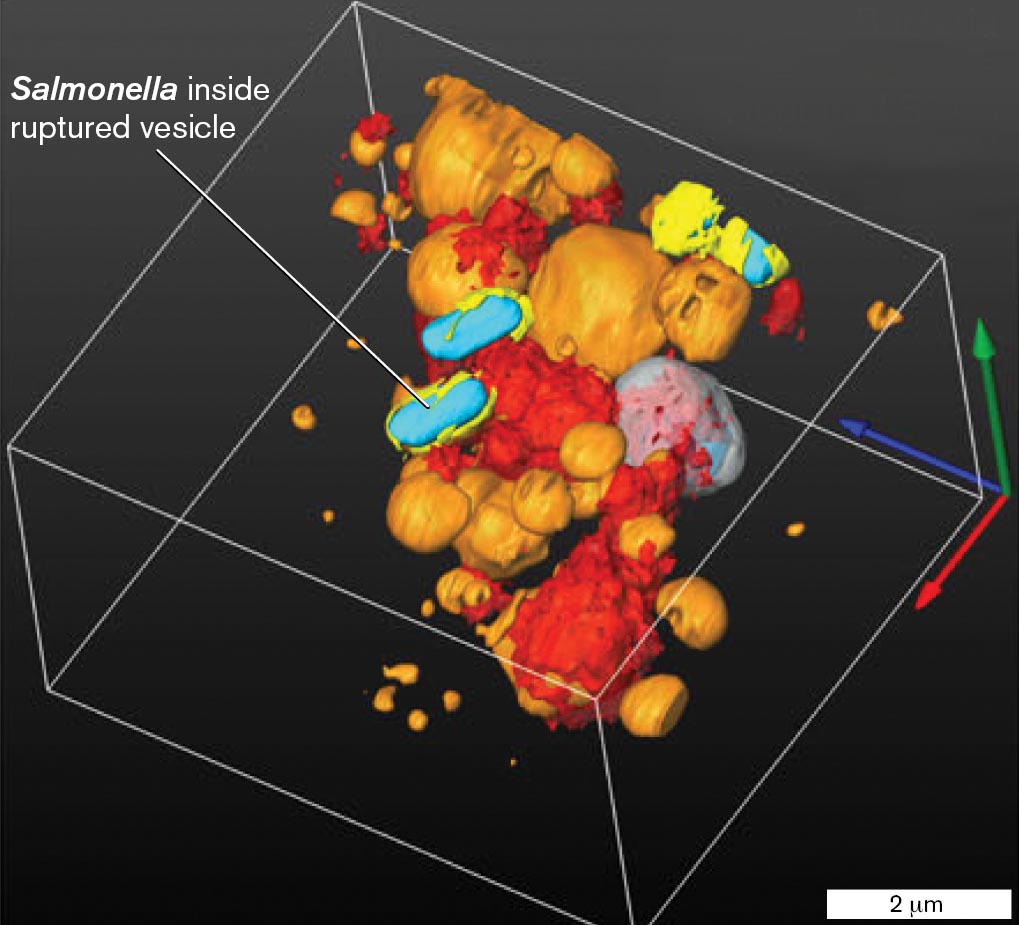
A scanning electron tomograph of Salmonella infecting a human cell. The Salmonella bacteria are identified within ruptured vesicles of the human cell. The bacteria are 1 micrometer long and 0.5 micrometer wide rod shaped organisms. The human cell appears as loosely connected spheres of varying sizes.
Scanning electron tomography, with focused ion beam tomography, reveals Salmonella bacteria infecting a human cell. The 3D image shows bacteria (cyan) inside ruptured cell vesicles (yellow). Large vesicles (orange) take up extracellular material by pinocytosis. Salmonella bacteria resist the cell’s defenses and multiply within the vesicles, thus evading the host immune system. J. FREDLUND ET AL. 2018. CELL MICROBIOL. 20 (4)
|
Chapter Sections |
Learning Objectives |
|
|
» |
Explain the distinction between detection and resolution, and summarize the use of microscopy across many scales of microbial size and shape. |
|
|
» |
Define the different ways that light interacts with an object. |
|
|
» |
Explain the function of bright-field microscopy and phase-contrast microscopy, and the factors that influence them. |
|
|
» |
Describe the use of fixation and staining in bright-field microscopy. |
|
|
2.5 Fluorescence Microscopy, FISH, and Chemical Imaging Microscopy |
» |
Describe the use of fluorescence microscopy, fluorescence in situ hybridization (FISH), and chemical imaging microscopy. |
|
2.6 Electron Microscopy, Scanning Probe Microscopy, and X-Ray Crystallography |
» |
Explain the use of transmission and scanning electron microscopy, and identify advanced methods of imaging cells and molecules. |
One definition of a microbe is an organism that requires a microscope to see. Microscopy reveals the vast realm of life that is invisible to the unaided eye. A microscope enables us to count the number of microbes in the human bloodstream or in natural environments such as the open ocean. It shows us how microbes swim and respond to signals such as a new food source. Microscopes reveal entire communities, such as the microbes that colonize the root of a tree or those that form plaque on the surface of our teeth.
More powerful microscopes reveal the parts of our cells and their function in real time. Fluorescence microscopy captures single molecules within a living cell. The fluorophores can label specific kinds of DNA or proteins. At higher resolution, electron microscopy explores the cell’s interior. Cryo-electron microscopy reveals the cell in three dimensions and models the shape of viruses such as coronaviruses, including their spike proteins that lead to deadly infections.
Chapter 2 begins with light microscopy, the essential tool for every student and professional in the field or clinic. We then explore fluorescence and super-resolution imaging, electron microscopy, and scanning probe microscopy, which push ever farther the frontiers of our ability to observe the microbial world. And we continue to invent new kinds of microscopy, such as chemical imaging microscopy, to reveal microbial metabolism at work.