Describe how we define a microbe, and explain why the definition is a challenge.
Explain how the first microscopes revealed microscopic organisms.
Describe the three major domains of life: Archaea, Bacteria, and Eukarya. Explain what the three domains have in common and how they differ.
Define viruses, and explain how they relate to living cells.
A microbe is commonly defined as a living organism that requires a microscope to be seen. Microbial cells range in size from millimeters (mm) down to 0.2 micrometer (μm), and viruses may be tenfold smaller (Figure 1.1). Some microbes consist of a single cell. A cell is the smallest unit of life, composed of a membrane-enclosed compartment of water solution containing molecules that carry out metabolism.
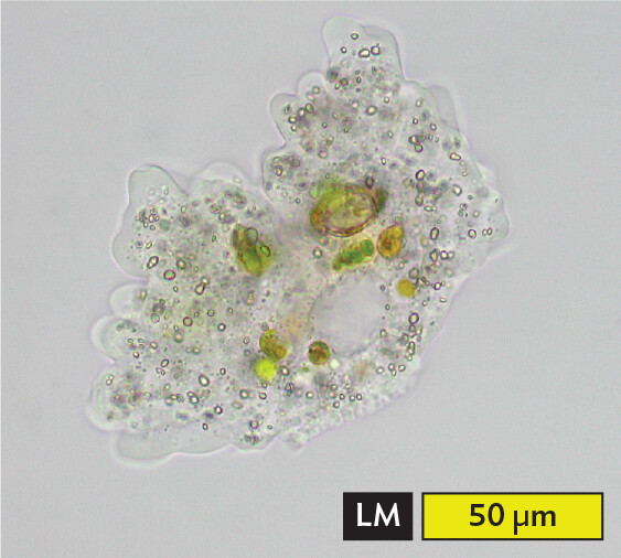
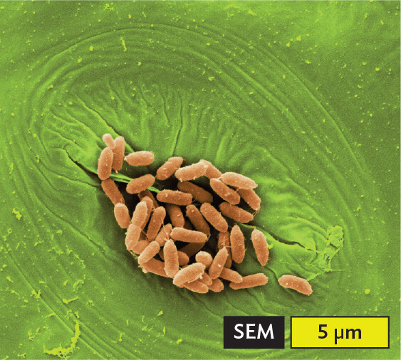
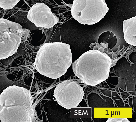
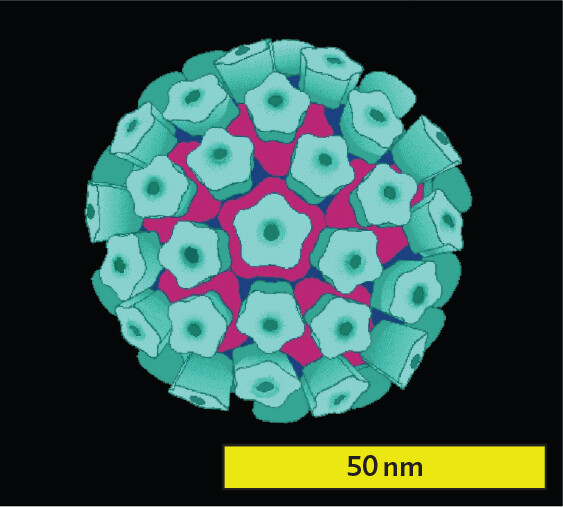
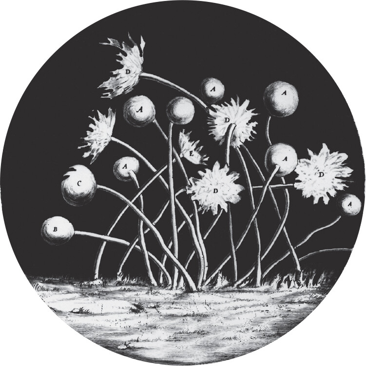
Figure 1.1Representative Microbes
More information
A light micrograph of Pelomyxa species, which is a large ameba with complex internal cell organelles. The amoeba is largely transparent with a few green circular organelles of different sizes seen within, as well as many small gray bubbles. The ameba is about 60 micrometers wide and 150 micrometers long.
A.Pelomyxa sp., a large ameba.More information
A scanning electron micrograph of Escherichia coli bacteria on a lettuce leaf. The cells are rod shaped and are colorized orange in this micrograph. Each cell is about 2 micrometers long and 0.5 micrometer wide. About 20 bacteria cluster over the green stomate cell.
B.Escherichia coli bacteria colonizing a stomate cell in a lettuce leaf (colorized scanning electron micrograph).
More information
A scanning electron micrograph of the archaeon Methanocaldococcus jannaschii. There are about 7 cells in the field of view, shown in white on a black background, with white web like tendrils connecting them to adjacent cells. Each cell is roughly spherical and has a roughened exterior surface. Each archaeon has a diameter of about 1 micrometer.
C.Methanocaldococcus jannaschii, an archaeon that produces methane.More information
A model based on electron microscopy of a human papillomavirus particle. The particle is roughly spherical. Numerous peg like proteins are embedded in the exterior surface of the particle. The virus particle has a diameter of about 45 micrometers.
D. Human papillomavirus, the cause of genital warts and cervical or penile cancer (model based on electron microscopy).
The earliest life on Earth, nearly 4 billion years ago, consisted of microbes—that is, microscopic organisms. Microbial life has since shaped our atmosphere, our geology, and the energy cycles of all ecosystems. Some early microbes eventually evolved into multicellular plants and animals, including ourselves. Today, microbes generate the very air we breathe, including nitrogen gas and much of the oxygen and carbon dioxide. They fix (or combine) nitrogen into forms used by plants, and they make essential nutrients that we consume, such as vitamin B12. Microbes are the primary producers of food webs, particularly in the oceans; when we eat fish, we indirectly consume tons of algae at the base of the food chain.
Each microbe contains in its genome the capacity to reproduce its own kind. Microbes are found throughout our biosphere, from the superheated black smoker vents at the depths of the ocean floor to the subzero ice fields of Antarctica. Bacteria such as Escherichia coli live in our intestinal tract, whereas algae and cyanobacteria turn ponds green. The human body hosts numerous bacteria that protect us and help our digestion and brain function.
Throughout history, humans have had a hidden partnership with microbes in activities ranging from food production and preservation to mining for precious minerals. Today, microbes serve as tools for biotechnology in fields from medicine to microscopic robots. Nevertheless, a small but critical proportion of all microbes are pathogens, the causative agents of disease. Diseases caused by pathogens, commonly called “germs,” remain the principal cause of human mortality.
Our simple definition of a microbe, however, leaves us with contradictions. Most single-celled organisms require a microscope to render them visible, and thus they fit the definition of “microbe.” Nevertheless, some protists and algae are large enough to be seen with the naked eye. An example is the ameba Pelomyxa, which can span several millimeters (Figure 1.1A). Some amebas can cause meningitis; others can harbor thousands of Legionella bacteria, the cause of legionellosis, a severe form of pneumonia.
Other kinds of microbes form complex multicellular assemblages, such as mycelia (multicellular filaments) and biofilms (discussed in Chapter 6). In a biofilm, cells are differentiated into distinct types that complement one another’s function as in multicellular organisms. On the other hand, some complex multicellular animals, such as mites and roundworms, require a microscope for us to see, but they are not considered microbes. Still, you may learn about parasitic invertebrates as part of “microbiology” because these infectious agents are transmitted in a manner similar to that of disease-causing microbes. For example, hookworms infect about 700 million people worldwide, including some in US communities with poor sanitation.
Microscopes Reveal the Microbial World
If microbes cannot be seen by the human eye, how were these “invisible” life forms discovered? Discovery of microbes required the microscope, an instrument that uses one or more lenses to magnify the image in our eyes. Microscopes were first devised during the seventeenth century, a time of growing inquiry and excitement about the “natural magic” of science such as the laws of physics and chemistry formulated by Isaac Newton (1642–1727) and Robert Boyle (1627–1691). Physicians attempted new treatments for disease involving the application of “stone and minerals” (that is, chemicals); today, we would call this chemotherapy. Minds were open to consider the astounding possibility that our surroundings, indeed our very bodies, were inhabited by tiny living beings.
Robert Hooke observes the microscopic world. The first microscopist to publish a systematic study of the world as seen under a microscope was Robert Hooke (1635–1703). As curator of experiments for the Royal Society of London, Hooke built a compound microscope, a magnifying instrument containing two or more lenses that multiply their magnification in series. With his microscope, Hooke observed biological materials such as nematodes (“vinegar eels”), mites, and mold filaments. He then published his illustrations of these materials in Micrographia (1665), the first publication that illustrated objects observed under a microscope (Figure 1.2).
Figure 1.2 Robert Hooke’s Micrographia
More information
A scan of Robert Hooke’s 1665 illustration of mold sporangia. The illustration is in greyscale. Several flower shaped structures with long thin stems sprout from the bottom surface. There are slight variations in the shapes of the flower like structures at the top of each stem, with some appearing spherical and others appearing open with spindle like structures extended out.
An illustration of mold sporangia, drawn by Hooke in 1665, from his observations of objects with a compound microscope.
Hooke was the first to observe distinct units of living material, which he called “cells.” The hollow shape of cell walls in a slice of cork reminded him of monks’ cells in a monastery. But his crude lenses achieved at best 30-fold power (30×), so he never observed single-celled microbes such as bacteria.
Antonie van Leeuwenhoek observes bacteria with a single lens. Robert Hooke’s Micrographia inspired other microscopists, including Antonie van Leeuwenhoek (1632–1723; Figure 1.3A). Leeuwenhoek became the first individual to observe single-celled microbes. As a young man, Leeuwenhoek lived in the Dutch city of Delft, where he worked as a cloth draper, a profession that introduced him to magnifying glasses. (The magnifying glasses were used to inspect the quality of the cloth, enabling the worker to count threads.) Later in life, Leeuwenhoek took up the hobby of grinding ever-stronger lenses to see into the world of the unseen.
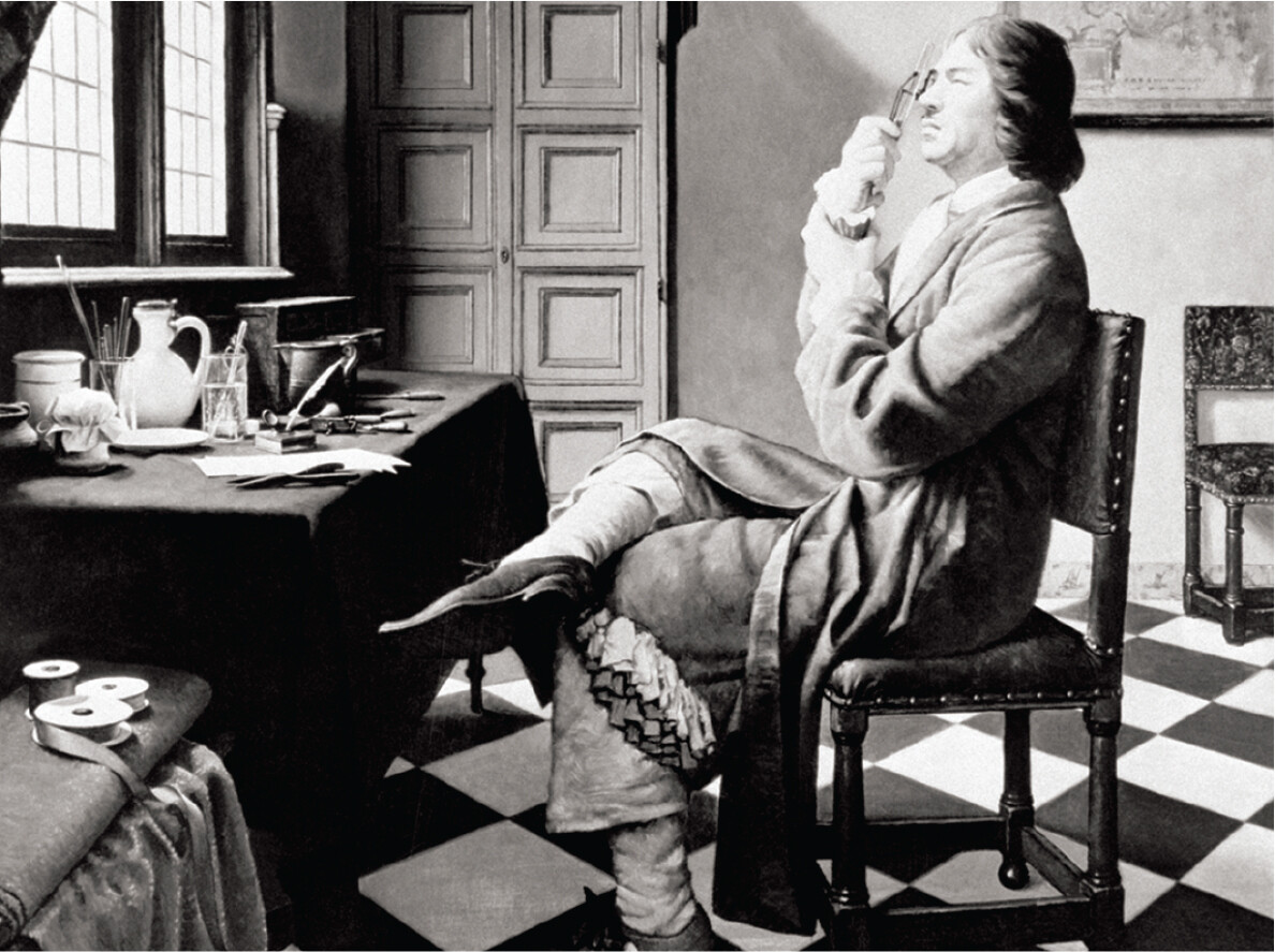
Figure 1.3 Antonie van Leeuwenhoek
More information
A portrait of Antonie van Leeuwenhoek seated on a chair and looking through his handheld microscope. Leeuwenhoek is wearing a long, tailored suit and fancy pointed shoes. His hair is shoulder length and neatly combed back. Leeuwenhoek is facing the windows as he looks through the microscope.
A. Portrait of Leeuwenhoek, the first person to observe individual microbes.
More information
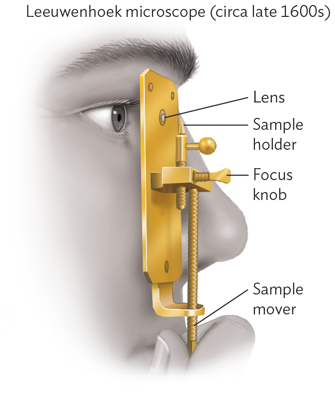
A diagram of the Leeuwenhoek microscope, circa late 1600s. There is a vertically arranged flat rectangular base structure with a lens near one end. The structure is held up to a person’s face so that they can look through the lens. Below the lens, a sample holder is attached to a pin that enters a center part that is jutting out of the flat rectangle. The sample holder holds the sample in line with the lens. A knob comes perpendicularly out of the center part of the microscope. This is the focus knob. A long, thin vertical tube from the center part extends to the bottom of the rectangle plate, and is connected to it by a curved piece of metal. The tube extends further into the observers hand, and is labeled the sample mover.
B. “Microscope” (magnifying glass) used by Leeuwenhoek.More information
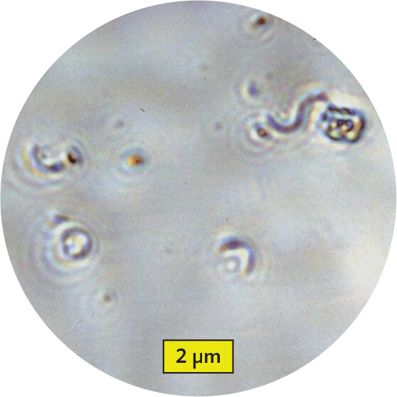
A micrograph of spiral bacteria viewed through a replica of the Leeuwenhoek microscope. The bacteria are visible, but only at low detail. The thin, curved shapes can be seen but they have blurry edges. Each bacterium is roughly 1.5 micrometer long and 0.1 micrometer wide.
C. Spiral bacteria viewed through a replica of Leeuwenhoek’s instrument.
Leeuwenhoek ground lenses stronger than Hooke’s, which he used to build single-lens magnifiers, complete with sample holder and focus adjustment (Figure 1.3B). Leeuwenhoek observed first insects, including lice and fleas; then the relatively large single cells of protists and algae; and finally bacteria. One day he used his microscope to observe matter extracted from between his teeth. He wrote, “To my great surprise [I] perceived that the aforesaid matter contained very many small living Animals, which moved themselves very extravagantly.”
Leeuwenhoek recorded page after page on the movement of microbes, reporting their size and shape so accurately that we can often determine the species he observed (Figure 1.3C). He performed experiments, comparing, for example, the appearance of “small animals” from his teeth before and after drinking hot coffee. The disappearance of microbes from his teeth after drinking a hot beverage suggested that heat kills microbes—a profoundly important principle for the study and control of microbes ever since.
Historians have often wondered why it took so many centuries for Leeuwenhoek and his successors to determine the link between microbes and disease. The very ubiquity of microbes—most of them harmless—may have obscured the deadlier roles of pathogens (disease-causing microbes). In addition, it was hard for the microscopist to distinguish between microbes and the single-celled components of the human body, such as blood cells and sperm. It was not until the nineteenth century that human tissues could be distinguished from microbial cells by the application of differential chemical stains. Microscopy is discussed further in Chapter 3.
Different Kinds of Microbes
Microbes, like other organisms, are classified as members of a species according to a shared set of genes and traits. The scientific name of the species, such Staphylococcus epidermidis for a common skin bacterium, consists of a capitalized genus name (Staphylococcus) and a lowercase species name (epidermidis), both italicized. In addition, members of a genus are often referred to informally by a romanized vernacular term, such as “staphylococci.” The names of some microbial species are sometimes changed to reflect our new understanding of genetic relationships. For example, the causative agent of bubonic plague was formerly called Bacillus pestis (1900) and Pasteurella pestis (1923), but it is now called Yersinia pestis (since 1944). The older names, however, still appear in the literature.
Microbes are classified according to their genetic relatedness. The more closely related two organisms are, the more recently they diverged from a common ancestor. Relatedness is important for understanding how microbes respond to treatment. For example, an antibiotic used against an intestinal pathogen will also kill many beneficial bacteria that normally live in the intestine; consequently, the antibiotic may cause digestive problems. The degree of genetic relatedness between microbes is calculated by comparing DNA sequences in the genome—the total DNA sequence content of each organism. Genome comparison is now the fundamental basis for classifying all life forms.
A major trait distinguishing microbes is possession or lack of a membrane-enclosed nucleus. Microbes that lack a nuclear membrane are called prokaryotes, which include bacteria and archaea. Microbial eukaryotes (cells with a nucleus) include fungi, protozoa, and algae. The genetic group of eukaryotes includes multicellular animals and plants. All life comprises three genetically distinct groups called domains: the Bacteria, the Archaea, and the Eukarya (Eukaryotes).
Bacteria (singular, bacterium) are prokaryotic cells, usually 0.2–20 μm in size—or one-tenth to one-hundredth the size of a sentence period. Different species may grow as single cells, as filaments (chains), or as communities with simple differentiated forms. An example is Escherichia coli (Figure 1.1B), a bacterial species that grows in the human intestine. Most strains of E. coli are harmless members of the microbial community that aid human digestion, but some strains cause acute gastroenteritis that may lead to kidney failure. Bacteria are found in every habitat of our biosphere, even several kilometers underground.
Archaea (singular, archaeon) are a domain that evolved by diverging from bacteria more than 3 billion years ago. Archaea are prokaryotic cells that produced the ancestor of all eukaryotes (cells with a nucleus), including humans. Some archaea are extremophiles that live in seemingly hostile environments, such as the boiling sulfur springs of Yellowstone. Other archaea are methanogens, whose metabolism releases methane (natural gas; Figure 1.1C). Methanogens are common in the gut of humans and animals, the source of the “gas” passed by one’s intestinal tract. Their metabolism increases the efficiency of digestion. A remarkable feature of archaea is that none cause disease. The absence of pathogenesis (disease causation) in archaea is of great interest to medical researchers studying the cause and prevention of disease.
Eukaryotes (Eukarya) are a domain of life that evolved from a form of Archaea. Eukaryotic microbes include protozoa (singular, protozoan), which are motile heterotrophs (consuming organic food), usually single-celled. A protozoan such as an ameba (Figure 1.1A) may be free-living or parasitic. Algae (singular, alga) are eukaryotic microbes containing chloroplasts that conduct photosynthesis. Algae form an essential base of the food web, although overgrowth causes algal blooms that poison fish. Protozoa and algae together are classified as protists. Distinct from protists are fungi (singular, fungus), heterotrophic organisms that are usually nonmotile and grow by absorbing nutrients from their surroundings. Fungi may grow as single cells (yeast) or as filaments (bread mold), or they may form complex structures such as mushrooms. Some fungi cause infections, especially in people with a depressed immune system.
Eukaryotic microbial pathogens are also called parasites. Parasites are organisms that live at the expense of a host they inhabit, debilitating the host. By convention, the word “parasite” is used for both single-celled and multicellular eukaryotes. Multicellular parasites include, for example, worms and mites (presented in Chapter 11).
Viruses, such as coronavirus and influenza virus, are noncellular microbes. Most viruses are too small to see even under a light microscope. A virus particle contains genetic material (DNA or RNA) that takes over the metabolism of a cell to generate more virus particles. Some viruses, such as papillomaviruses (Figure 1.1D), consist of only a few molecular parts. Others, such as herpesviruses, show complexity approaching that of a cell, although no virus is a fully functional cell. Engineered viruses are used as tools for gene therapy. For example, in 2017 the Food and Drug Administration (FDA) approved a nonpathogenic derivative of the human immunodeficiency virus (HIV) for gene delivery to the white blood cells of a child, enabling the child’s immune system to overcome leukemia.
SECTION SUMMARY
Microbes are microscopic; that is, they are organisms too small to be seen without a microscope. Different species of microbes grow as single cells, in filaments, in biofilms, or in simple differentiated structures.
The light microscope is an instrument that uses a series of lenses to magnify the image of microscopic objects such as microbes.
Robert Hooke and Antonie van Leeuwenhoek were the first to record observations of microbes through simple microscopes.
Bacteria are cells lacking a nucleus (prokaryotes). Bacteria grow in all habitats. Most human-associated species are harmless, but some cause disease.
Archaea are nonbacterial cells that lack a nucleus (prokaryotes) and are distantly related to bacteria and eukaryotes. Methanogens live in the human intestine (among other places), where their metabolism releases methane. No archaea cause disease.
Eukaryotic microbes include protists (protozoa and algae) and fungi. Parasitic protozoa and fungi may infect humans.
Viruses are noncellular microbes that must infect a host cell. Some viruses of humans can cause pandemic disease; for example, the virus SARS-CoV-2 causes COVID-19.
A bacterial, viral, fungal, protozoan, or helminthic agent of disease; among health professionals, pathogens typically are limited to bacteria, viruses, and fungi.
Any bacterium, virus, fungus, protozoan (protist), or helminth that colonizes and harms its host; the term commonly refers to protozoa and to invertebrates.