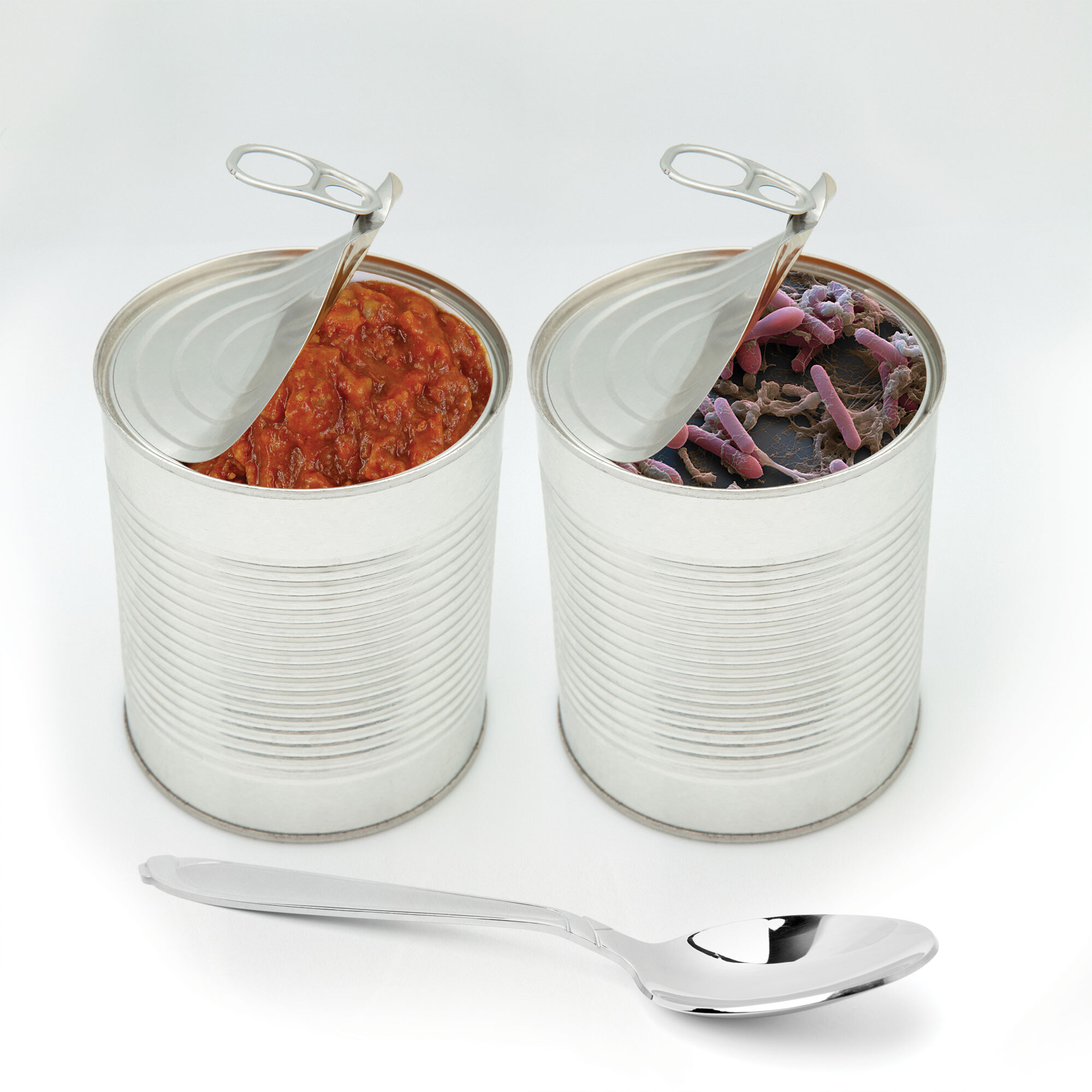
A photo of two aluminum cans followed by a case study on food poisoning in two children. Both of the cans in the photo are partially opened revealing the contents within. One can contains a red chili sauce. The other can contains an electron micrograph of rod shaped bacteria. A spoon rests on the surface in front of the cans indicating the contents will be ingested. The case study that follows is titled Pathogen in a Can. There is an illustrated silhouette of two children holding hands at the center of the case study. There is also a light micrograph of Clostridium botulinum bacteria. The case study reads as follows. Scenario. Marianna and Julio, two children in Oklahoma aged 8 and 11, fell ill with food poisoning after consuming commercially canned hot dog chili sauce. The children complained of double vision and an inability to move their facial muscles. Signs and Symptoms. Examination showed normal vital signs, symmetrical weakness open parenthesis equal on right and left sides close parenthesis, and fixed pupils due to cranial nerve palsy or paralysis. Upper body paralysis gradually spread downward and breathing became labored. Testing. Botulinum toxin, a bacterial neurotoxin, was identified in their blood by fluorescent antibodies. Treatment. The children were placed on a mechanical ventilator, which is a machine that assists breathing, and treated with botulinum antitoxin. After several days, the children were removed from mechanical ventilation. They underwent physical rehabilitation for a full recovery. Follow Up. The chili sauce was traced to a canning facility where six swollen cans tested positive for botulinum toxin A. From the cans, inspectors cultured a bacterium that grew only without oxygen, as in a closed can. A microscope showed that the bacterium had a distinctive club shaped appearance. The bacterium was Clostridium botulinum, which produces botulinum toxin, the cause of botulism. One end of the club shaped cell contains a developing endospore, an inert form of the cell that can germinate and grow in a closed container of food. Growing cells produce botulinum toxin, leading to botulism, a life threatening form of paralysis. Microscopy of the pathogens unique form confirms the identification. The micrograph shows short arrow heads in purple with a small pink circle within the arrow head.
Pathogen in a Can
More information
An illustrated silhouette of a boy and a girl holding hands. They appear to face the viewer. The children are barefoot and wearing t shirts with shorts. Their free hands are held by their sides.
SCENARIO Marianna and Julio, two children in Oklahoma aged 8 and 11, fell ill with food poisoning after consuming commercially canned hot dog chili sauce. The children complained of double vision and an inability to move their facial muscles.
SIGNS AND SYMPTOMS Examination showed normal vital signs, symmetrical weakness (equal on right and left sides), and fixed pupils due to cranial nerve palsy (paralysis). Upper body paralysis gradually spread downward, and breathing became labored.
TESTING Botulinum toxin, a bacterial neurotoxin, was identified in their blood by fluorescent antibodies.
TREATMENT The children were placed on a mechanical ventilator (a machine that assists breathing) and treated with botulinum antitoxin. After several days, the children were removed from mechanical ventilation. They underwent physical rehabilitation for a full recovery.
FOLLOW-UP The chili sauce was traced to a canning facility where six swollen cans tested positive for botulinum toxin A. From the cans, inspectors cultured a bacterium that grew only without oxygen, as in a closed can. A microscope showed that the bacterium had a distinctive club-shaped appearance. The bacterium was Clostridium botulinum, which produces botulinum toxin, the cause of botulism. One end of the club-shaped cell contains a developing endospore, an inert form of the cell that can germinate and grow in a closed container of food. Growing cells produce botulinum toxin, leading to botulism, a life-threatening form of paralysis. Microscopy of the pathogen’s unique form confirms the identification.
More information
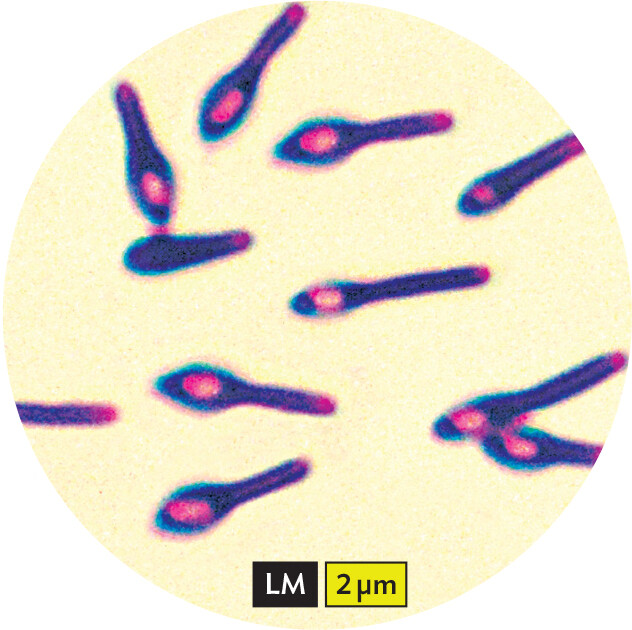
A light micrograph of Clostridium botulinum bacteria. The bacteria are club shaped, purple cells, with oval pink endospores contained in the clubbed ends. Each cell is about 4 micrometers in length. The cells range from about 1 micrometer in width at their clubbed end to 0.5 micrometer in width at the other pole. A caption reads, The distinctive club shaped cells, stained purple, each show the endospore, which is the pink stained oval, near one end. The micrograph was obtained via bright field microscopy with Gram stain.
Clostridium botulinum The distinctive club-shaped cells (purple) each show the endospore (pink oval) near one end. Bright-field microscopy with Gram stain.
CHAPTER OBJECTIVES
After reading this chapter, you will be able to:
Explain how light microscopy reveals information about pathogenic microbes isolated from patients.
Explore a variety of microbial cells under a microscope.
Identify the types of microscopy used to obtain images of microbes.
Explain the information provided by images from light microscopy, fluorescence microscopy, and electron microscopy.
A can of food that looks harmless can hide deadly life forms, too small for you to see. But a microscope reveals the food-borne contaminant Clostridium botulinum, the cause of botulism. Food-borne botulism has low incidence in the United States (about 28 cases per year), but its onset is rapid and it can be fatal without a prompt diagnosis.
The bacterium C. botulinum has a distinctive club-shaped cell. The swollen end of the “club” contains an endospore, a dormant particle in which the cell’s DNA is condensed with a few enzymes protected by a thick coat. Endospores of certain types of bacteria can survive many years until they encounter the right conditions for growth—for this species, access to food and moisture in the absence of oxygen. The club-shaped form of the sporulating cell is unique to C. botulinum. You can see C. botulinum by using bright-field microscopy with the simple technique of the Gram stain, which we will discuss later in the chapter.
Microscopy reveals the vast realm of bacteria, fungi, and protozoa invisible to our unaided eyes. The microbial world spans a wide range of sizes over several orders of magnitude. For different size ranges, we use different instruments, from the simple bright-field microscope to the electron microscope. The microscope enables us to count the microbes in the human bloodstream or in natural environments like the ocean. It detects emerging pathogens, such as the Salmonella transmitted from jars of peanut butter in 2022, a multistate epidemic in the United States. The microscope can even follow pathogenic bacteria as they attach, invade, and move through a human host cell.
In this chapter, you will learn how to use the light microscope to observe and distinguish different microbes. Microscopes are used in many settings, from hospitals and veterinary clinics to industrial plants and wastewater treatment facilities. You will also learn about the capabilities of more advanced techniques, such as fluorescence and electron microscopy.