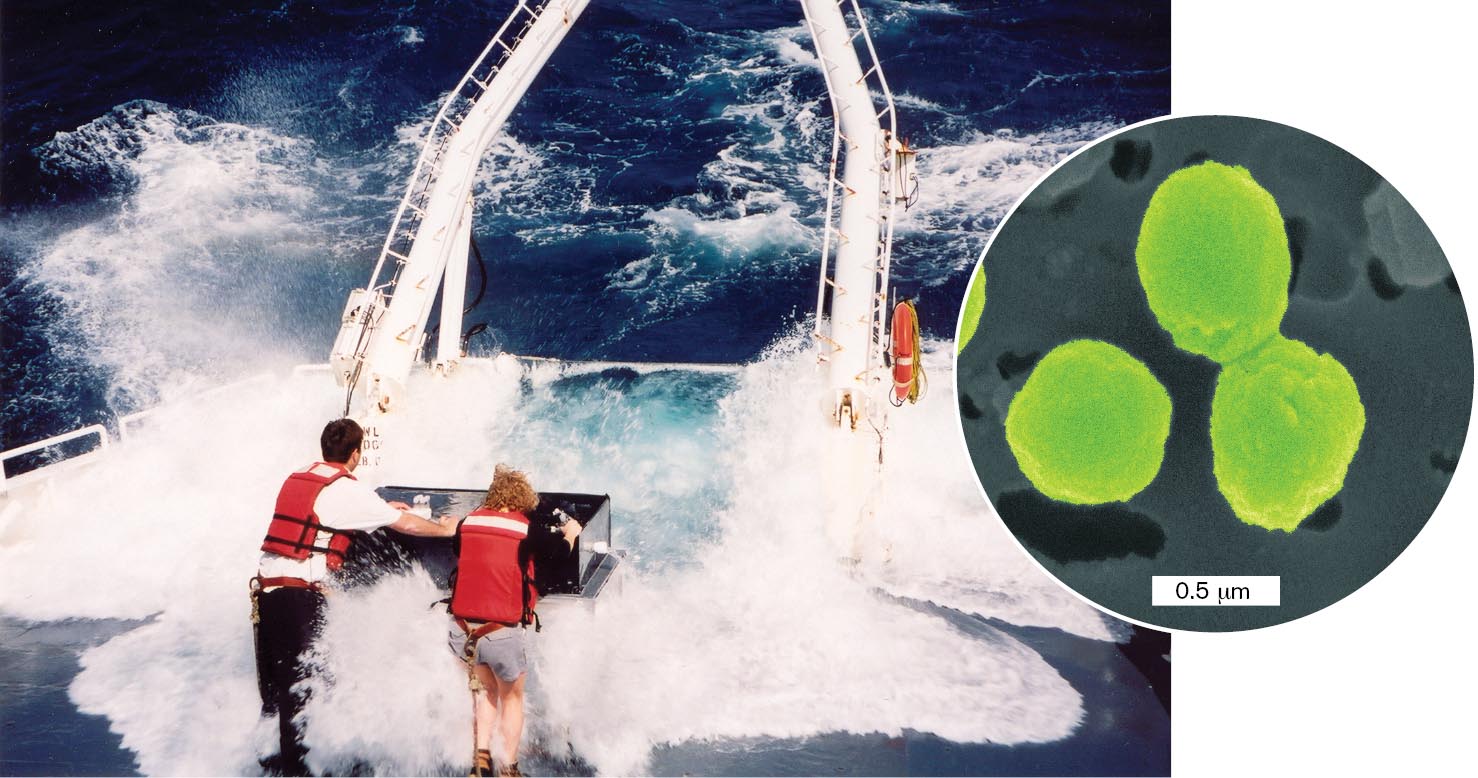
Life began early in the history of planet Earth in the form of microscopic organisms, or “microbes.” Over the eons, those microbes evolved to shape our atmosphere, our geology, and the energy cycles of all ecosystems. For the first 2 billion years, all life was microbial. Then, as now, a vast realm of that microbial diversity resided in Earth’s oceans. Oceans constitute the largest biome on Earth’s surface, a community where the biomass is dominated by microbes (Fig. 1.1). The waves pounding the research vessel in the photograph contain trillions of photosynthetic Prochlorococcus bacteria, tiny cells that make much of the oxygen gas we breathe.
More information
Scientists operating a boat in rough water. An inset indicates the cells present in the water. The inset is a colorized micrograph of circular cells, each about 0.75 micrometers in diameter.
FIGURE 1.1 ■Marine microbiologists sample bacteria such as the phototroph Prochlorococcus.Inset: Colorized scanning electron micrograph of Prochlorococcus. ERIK ZINSER (AUTHOR)ANNE THOMPSON, CHISHOLM LAB, MIT
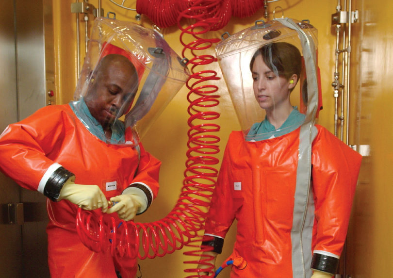
Yet, before we devised microscopes in the seventeenth century, we humans were unaware of the unseen living organisms that surround us, that float in our air and water, and that inhabit our own bodies. Microbes fix nitrogen for plants, and they make vitamins, such as vitamin B12. In the ocean, microbes produce biomass for the food web that feeds the fish we eat, and microbes consume toxic wastes such as the oil from the Deepwater Horizon spill in the Gulf of Mexico in 2010. At the same time, pathogens (agents of disease) such as SARS-CoV-2 take our lives—and researchers risk their lives to study them. Working with deadly pathogens requires containment at biosafety level 3 or 4 (BSL-3 or BSL-4). The highest containment level, BSL-4 requires sealed suits and respiratory equipment (Fig. 1.2). BSL-4 is required, for example, to study Ebola virus infection of cells in tissue culture. Despite all our advances in medicine and public health, humans continue to die of microbial diseases.
More information
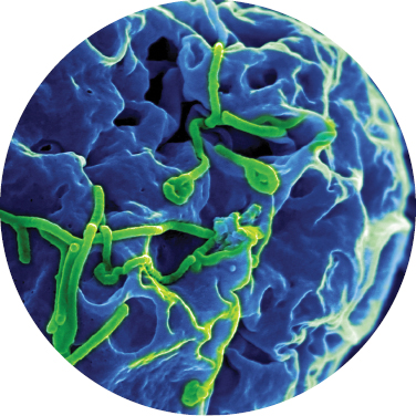
A photograph shows two laboratorians in inflated body suits. An inset depicting Ebola virus is linked to the photograph. The laboratorians are wearing thick orange suits, transparent head cases, and gloves that are securely attached to the suits. They wear headphones with a microphone on their heads, inside the suit. The inset of Ebola shows a digital rendering of cells with viral particles budding from the cell surfaces.
More information
A photograph shows two laboratorians in inflated body suits. An inset depicting Ebola virus is linked to the photograph. The laboratorians are wearing thick orange suits, transparent head cases, and gloves that are securely attached to the suits. They wear headphones with a microphone on their heads, inside the suit. The inset of Ebola shows a digital rendering of cells with viral particles budding from the cell surfaces.
FIGURE 1.2 ■Researching deadly pathogens. Biosafety level 4 containment is required to study the most dangerous pathogens. Inset: Ebola virus (green) budding from monkey cells in tissue culture; requires BSL-4 containment.H. S. PHOTOS/ALAMY STOCK PHOTOSCIENCE SOURCE/SCIENCE SOURCE
Today, microbes provide new tools that affect human society. For example, the use of heat-stable bacterial DNA polymerase (a DNA-replicating enzyme) in a technique called the polymerase chain reaction (PCR; see eAppendix 3) enables us to detect minute amounts of DNA in traces of blood or fossilized bone. Microbial technologies led us from the discovery of the double helix to the sequence of the human genome, the total genetic information that defines our species.
A Microbe Is a Microscopic Organism
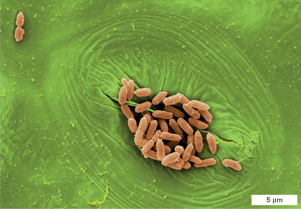
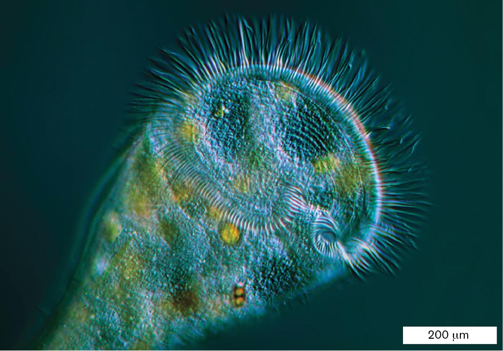
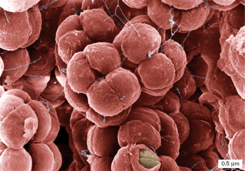
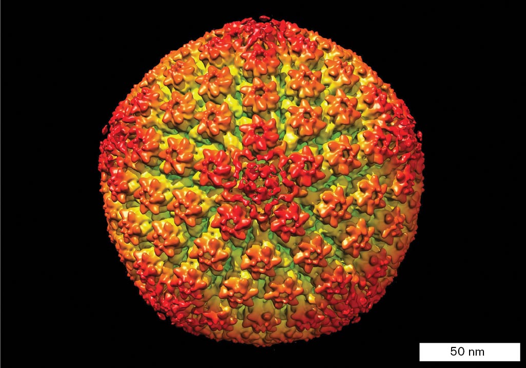
A microbe is commonly defined as a living organism that requires a microscope to be seen. All around us, microbes exhibit diverse forms and lifestyles (Fig. 1.3). Rod-shaped bacteria such as Escherichia coli are among the 100 trillion inhabitants of our intestines, where they help digest our food. Alternatively, E. coli may colonize the plants we eat (Fig. 1.3A). Microbial eukaryotes (cells with nuclei) such as the voracious Stentor engulf aquatic prey (Fig. 1.3B). Archaea are a life form distinct from both bacteria and eukaryotes. Some archaea grow in extreme environments, such as concentrated salt (Fig. 1.3C). And all kinds of life host viruses. For example, herpes simplex virus infects human cells (Fig. 1.3D). Microbial cells range in size from 5 millimeters (mm) down to less than 0.2 micrometer (µm), and viruses may be tenfold smaller (Table 1.1).
A. Bacteria:E. coli
More information
Four colorized micrographs of different kinds of microbes are shown. The first micrograph shows the bacterium E. coli. The second micrograph shows the eukaryote Stentor. The third micrograph shows the archaeon Halococcus. The fourth micrograph shows Herpes simplex virus.
A micrograph shows rod shaped cells of the bacterium E. coli on a leaf stomate. The E. coli cells are each about 2 micrometers long and 0.5 micrometers wide, and look similar to rice grains. They are clustered together over a wrinkled portion of the leaf stomate.
B. Eukaryote:Stentor
More information
A micrograph shows the eukaryote Stentor, which has a long body and flattened top lined with thin hairs. The body of Stentor is 400 micrometers wide at it’s broadest point and greater than 400 micrometers long. The circular, flattened top of Stentor is about 300 micrometers in diameter. The top is lined with thin hairs of even length that fan away from the protist’s body structure.
C. Archaea: Halophiles
More information
A micrograph shows the coccoid cells of the archaeon Halococcus, arranged in tetrads. Each cell is about 0.5 micrometer in diameter; tetrads are 1 micrometer in length and width. The tetrad formations are shown packed closely together and overlapping in space.
D. Virus: Herpes simplex virus
More information
A micrograph shows Herpes simplex virus, which is a sphere of highly organized, complex structures. The virus has a diameter of 150 nanometers.
FIGURE 1.3 ■Representative kinds of microbes.A. Bacterium: E. coli on leaf stomate. B. Eukaryote: Stentor, a predatory protist. C. Archaeon: Halococcus. D. Virus: Herpes simplex virus (with envelope removed). Colorized. SCIMAT/SCIENCE SOURCEBLICKWINKEL, ALAMY STOCK PHOTOEYE OF SCIENCE/SCIENCE SOURCELOUISE HUGHES/SCIENCE SOURCE
TABLE 1.1
Sizes of Some Microbes
Microbe
Description
Approximate size
Varicella-zoster virus 1
Virus that causes chickenpox and shingles
100 nanometers (nm) = 10−7 meter (m)
Prochlorococcus
Photosynthetic marine bacterium
500 nm = 5 × 10⁻⁷ m
Escherichia coli
Bacterium growing within human intestine
2 micrometers (µm) = 2 × 10⁻⁶ m
Spirogyra
Aquatic alga that forms long filaments of cells
5 cm = 0.05 m (chain of cells)
Pelomyxa
Ameba (a protist) that consumes bacteria in soil or water
5 millimeters (mm) = 5 × 10⁻³ m
Some microbes consist of a single cell, the smallest unit of life, a membrane-enclosed compartment of water solution containing molecules that carry out metabolism. Each microbe contains a genome used to reproduce its own kind. Microbial cells acquire food, gain energy to build themselves, and respond to environmental change. Microbes evolve at rapid rates—often fast enough to see evolution occur in the laboratory (discussed in Chapter 17). Much of Earth’s microbial inventory remains a mystery. Barely 0.1% of the microbes in our biosphere can be cultured in the laboratory; even the digestive tract of a newborn infant contains species of bacteria unknown to science.
The more diverse kinds of microbes we discover, the more contradictions we find with our definitions of a microbe. For example:
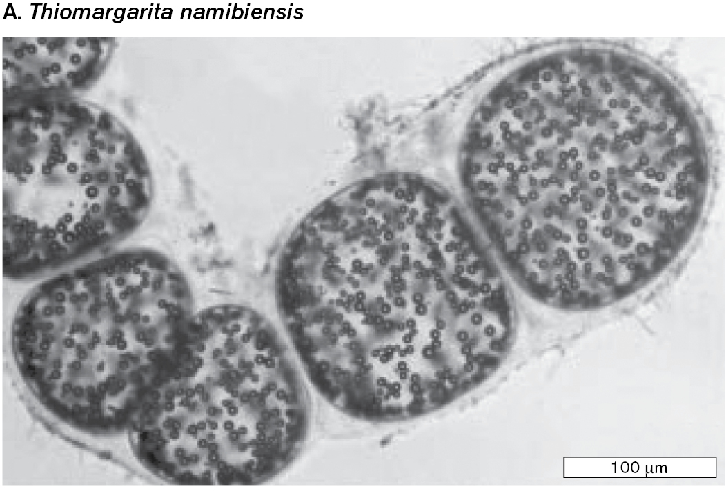
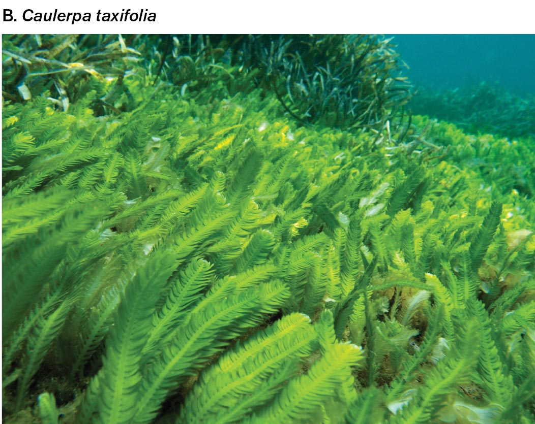
Super-size microbial cells. Most single-celled organisms require a microscope to render them visible, and thus they fit the definition of a microbe. Nevertheless, some bacteria grow to sizes large enough to see with the unaided eye. The marine sulfur bacterium Thiomargarita namibiensis, called the “sulfur pearl of Namibia,” grows to a thousand times larger than Prochlorococcus (Fig. 1.4A). The related bacterium Thiomargarita magnifica grows a thread-like cell nearly a centimeter long. Even more surprising, a single cell of the “killer alga” Caulerpa taxifolia grows feather-like fronds from stalks up to several meters long (Fig. 1.4B). This alga forms multiple nuclei and extends without cell division. Caulerpa now covers many acres beneath the coastal waters of California.
Microbial populations and communities. Many microbes form complex multicellular assemblages such as biofilms and fruiting bodies. Within a microbial population, or a community of multiple species, microbial cells may be differentiated into distinct types that complement each other’s functions, as in multicellular organisms. Yet, multicellular animals such as mites and tardigrades require a microscope for us to see but are not considered microbes.
Viruses. A virus is a noncellular particle containing genetic material that takes over the metabolism of a cell to generate more virus particles (see Chapter 6). Some viruses consist of only a short chromosome packed in protein. Other kinds of viruses, such as pandoraviruses that infect amebas, have the size and complexity of a cell. Although viruses are not fully functional cells, some viral genomes may have evolved from cells.
More information
Two examples of giant microbial cells are shown. The first image is a micrograph of the bacterium Thiomargarita namibiensis. The second image is a photograph of the algae Caulerpa taxifolia.
A micrograph of the bacterium Thiomargarita namibiensis shows coccoid cells of 150 micrometers in diameter. The cells are linked together in a chain. Granules are visible within the cells.
More information
A photograph of the algae Caulerpa taxifolia shows tall, thin, feather like algal strands along the seafloor.
FIGURE 1.4 ■A giant microbial cell.A.Thiomargarita namibiensis, a marine sulfur bacterium (light microscopy). B.Caulerpa taxifolia, an invasive alga that consists of a single multinucleated cell.H. N. SCHULZ ET AL., 1999. SCIENCE284:493–495O.DIGOIT/ALAMY STOCK PHOTO
Note: Each section of text contains Thought Questions that may have various answers. Possible responses are provided in the ebook and at the back of the printed book.
Thought Questions
1.1 The minimum size of most microbial cells is about 0.2 µm. How can even smaller cells be discovered? What factors may determine the minimum size of a cell?
ANSWER ANSWER
The smallest cells that are well studied, about 0.2 µm in length, are cell wall–less bacteria called mycoplasmas; for example, Mycoplasma pneumoniae, a causative agent of pneumonia. Bacteria smaller than 0.2 µm have been discovered by passing stream water through a filter of that pore size. It is hard to see how their cell components, such as ribosomes (about a tenth this size), could fit inside such a small cell. The volume required for DNA and the apparatus of transcription and translation probably sets the lower limit on cell size.
1.2 If viruses are not functional cells, are they “alive”?
ANSWER ANSWER
A traditional definition of a life form includes the capability for metabolism and homeostasis (maintaining internal conditions of its cytoplasm), as well as reproduction and response to its environment. Viruses direct their own replication and respond to the environment of the host cell, but they lack both metabolism and homeostasis outside their host cell. Nevertheless, viruses such as herpesviruses contain numerous metabolic enzymes that participate in the metabolism of the host. Certain large viruses, such as the mimivirus, appear to have evolved from cells. Some microbiologists argue that viruses should be considered alive if reproduction is the main criterion and if the viral “environment” is considered the inside of the host cell.
In practice, our definition of a microbe derives from tradition as well as genetic considerations. In this book we consider microbes to include prokaryotes (cells lacking a nucleus, including bacteria and archaea), as well as certain classes of eukaryotes (cells with a nucleus), such as algae, fungi, and protozoa (discussed in Chapter 20). The bacteria, archaea, and eukaryotes—known as the three “domains”—evolved from a common ancestral cell (see Section 1.5 and Chapter 21). Viruses and even smaller infectious particles are discussed in Chapters 6 and 11.
Note: The formal names of the three domains are Bacteria, Archaea, and Eukarya. Members of these domains are called bacteria (singular, bacterium), archaea (singular, archaeon), and eukaryotes (singular, eukaryote), respectively. The microbiology literature includes alternative spellings for some of these terms, such as “archaean” and “eucaryote.”
Microbial Genomes Are Sequenced
Our modern concept of a microbe has deepened through the use of two major research tools: advanced microscopy (see Chapter 2) and the sequencing of genomes (see Chapter 7). A genome is the total genetic information contained in an organism’s chromosomal DNA. The genes in a microbe’s genome and the sequence of DNA tell us a lot about how that microbe grows and associates with other species. For example, if a microbe’s genome includes genes for nitrogenase, a nitrogen-fixing enzyme, that microbe probably can fix nitrogen from the atmosphere into proteins—its own proteins and those of associated plants. And by comparing DNA sequences of different microbes, we can figure out how closely related they are and how they evolved.
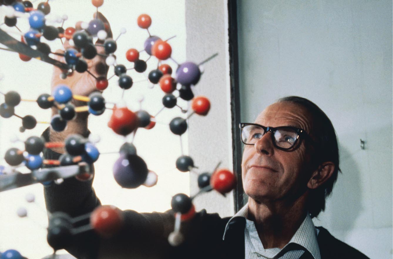
The first method of DNA sequencing that was fast enough to sequence large genomes was developed by Fred Sanger (1918–2013) at the University of Cambridge (Fig. 1.5). This achievement—which jump-started the study of molecular biology—earned Sanger the 1980 Nobel Prize in Chemistry, together with Walter Gilbert and Paul Berg. Sanger and his colleagues used the new method to sequence DNA containing tens of thousands of base pairs, such as the DNA of the human mitochondrion (plural, mitochondria).
More information
A photo shows Fred Sanger analyzing the structure of a complex ball-and-stick model.
FIGURE 1.5 ■Fred Sanger devised the method of DNA sequence analysis used to sequence the first genomes.BETTMANN/GETTY IMAGES
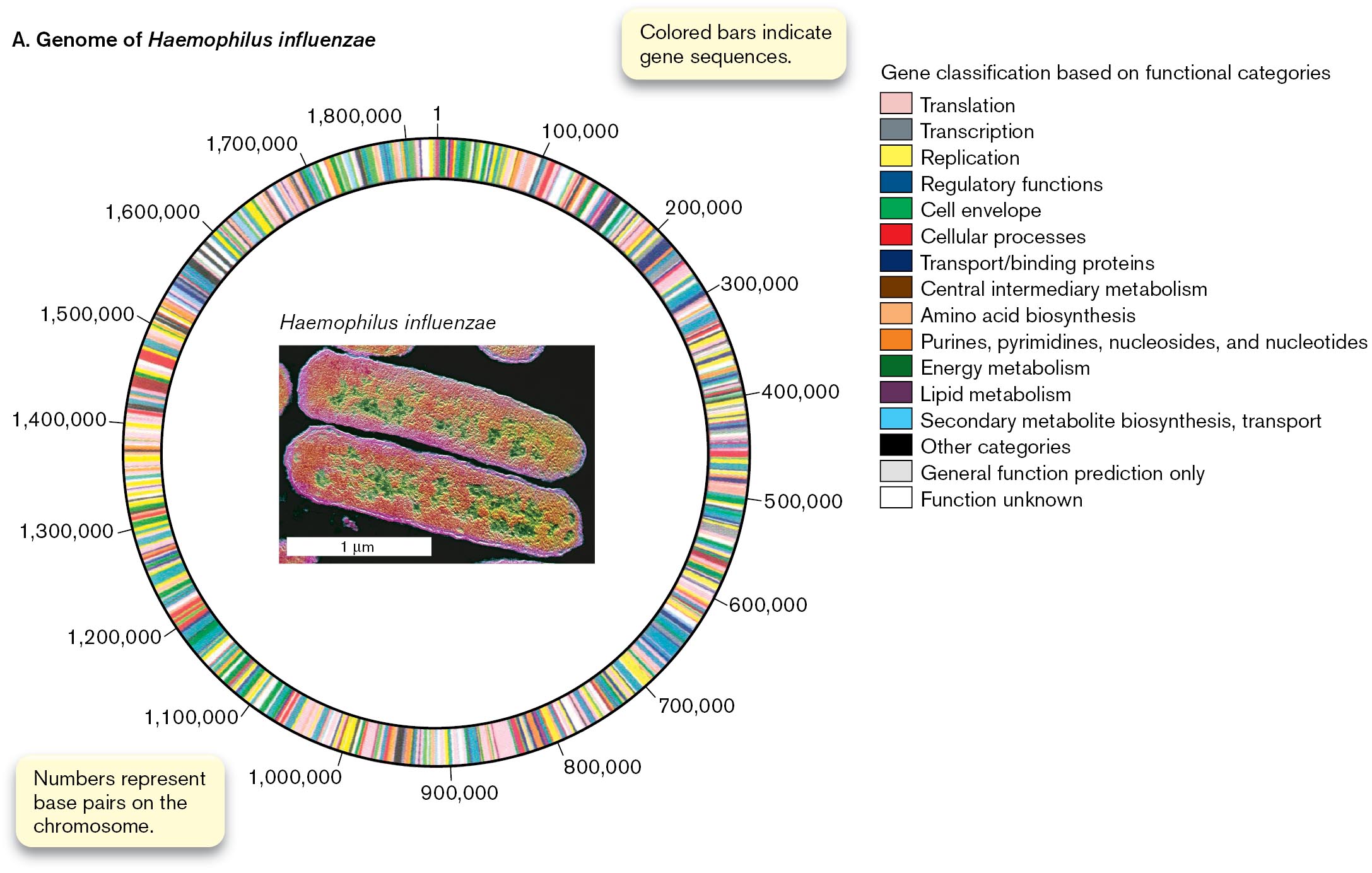
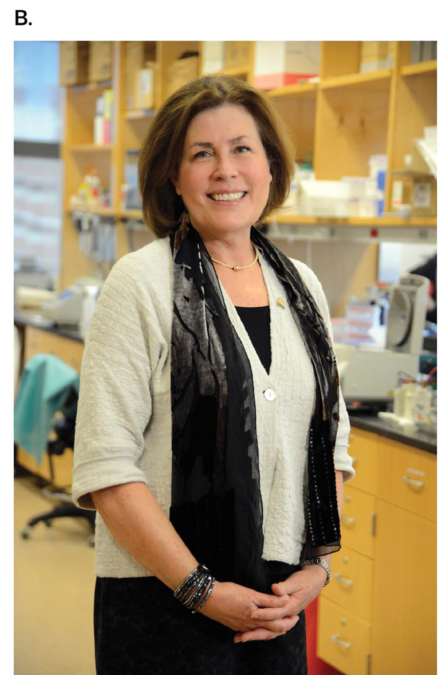
But most genomes of cells contain millions or even billions of base pairs (complementary bases paired on two strands of DNA helix). In 1995, scientists completed the first genome sequence of a cellular microbe, the bacterium Haemophilus influenzae (Fig. 1.6A). H. influenzae causes several diseases including meningitis in children, a disease now prevented by the Hib vaccine. The H. influenzae genome has nearly 2 million base pairs, which specify about 1,700 genes. The sequence was determined by a large team of scientists led by Craig Venter, Hamilton Smith, and Claire Fraser (Fig. 1.6B) at The Institute for Genomic Research (TIGR). The TIGR team devised a computational strategy for assembling large amounts of sequence data, which Fraser used to sequence many microbial genomes. This strategy was later used to sequence the human genome.
More information
A color-coded wheel used to represent the genome of Haemophilus influenzae and photo of Claire Fraser.
A wheel representing the genome of Haemophilus influenzae is color-coded for gene functional classifications. Thin bars within the wheel symbolize gene sequences, no particular color is predominant. The different colors of the bars represent different functional categories of the gene sequences. The categories are as follows, translation, transcription, replication, regulatory functions, cell envelope, cellular processes, transport, and binding proteins, central intermediary metabolism, amino acid biosynthesis, purines, pyrimidines, nucleosides, nucleotides, energy metabolism, lipid metabolism, secondary metabolite biosynthesis, transport, and other. Two additional categories are titled, general function prediction only, and function unknown. The bars on the wheel are labeled 1 to 1,800,000 with intervals at each 100,000. The number labels represent base pairs on the chromosome. An inset of a micrograph of Haemophilus influenzae is located at the center of the wheel.
An inset of a micrograph of Haemophilus influenzae shows two rod-shaped bacteria. The bacteria are about 2 micrometers long and 0.5 micrometer wide.
More information
A photo of Claire Fraser standing in a laboratory. She has short brown hair.
FIGURE 1.6 ■Bacterial genomes were sequenced.A. The genome of Haemophilus influenzae, a bacterium that causes ear infections and meningitis, was the first DNA sequence completed for a cellular organism. Inset: Colorized electron micrograph of H. influenzae.B. Claire Fraser, past president of The Institute for Genomic Research (TIGR), sequenced numerous microbial genomes.CNRI/SCIENCE SOURCEINSTITUTE OF GENOME SCIENCE, UNIVERSITY OF MARYLAND SCHOOL OF MEDICINE
Today we sequence new bacterial genomes daily. In addition to sequencing individual genomes, computational strategies are used to sequence thousands of genomes of microbes sampled from a natural environment, such as the ocean or soil, or the intestinal tract of a cow. The collection of sequences taken directly from the environment is called a metagenome. Now, metagenomes are sequenced for microbial communities of medical interest, such as that of the human colon. Human gut microbes contain 100 times more genes in their metagenome (DNA of all microbes in a community) than the human genome contains—and many of these microbial genes contribute to our health!
Comparing genomes has revealed a set of core genes shared by all organisms. These core genes add further evidence that all living beings on Earth, including humans, share a common ancestry. Genomes are discussed further in Chapter 7, and the evolution of genomes and metagenomes is discussed in Chapters 17 and 21.
To Summarize
A microbe is a living organism that requires a microscope to be seen. Some organisms exist in both microscopic and macroscopic forms.
Microbes are found all around us, as part of human bodies and throughout our environment.
Major categories of microbes include bacteria, archaea, microbial eukaryotes, and viruses. Viruses are noncellular and require replication within a host cell.
Microbes may grow in communities such as a biofilm. A community with a shared habitat may include more than one species of organism, both microscopic and macroscopic.
Microbial capabilities are defined by their genome sequences.
One of the three domains of life, consisting of organisms with a last common ancestor not shared with members of Archaea or Eukarya. Organisms are prokaryotic (lacking nuclei, unlike eukaryotes) and possess primarily ester-linked phospholipid membranes (like eukaryotes, unlike archaea).
One of the three domains of life, consisting of organisms with a last common ancestor not shared with members of Bacteria or Eukarya. Organisms are prokaryotic (lacking nuclei, unlike eukaryotes) and possess ether-linked phospholipid membranes (unlike bacteria).
One of the three domains of life, consisting of organisms with a last common ancestor not shared with members of Archaea or Bacteria. Cells possess nuclei, unlike cells of bacteria and archaea.

 ANSWER
 ANSWER