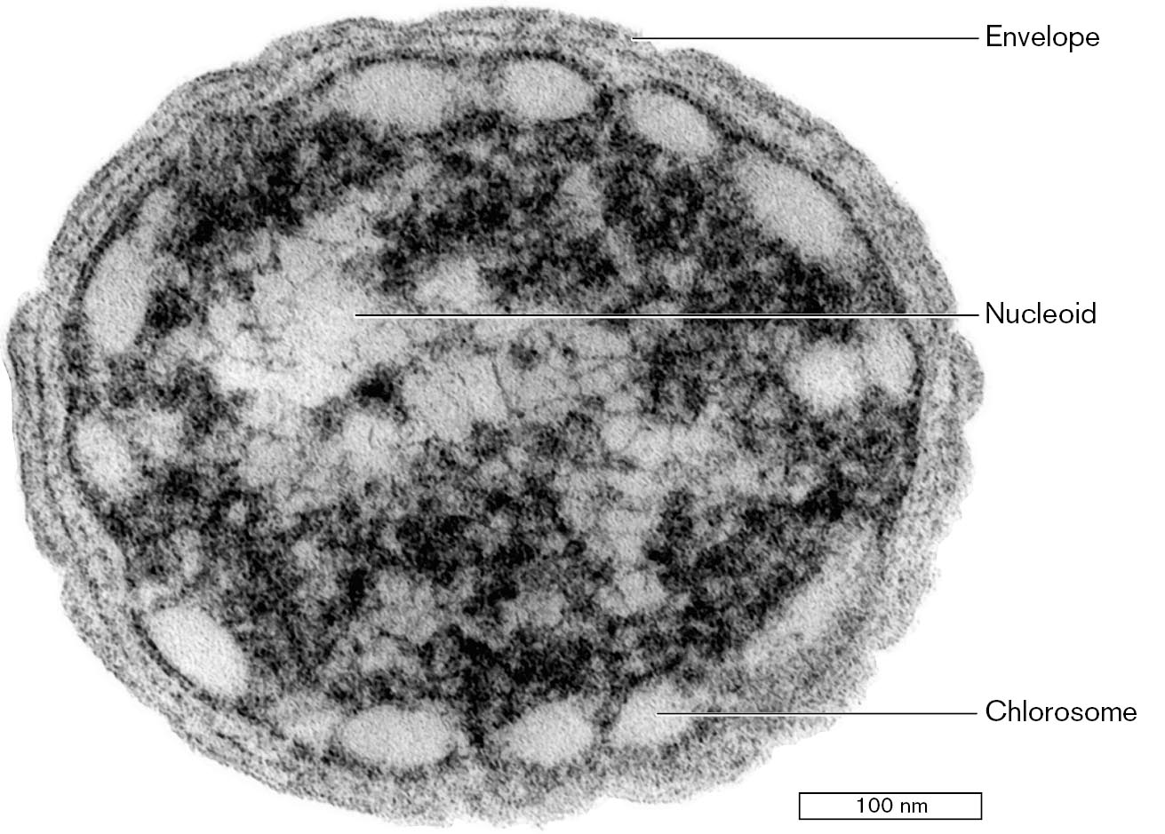
During the twentieth century, amid world wars and societal transformations, the field of microbiology exploded with new knowledge (see Table 1.2). More than 99% of what we know about microbes today was discovered after 1900 by scientists too numerous to cite in this book. New tools of microscopy and genomic analysis offered unprecedented applications for human medicine and industry (discussed in Chapters 7–12). Electron microscopy and biochemistry revealed the fundamental structure and function of cell membranes and proteins. For example, an electron micrograph of a stained section of Chlorobium bacteria reveals unique light-harvesting organelles called “chlorosomes” (Fig. 1.32). The chlorosomes conduct photosynthesis at extremely low light levels, such as that of long-wavelength energy radiated from thermal vents at the ocean floor.
More information
An electron micrograph of Chlorobium species. The bacterium has a circular shape, a few structures are labeled. The outer layer is labeled as the envelope. A labeled nucleoid is partially visible in the center. Oval-shaped structures that sit close to the cell membrane are labeled as chlorosomes. The bacterium is about 400 nanometers in diameter.
FIGURE 1.32 ■Electron micrograph of Chlorobium species, a photosynthetic bacterium. The thin section reveals the nucleoid (containing DNA), the light-harvesting chlorosomes, and envelope membranes.NIELS-ULRIK FRIGAARD ET AL. 2002 J. BACTERIOL. 184:3368
Cell Membranes and Macromolecules
In 1900, the study of cell structure was still limited by the resolution of the light microscope and by the absence of tools that could take apart cells to isolate their components. Both of these limitations were overcome by the invention of powerful instruments. Just as society was being transformed by machines ranging from jet airplanes to vacuum cleaners, the study of microbiology was also being transformed by machines. Two instruments had exceptional impact: The electron microscope revealed the internal structure of cells (see Chapter 2), and the ultracentrifuge enabled isolation of subcellular parts (see Chapter 3).
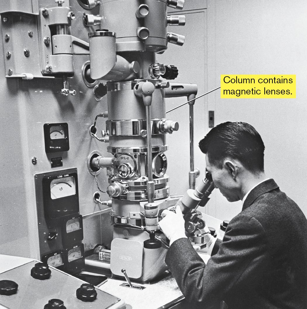
The electron microscope. In the 1920s, at the Technical University in Berlin, student Ernst Ruska (1906–1988) was invited to develop an instrument for focusing rays of electrons. Ruska recalled, from his childhood, that his father’s microscope could magnify fascinating specimens of plants and animals, but that its resolution was limited by the wavelength of light. He was eager to devise lenses that could focus beams of electrons, with wavelengths far smaller than that of light, to reveal living details never seen before. Ultimately, Ruska built lenses to focus electrons using specially designed electromagnets. Magnetic lenses were used to complete the first electron microscope in 1933 (Fig. 1.33). Early transmission electron microscopes achieved about tenfold greater magnification than the light microscope, revealing details such as the ridged shell of a diatom. Further development steadily increased magnification, to as high as a millionfold. Today’s electron microscopes can visualize phospholipid membranes and protein complexes such as individual antibodies. Exciting kinds of microscopy are presented in Chapter 2.
More information
A photo of a scientist looking into the transmission electron microscope in a laboratory. The large cylindrical column of the microscope is labeled to contain magnetic lenses.
FIGURE 1.33 ■An early transmission electron microscope.HAGLEY MUSEUM AND ARCHIVE/SCIENCE SOURCE
Subcellular structures, however, raised many questions about cell function that visualization alone could not answer. Biochemists showed that cell function involves numerous chemical transformations mediated by enzymes. A milestone in the study of metabolism was the elucidation by German biochemist Hans Krebs (1900–1981) of the tricarboxylic acid cycle (TCA cycle, or Krebs cycle), by which the products of sugar digestion are converted to carbon dioxide. The TCA cycle provides energy for many bacteria and for the mitochondria of eukaryotes. But even Krebs understood little of how metabolism is organized within a cell; he and his contemporaries considered the cell a “bag of enzymes.” The full understanding of cell structure required experiments on isolated parts of cells.
The ultracentrifuge. Centrifugation can separate whole cells from the fluid in which they are suspended. The first centrifuges spun samples in a rotor with centrifugal force of a few thousand times that of gravity. In the nineteenth century, biochemists proposed that even greater centrifugal forces could separate components of lysed cells, even macromolecules such as proteins. The Swedish chemist Theodor Svedberg (1884–1971), at the University of Uppsala, built such a machine: the ultracentrifuge. By the twentieth century, ultracentrifuges had achieved rotation rates so high that they required a vacuum to avoid burning up like a space reentry vehicle. Ultracentrifuges isolated protein complexes such as ribosomes and DNA molecules such as plasmids (small circular pieces of DNA).
Experiments combining electron microscopy and ultracentrifugation revealed how membranes govern energy transduction within bacteria and within organelles such as mitochondria and chloroplasts. In the 1960s, English biochemists Peter Mitchell (1920–1992) and Jennifer Moyle (1921–2016) proposed and tested a revolutionary idea called the chemiosmotic theory. The chemiosmotic theory states that the reduction-oxidation (redox) reactions of the electron transport system store energy in the form of a gradient of protons (hydrogen ions) across a membrane, such as the bacterial cell membrane or the inner membrane of the mitochondrion. The energy stored in the proton gradient, in turn, drives the synthesis of ATP (discussed in Chapter 14).
Microbial Genetics Leads the DNA Revolution
As the form and function of living cells emerged in the early twentieth century, a largely separate line of research revealed patterns of heredity of cell traits. In eukaryotes, the Mendelian rules of inheritance were rediscovered and connected to the behavior of subcellular structures called chromosomes. Frederick Griffith (1879–1941) showed in 1928 that an unknown substance from dead bacteria could carry genetic information into living cells, transforming harmless bacteria into a strain capable of killing mice—a process called transformation. Some kind of “genetic material” must be inherited to direct the expression of inherited traits. Biochemists thought the inherited material might be protein, because of the tremendous variety of amino acid sequences.
Then, in 1944, Oswald Avery (1877–1955) and colleagues showed that the genetic material for transformation is deoxyribonucleic acid, or DNA. An obscure acidic polymer, DNA had been previously thought too uniform in structure to carry information; its precise structure was unknown. As World War II raged among nations, scientists embarked on an epic struggle: the quest for the structure of DNA.
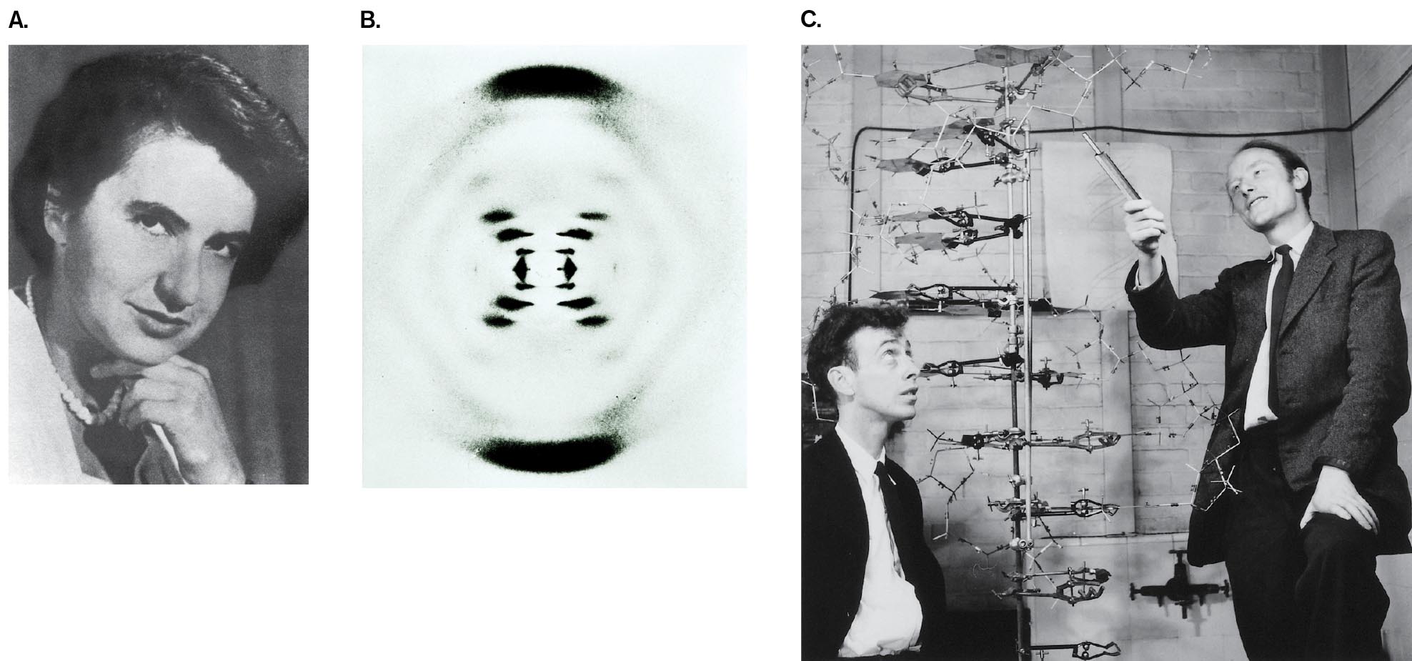
The double helix. The tool of choice to discover the structure of molecules was X-ray crystallography, a method developed by British physicists in the early 1900s. The field of X-ray analysis included an unusual number of women, including Dorothy Hodgkin (1910–1994), who later won a Nobel Prize for the structures of penicillin and vitamin B12 (discussed in Chapter 2). In 1953, crystallographer Rosalind Franklin joined a laboratory at King’s College London to study the structure of DNA (Fig. 1.34A). As a woman and as a Jew who supported relief work in Palestine, Franklin felt socially isolated at the male-dominated Protestant university; her work was disparagingly called “witchcraft.” Nevertheless, her exceptional X-ray micrographs (Fig. 1.34B) revealed for the first time that the common form of DNA was a double helix.
Without Franklin’s knowledge, her colleague Maurice Wilkins (1916–2004) showed her data to a competitor, James Watson at the University of Cambridge. The pattern led Watson and Francis Crick (1916–2004) to propose that the four bases of the DNA “alphabet” were paired in the interior of Franklin’s double helix (Fig. 1.34C). They published their model in the journal Nature, while denying that they had used Franklin’s data. The discovery of the double helix earned Watson, Crick, and Wilkins the 1962 Nobel Prize in Physiology or Medicine. Franklin died of ovarian cancer before the prize was awarded. Before her death, however, she had turned her efforts to the structure of ribonucleic acid (RNA). She determined the helical form of the RNA chromosome within tobacco mosaic virus, the first viral RNA to be characterized.
More information
A photo of Rosalind Franklin, an x-ray diffraction pattern, and a photo of James Watson and Francis Crick are shown.
A headshot of Rosalind Franklin.
An x-ray diffraction image of an hourglass-shaped pattern of D N A.
A photograph of James Watson and Francis Crick examining a life-size model of the D N A double helix.
FIGURE 1.34 ■The DNA double helix.A. Rosalind Franklin discovered that DNA forms a double helix. B. X-ray diffraction pattern of DNA, obtained by Franklin. C. James Watson (left) and Francis Crick discovered the complementary pairing between bases of DNA and the antiparallel form of the double helix.SCIENCE SOURCEOMIKRON/SCIENCE SOURCEA. BARRINGTON BROWN/SCIENCE SOURCE
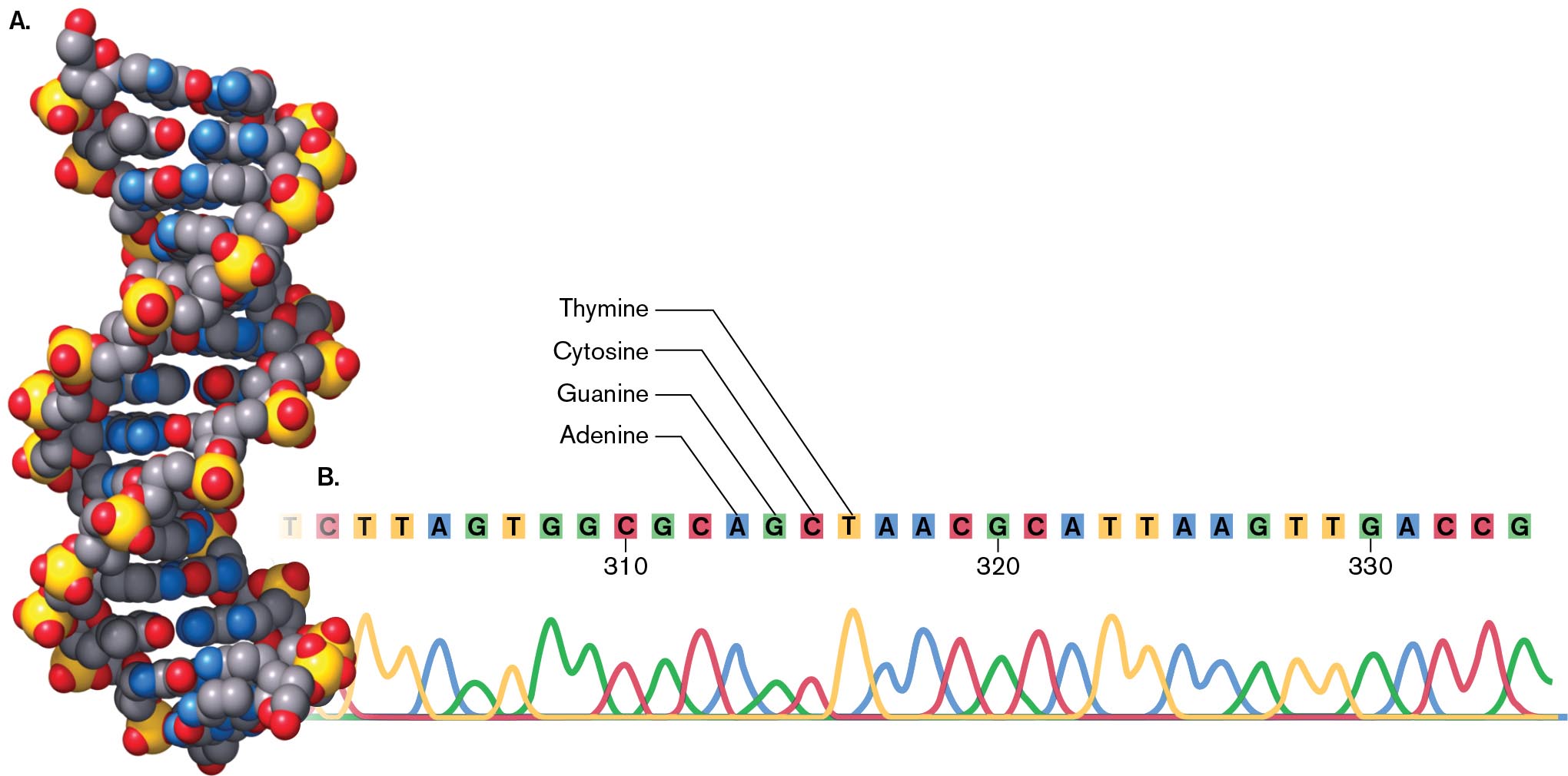
Modern X-ray crystallography (discussed in Chapter 2) reveals with atomic precision the structure of DNA, including its complementary base pairs (Fig. 1.35A). The complementary pairing of DNA bases led to the development of techniques for DNA sequencing, the reading of a sequence of DNA base pairs. Figure 1.35B shows a portion of the DNA sequence from bacterial DNA isolated by an undergraduate student. (The sequencing process is described in Chapter 7.) In the data, each color represents a fluorescent signal from one of the four bases: adenine (A), guanine (G), cytosine (C), or thymine (T). Each peak represents a DNA fragment terminating in that particular base. The order of fragment lengths yields the sequence of bases in one strand. Reading the DNA sequence enabled microbiologists to determine the beginning and endpoint of microbial genes, and ultimately entire genomes, as discussed in Section 1.1.
More information
A model and an illustration of D N A are shown.
A space filling model of the D N A double helix.
The sequence reads as follows: C, T, T, A, G, T, G, G, C, G, C, A, G, C, T, A, A, C, G, C, A, T, T, A, A, G, T, T, G, A, C, C, G. The base pairs are labeled as T for Thymine, C for Cytosine, G for Guanine, and A for Adenine. Every 10 bases is marked, starting with the second C in the sequence at 310. Four different colors of curves in sequence intersect each other at the bottom.
FIGURE 1.35 ■DNA.A. The structure of DNA, based on modern X-ray crystallography. B. A DNA sequence fluorogram obtained from bacterial genomic DNA. Each colored trace represents the fluorescence of one of the four bases terminating a fragment of DNA. Units represent number of DNA bases.
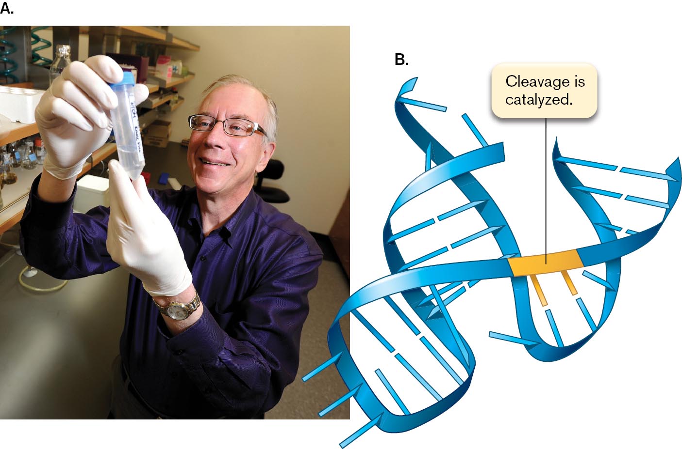
Reading the genomes enabled microbiologists to see the history of microbial evolution, reaching back to a time even before the advent of DNA—to a pre-DNA world when the cell’s chromosomes were actually composed of ribonucleic acid (RNA). This hypothetical world without DNA is called the RNA world. How did life function in the RNA world? We hypothesize that cells used RNA for all the functions of DNA and protein, including information storage and replication, and for biochemical catalysis. RNA molecules capable of catalysis, called ribozymes, were discovered in 1982 by Thomas Cech at the University of Colorado and Sidney Altman at Harvard University, who jointly earned the Nobel Prize in Chemistry in 1989 (Fig. 1.36). That same year, Jennifer Doudna and Jack Szostak at Harvard showed how an RNA molecule from the protozoan Tetrahymena could catalyze its own replication. These achievements support the theory that early organisms were composed primarily of RNA.
More information
A photograph of Tom Cech holding a test tube and a diagram of catalytic RNA are shown.
A photo shows Tom Cech holding a test tube containing liquid.
A model shows the structure of R N A, and a segment in the center is labeled as cleavage is catalyzed.
FIGURE 1.36 ■Discovery of catalytic RNA.A. Tom Cech holds a flask containing protists that make catalytic RNA, the kind of molecule that in early cells may have served both genetic and catalytic functions. B. Diagram of a catalytic RNA, where horizontal bars represent bases. The RNA catalyzes cleavage of itself.JEREMY PAPASSO/DIGITAL FIRST MEDIA/BOULDER DAILY CAMERA VIA GETTY IMAGES
How do DNA and RNA sequences convey information in the cell? To read the DNA language required deciphering the genetic code—how triplets of DNA “letters” specify the amino acid units of proteins. This story is discussed in Chapter 8.
The DNA revolution began with bacteria. What amazed the world about DNA was that such a simple substance, composed of only four types of subunits, is the genetic material that determines all the different organisms on Earth. The promise of this insight was first fulfilled in bacteria and bacteriophages, whose small genomes and short generation times made key experiments possible (see Chapters 6–9). Bacterial tools were later extended to animals and plants; for example:
Bacteria readily recombine DNA from unrelated organisms. The mechanisms of bacterial recombination led to construction of artificially recombinant DNA, or “gene cloning.” Recombinant DNA ultimately enabled us to transfer genes between the genomes of virtually all types of organisms.
Bacterial DNA polymerases are used for polymerase chain reaction (PCR) amplification of DNA. A hot spring in Yellowstone National Park yielded the bacterium Thermus aquaticus, whose DNA polymerase could survive many rounds of cycling to near-boiling temperature. The Taq polymerase formed the basis of a multibillion-dollar industry of PCR amplification of DNA, with applications ranging from genome sequencing to forensic identification.
Gene regulation discovered in bacteria provided models for animals and plants. The first key discoveries of gene expression were made in bacteria and bacteriophages. Regulatory DNA-binding proteins were discovered in bacteria and then subsequently found in all classes of living organisms.
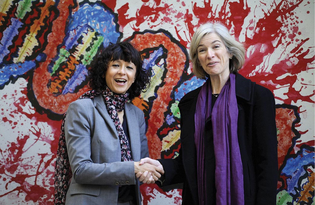
CRISPR-Cas9 is a molecular mechanism of bacterial defense against bacteriophages. This mechanism was developed as a means of editing human genomes for gene therapy. The use of CRISPR-Cas9 for human genome editing earned the 2020 Nobel Prize in Chemistry for Emmanuelle Charpentier (currently at the Max Planck Unit for the Science of Pathogens) and Jennifer Doudna, at UC Berkeley (Fig. 1.37). CRISPR-Cas9 is described in Chapter 12.
More information
A photograph of Emmanuelle Charpentier and Jennifer Doudna shaking hands.
FIGURE 1.37 ■Nobel Prize for CRISPR-Cas9. Emmanuelle Charpentier and Jennifer Doudna won the 2020 Nobel Prize in Chemistry for CRISPR-Cas9 discovery and application for human genome editing.REUTERS/ALAMY STOCK PHOTO
In the 1970s, when the DNA revolution began, its implications drew public concern. The use of recombinant DNA to make hybrid organisms—organisms combining DNA from more than one species—seemed “unnatural.” We now know that in natural environments, genes frequently move between species. Furthermore, recombinant DNA technology raised the specter of placing deadly genes that produce toxins such as botulin into innocuous human-associated bacteria such as E. coli.
The unknown consequences of recombinant DNA so concerned molecular biologists that in 1975 they held a conference to assess the dangers of and restrict recombinant DNA experimentation. The conference, led by Paul Berg and Maxine Singer at Asilomar (Pacific Grove, California), was possibly the first time in history that a group of scientists organized and agreed to regulate and restrict their own field.
On the positive side, the emerging world of molecular biology excited the imagination of young scientists and entrepreneurs. The pioneering biotechnology company Genentech was founded in 1976 by Robert Swanson and bacterial geneticist Herbert Boyer, from UC San Francisco. Growing numbers of students entered the field of molecular biology, seeking to invent medical cures—or even to clone dinosaurs, as in Michael Crichton’s novel and film Jurassic Park (1993). While the idea of cloning a dinosaur remains science fiction, the tools of microbial genetics opened a window into the past by letting us read the DNA of long-dead organisms preserved in museums. In 1980, the first patent was upheld for a living organism—a bacterium genetically modified to catabolize oil components from petroleum spills. Since then, it has become accepted to patent modified mice for cancer studies.
Thought Question
1.11 State an argument in favor of patenting a microbial isolate or a gene sequence. What argument can be made against patenting microbes or genes?
ANSWER ANSWER
A microbe can be patented if it is genetically modified from the “natural” state, and if it has a commercial use or application. Mere discovery of a pollutant-eating organism from nature is not patentable, but modifying an organism for commercial use constitutes technology. If a microorganism consists of a complex of molecules, then modifying the organism is legally equivalent to modifying a drug molecule or other industrial chemical. An argument against patenting organisms might be that life has a special status, in that organisms have their own agency to proliferate. Living organisms have a special spiritual status in the worldview of most major religions. Therefore, it would be inappropriate to patent a microbe or a mouse—or a human being treated by gene modification therapy.
Microbial Discoveries Transform Medicine and Industry
Twentieth-century microbiology transformed the practice of medicine and generated entire new industries of biotechnology and bioremediation. After the discovery of penicillin, Americans poured millions of dollars of private and public funds into medical research. In 1938, the March of Dimes was founded to discover a polio vaccine; today, polio has been nearly eliminated. Research on microbes and other aspects of biology has grown with support from U.S. government agencies, such as the National Institutes of Health and the National Science Foundation, as well as from governments of other countries, particularly the European nations, Japan, and China. Further support comes from private foundations, such as the Pasteur Institute, the Wellcome Trust, and the Howard Hughes Medical Institute. Medical research generates astonishing advances, such as the use of human immunodeficiency virus (HIV, the cause of AIDS) to devise gene therapy agents that cure cancer (discussed in Chapter 11).
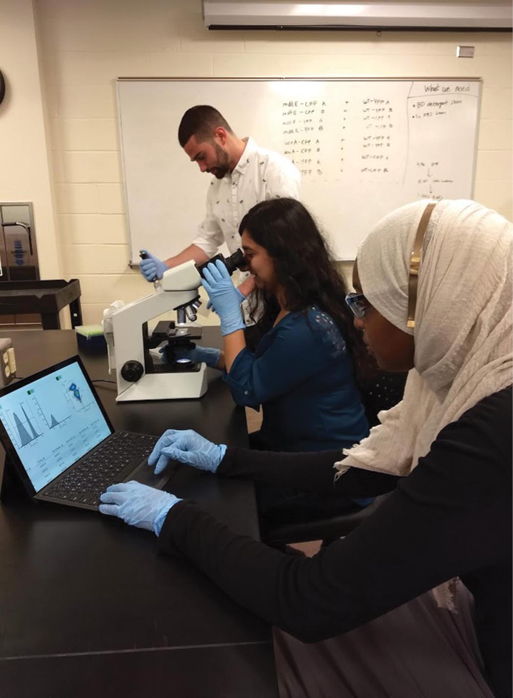
Research in microbiology includes fields as diverse as medicine and space science (Table 1.3). These fields all recruit microbiologists (Fig. 1.38). Industrial and applied biology (see Chapter 16) use bacteria to clone and produce therapeutic proteins, such as insulin for diabetics. Recombinant viruses make safer vaccines. At the frontiers of science, we use microbes for “synthetic biology,” the construction of novel organisms with useful functions (see Chapter 12). For example, synthetic biology may design bacteria with an on/off switch to report the presence of arsenic in environmental samples.
More information
A photo shows two women and a man in a research room. All three wear a pair of gloves and work on a table. One of the women works on a laptop, while the other woman uses a microscope. The man holds a pipette.
FIGURE 1.38 ■Microbiologists at work. Students at Kenyon College conduct research on bacterial gene expression.JOAN SLONCZEWSKI, KENYON COLLEGE
On a global level, the management of our planet’s biosphere, with the challenges of pollution and global warming, increasingly depends on our understanding of microbial populations. For example, as Earth heats up, frozen soil at the polar regions activates microbial decomposition, releasing carbon dioxide and methane gases that accelerate global warming. Microbiologists play critical roles in the field of climate science (see Chapter 22).
TABLE 1.3
Fields of Research in Microbiology
Field
Subject of study
Experimental microbiology
Fundamental questions about microbial form and function, genetics, and ecology
Medical microbiology
The mechanism, diagnosis, and treatment of microbial disease
Epidemiology
Distribution and causes of disease in humans, animals, and plants
Immunology
The immune system and other host defenses against infectious disease
Food microbiology
Fermented foods and food preservation
Industrial microbiology
Production of drugs, cloned gene products, and biofuels
Environmental microbiology
Microbial diversity and microbial processes in natural and artificial environments
Bioremediation
The use of microbial metabolism to remediate human wastes and industrial pollutants
Forensic microbiology
Analysis of microbial strains as evidence in criminal investigations
Climate science
The study of weather patterns and temperature changes over time, including processes mediated by microbial populations
To Summarize
Cell structure was revealed by new tools such as the electron microscope and the ultracentrifuge.
Genetics of bacteria, bacteriophages, and fungi in the early twentieth century revealed fundamental insights about gene transmission that apply to all organisms.
Structure and function of the genetic material, DNA, emerged from a series of experiments in the twentieth century.
Molecular microbiology generated key advances, such as the cloning of the first recombinant molecules and the invention of DNA sequencing technology.
Genome sequence determination and bioinformatic analysis became the tools that shape the study of biology in the twenty-first century.
Microbial discoveries transformed medicine and industry. Biotechnology produces new kinds of pharmaceuticals and industrial products. Synthetic biology engineers new kinds of organisms with useful functions.
A theory stating that the products of oxidative metabolism store their energy in an electrochemical gradient that can drive cellular processes such as ATP synthesis.
 ANSWER
 ANSWER