Who Were They? What Were They Like? The Bioarchaeology of People
One of archaeology’s principal aims is to recreate the lives of the people who produced the archaeological record, and what more direct evidence can there be than the physical remains of past humanity? Archaeology draws on the skills of a great variety of scientists, from radiocarbon experts to botanists, and the role of the modern archaeologist is to learn how best to use and interpret the lives of people from the archaeological point of view. Biological anthropology yields a wealth of evidence to enrich the archaeologist’s understanding of the past. Dealing with human remains also raises ethical issues (see Chapter 14). The word “bioarchaeology,” first coined in the 1970s by Grahame Clark to mean the study of animal bones, has now been applied instead to the study of human remains from archaeological sites (although in the Old World it still encompasses other organic material). When possible human remains are encountered and excavated by archaeologists (or, indeed, by the public or police), forensic anthropologists are usually brought in to examine them. Having established that the remains are indeed human, their task is to build up a biological profile of the deceased.
One of the most interesting and fast-developing fields of study in archaeology is in the origins of the human species, and in the origins and migrations of later populations anywhere on the planet. When and how did the uniquely human abilities emerge? What were the processes that led to the development of the first hominins? What did the first humans look like, and how did they adapt as they traveled across the globe? The use of genetic techniques such as ancient DNA are helping to answer these questions, but they are also working to challenge preconceptions of race and ethnic distinctiveness that many still hold today—highlighting, yet again, the value of archaeology.
A major reason for the lack of integration between archaeology and biological anthropology in the decades immediately after World War II was the question of “race.” During the nineteenth and early twentieth centuries, some scholars (and many politicians) attempted to use biological anthropology to help prove their theories of white “racial” superiority in the African and American continents. This stemmed from their belief that local, indigenous people were incapable of constructing impressive monuments, for instance the burial mounds of the eastern United States. As recently as the 1970s, the white government of Rhodesia maintained that the great monument that today gives the nation its name—Zimbabwe—could not have been the unaided work of the indigenous black population (see box, pp. 482–83). Today, biological anthropologists are much less willing to recognize supposedly different human populations on the basis of a few skeletal measurements. That does not mean that physical distinctions cannot be looked for and studied, but a more robust methodology is needed, supported by well-conceived statistical methods to ensure that any variations observed are not simply of a random nature.
Developments in biochemistry and genetics are now allowing much more work to be done at the molecular level, although the osteology—the study of bones—remains fundamental. These combined methods are discovering the genetic traces of otherwise unknown past populations, and of previously underestimated levels of population admixture, which are fueling new critiques on concepts of “race,” and showing that the ethnic fault lines drawn today are relatively recent, and were constantly changing.
The Variety of Human Remains
In order to answer questions about human populations and human physical characteristics in the past, an initial step is to establish that human remains are present, and in what number. This is relatively easy where intact bodies, complete skeletons, or skulls are available. Individual bones and large fragments should be recognizable to competent archaeologists. Even small fragments may include diagnostic features by which human beings can be recognized. In some careful excavations, individual hairs have been recovered that can be identified under the microscope as human. In cases of fragmentary multiple burials or cremations, the minimum number of individuals (see box, pp. 288–89) can be assessed from the part of the body that is most abundant. A new technique has been applied at the Scottish Mesolithic site of Cnoc Coig, on the island of Oronsay, where small unidentifiable scraps of bone were proved to be human by means of paleoproteomics (see p. 300)—other fragments were seal and pig.
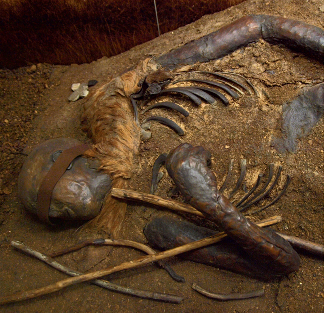
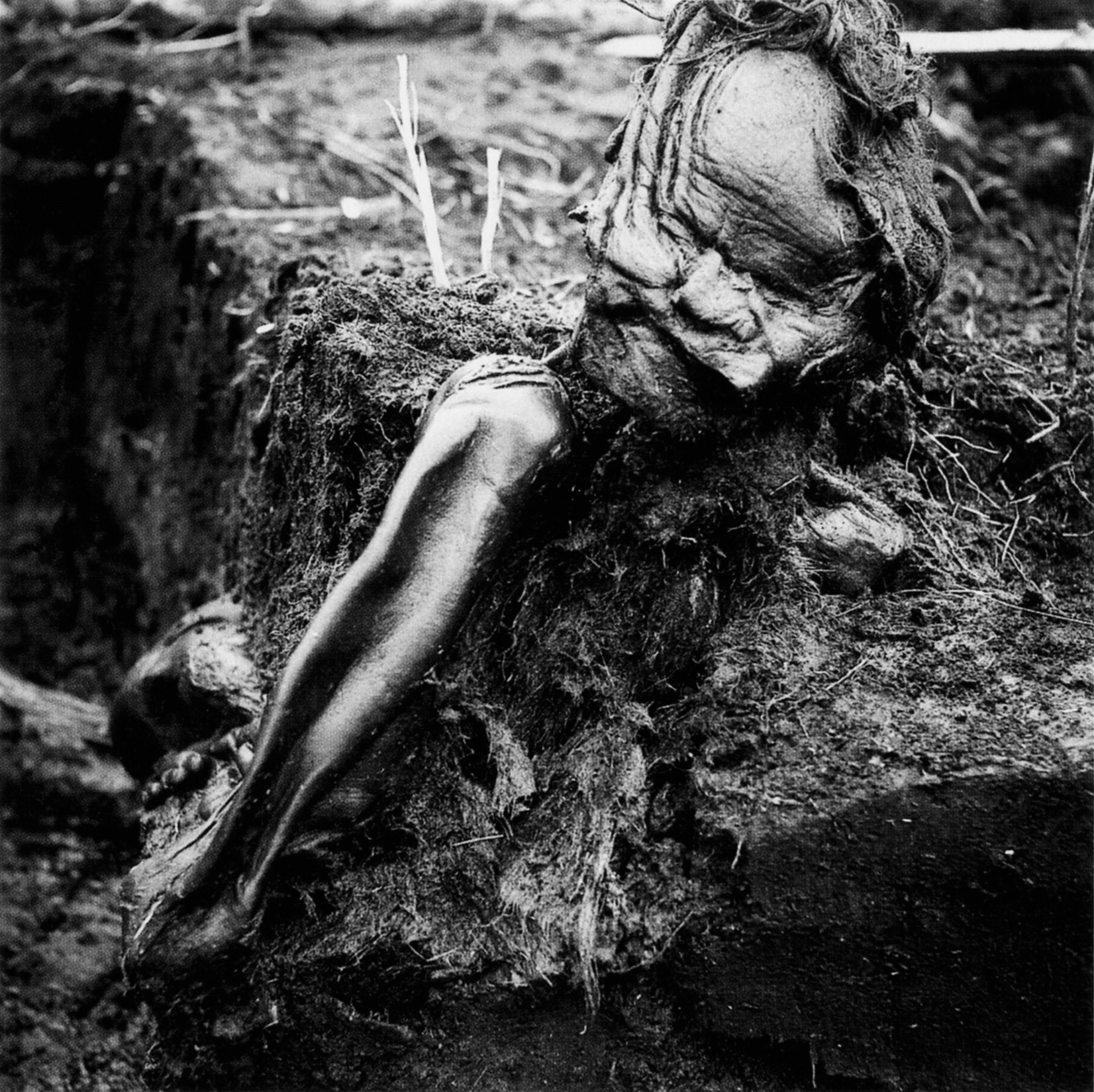
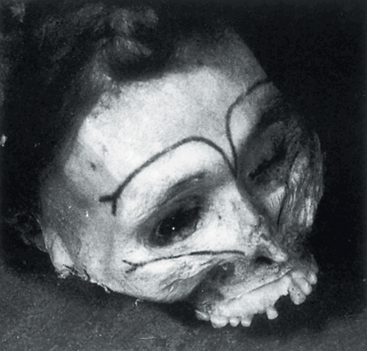
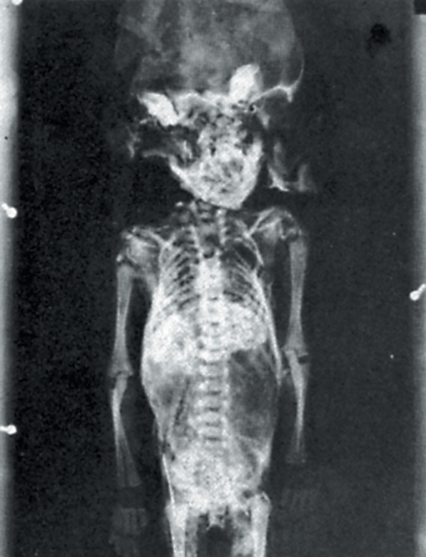
11.1 The variety of human remains. The wellpreserved body of a blindfolded boy, drowned in a bog pool at Windeby, northern Germany, about 2,000 years ago.
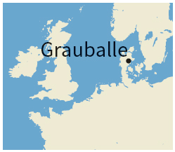
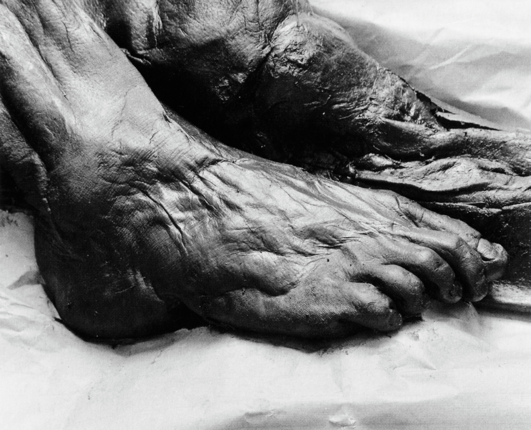
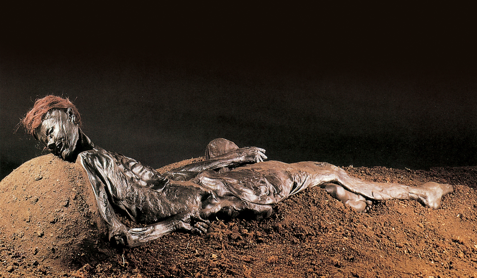
As we saw in Chapter 2, humanly created mummies are by no means the only bodies to have survived intact: others have become naturally desiccated, freeze-dried, or preserved in peat. Since so much of our appearance lies in the soft tissues, such corpses can reveal what mere skeletons cannot, namely features such as the length, style, and color of hair; skin color, and marks on the skin such as wrinkles, scars, and tattoos (some very clear, as in the fifth-century BCE frozen body of a Scythian chieftain); and details such as whether the penis is circumcised. In exceptional circumstances, the lines on fingertips that produce fingerprints, and the corresponding lines on the soles of the feet, may survive—the most famous example being the Iron Age Grauballe Man from Denmark (see box, pp. 454–55). Sometimes chemical action will alter original hair color but, observing material under ultraviolet light using a fluorescence microscope can often help to establish what that original color was.
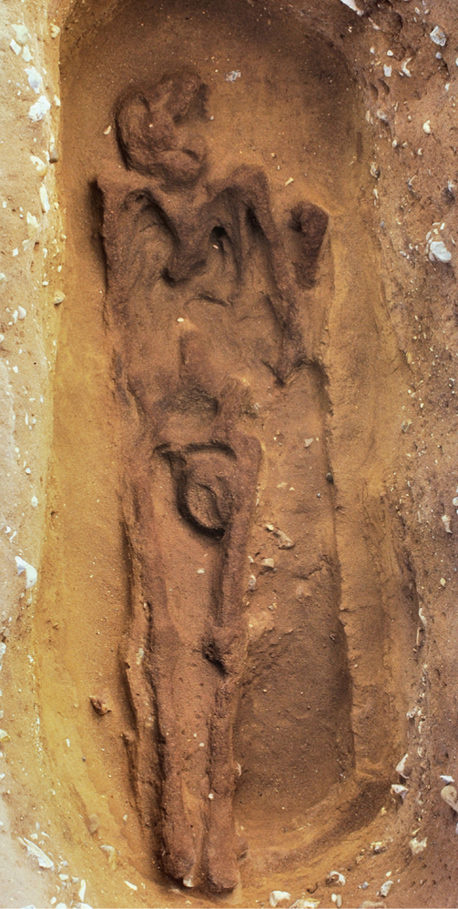
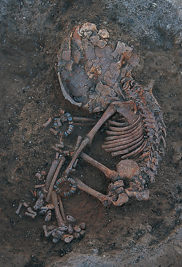
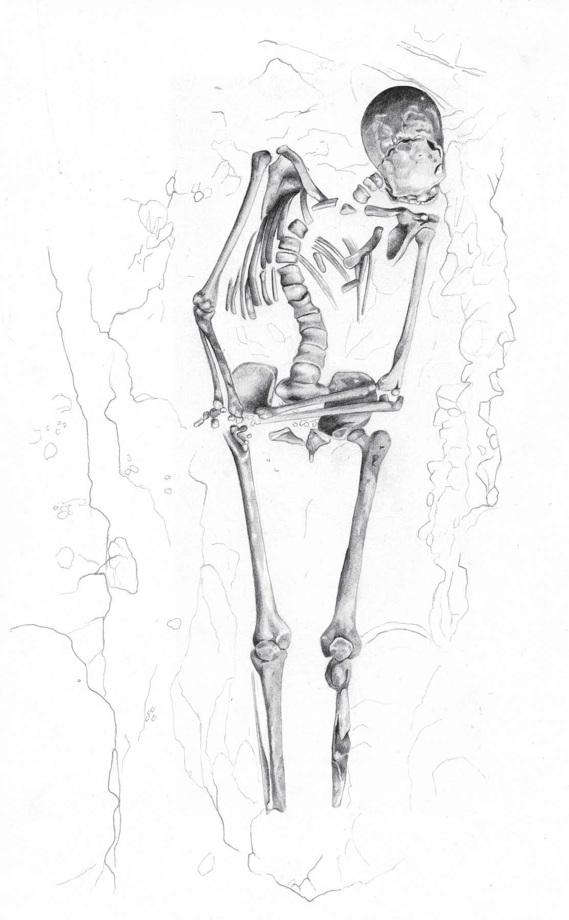
11.2 At Sutton Hoo, eastern England, the early medieval burials could be recovered only as outlines in the acid sandy soil.11.3 An Early Neolithic skeleton of a small child from the site of Çatalhöyük in Turkey, some 8,500 years old, wearing anklets and bracelets. Large numbers of beads are often found associated with child burials at the site.
Even where the body has disappeared, evidence may sometimes survive. The best-known examples are the hollows left by the bodies of the people of Pompeii as they disintegrated inside their hardened casing of volcanic ash (see box, pp. 24–25). Modern plaster casts of these bodies show not just general physical appearance, hairstyles, clothing, and posture but even such fine and moving detail as the facial expression at the moment of death. Footprints and handprints are a different kind of “hollow” in the archaeological record, and will be examined later.
Disappeared bodies can also be detected by other means. At Sutton Hoo, eastern England, the acid sandy soil has destroyed most remains, usually leaving only a shadowy stain in the soil—a kind of sand silhouette. If such traces are flooded with ultraviolet light, the “bone” in them fluoresces, and can be recorded photographically. Amino acids and other products of organic decay in the soil may help identify the sex and blood groups of such “invisible” corpses.
In Germany, numerous intact empty pots, buried in the cellars of houses between the sixteenth and nineteenth centuries CE, were tested by archaeologist Dietmar Waidelich. Samples of sediment from inside them were found, through chromatography, to contain cholesterol, which pointed to human or animal tissue, and steroid hormones such as estrone and estradiol. It is virtually certain, therefore, that the pots had been used to bury human placenta (afterbirth)—according to local folklore, this ensured the children’s healthy growth.
Finally, even where no skeletal remains have been found at all, it has now become possible to detect human presence in sites simply from their DNA. It has been known since 2003 that DNA can survive in ancient sediments, and in a recent study Neanderthal DNA, dating between 400,000 to 40,000 years ago, has been detected in sediments from four caves in Eurasia.
Nevertheless, the vast majority of human remains are in the form of actual skeletons and bone fragments. Indirect physical evidence about people also comes from ancient art and assumes great importance when we try to reconstruct what people looked like.
Identifying Physical Attributes
Once the presence and abundance of human remains have been established, how can we attempt to reconstruct physical characteristics—sex, age at death, build, appearance, and relationships?
Which Sex?
In the case of intact bodies and artistic depictions, sexing is usually straightforward from the genitalia. If these are not present, secondary characteristics such as breasts and beards and moustaches provide fairly reliable indicators. Without such features, the task is more of a challenge—length of hair is no guide, but associated clothing or artifacts may be of help. With depictions, one can go no further—for example, among the late Ice Age human figures from La Marche, France, the only definite females have vulvas or breasts, the definite males have male genitalia or beards/moustaches, and the rest of the figures have to be left unsexed. Claims that it is possible to distinguish male and female hand stencils in Europe’s Ice Age caves through measurements conflict with data from stencils in Australia where such distinctions are reportedly unreliable.
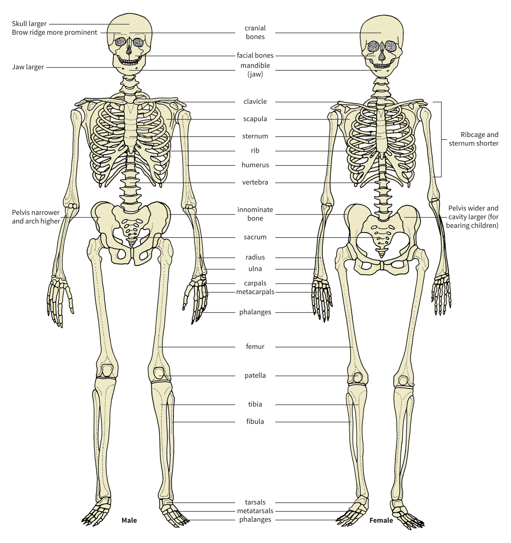
Where human skeletons and bone remains without soft tissue are concerned, however, one can go a great deal further owing to sexual dimorphism. The best indicator of sex is the shape of the pelvis, since males and females have different biological requirements. Other parts of the skeleton can also be used in sex differentiation. Male bones are generally bigger, longer, more robust, and have more developed muscle markings than those of females, which are usually slighter and more gracile. The proximal ends of male arm and thigh bones have bigger joint surfaces; and males have bigger skulls, with more prominent brow-ridges and mastoid processes (the bump behind the ear), a sloping forehead, a more massive jaw and teeth, and, in some populations, a bigger cranial capacity (in Europeans, above 1,450 cc, or 90 cu. in., tends to indicate a male, below 1,300 cc, or 80 cu. in., a female). These criteria, used in blind tests on modern adult bones, can achieve 85 percent or more accuracy—but females in certain parts of the world, such as some Polynesians and Australian Aborigines, often have very large skulls and large robust bones.
We should not rely on measurements of any one bone, but combine results from as many as possible. The objective is to assess variation in both size and shape. Single dimensions, such as the diameter of the round proximal head of the thigh bone (femur), can only indicate size, with one sex being larger, on average, than the other. Multiple measurements, especially when combined in multivariate analyses, permit the characterization of shape, which often provides better separation of the two sexes than size alone.
For children, it is worth noting that, with the exception of preserved bodies and artistic depictions, their remains cannot be sexed with the same degree of reliability as adults, although dental measurements have had some success. Progress has been made in sexing them by examining common physical differences in juvenile skeletons from Spitalfields, London (see box, p. 435), the sex and age of which are known from coffin labels.
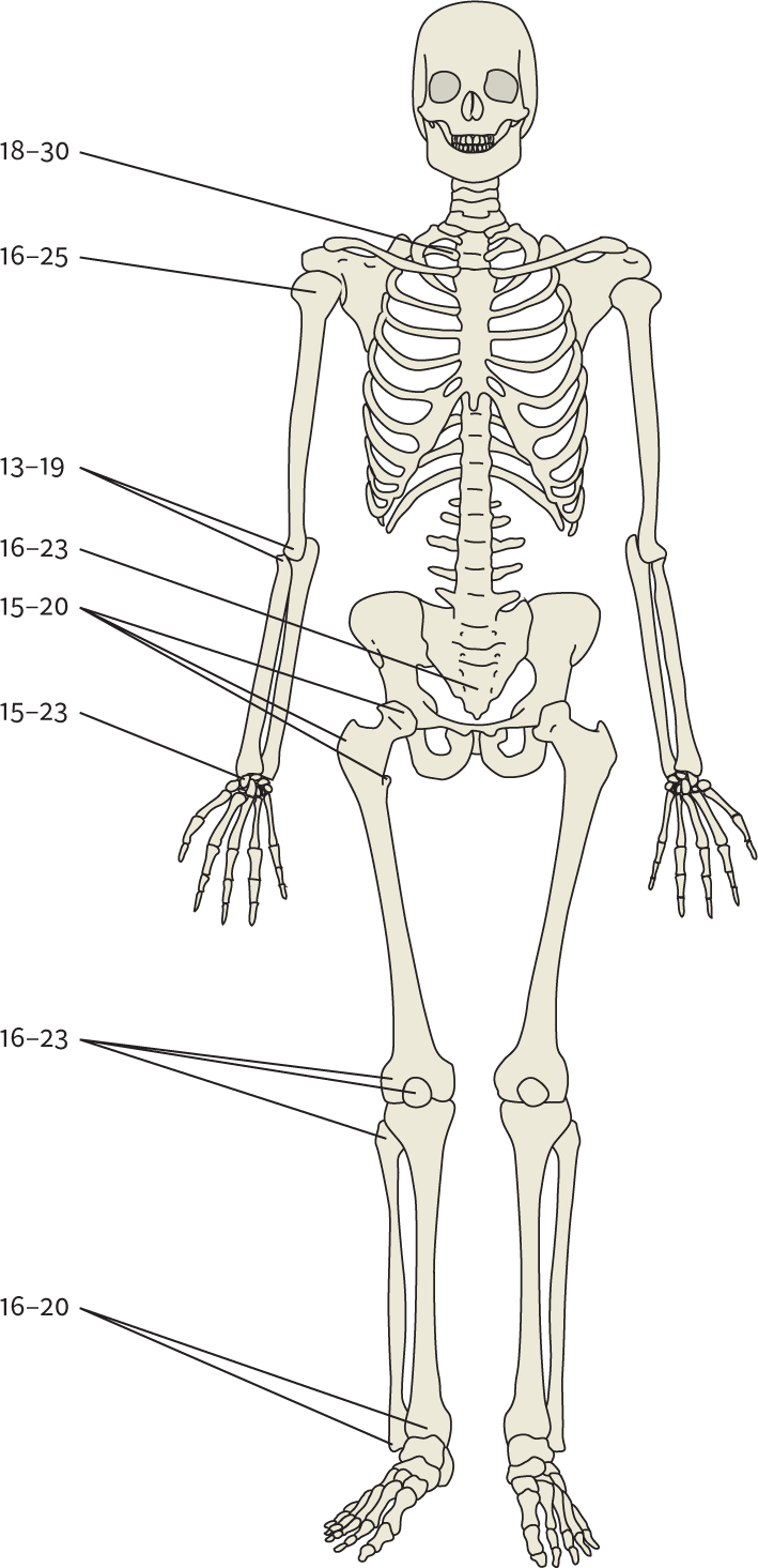
11.4 Bones of the human skeleton, with salient differences between the sexes.
Recently, a new technique has been developed for determining the sex of fragmentary or infant skeletal remains from DNA analysis (see below, p. 440–43). For example, skeletons of one hundred newborns have been recovered from a sewer beneath a Roman bathhouse (and probable brothel) at Ashkelon, Israel, most likely the victims of infanticide. Out of forty-three left femurs tested for DNA, nineteen produced results: fourteen were male and five female. A new DNA sequencing method has likewise successfully sexed ancient humans up to 70,000 years old, and has proven invaluable for determining the sex of juvenile or highly degraded remains. DNA can also be extracted from ancient feces, thanks to cells being sloughed off from the intestines during defecation, and can therefore determine the sex of the person who produced them—information that could eventually elucidate gender-based differences in diet. For instance, four feces from the La Quinta site, California, and Lovelock Cave, Nevada, were analyzed, and the originators of two were identified as female, one as male, and one remained indeterminate. Experiments on sex determination in excrement have also been carried out through an analysis of hormones and steroids such as estradiol and testosterone in feces from Salts Cave and Mammoth Cave in Kentucky, which, it turned out, had all been left by men.
How Long Did They Live?
As will been seen below, some scholars feel able to establish an exact age at death, but it should be stressed that all we can usually assign with any certainty is biological age at death—young, adult, old—rather than any accurate chronometric measurement in years and months. The best indicators of age for juveniles, as with fauna, are the teeth. Here one studies the calcification, eruption, and replacement of the milk teeth; the sequence of eruption of the permanent dentition; and finally the degree of wear, allowing as best one can for the effects of diet and method of food preparation.
A timescale for age at death derived from this kind of dental information in people works reasonably well for recent periods, despite much individual variation. But can it be applied to the dentition of our remote ancestors? Work on the microstructure of teeth suggests that old assumptions need to be tested afresh. Tooth enamel grows at a regular, measurable rate, and its microscopic growth lines form ridges that can be counted from epoxy resin replicas of the tooth placed in a scanning electron microscope. In modern populations a new ridge grows approximately each week, and analysis of molar structure in Neanderthals has shown that they had a very similar rate of growth to that of modern humans. The method has also been shown to be accurate on the Spitalfields juveniles (see box below).
Spitalfields: Determining Biological Age at Death
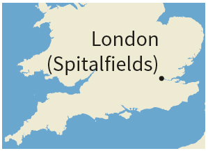
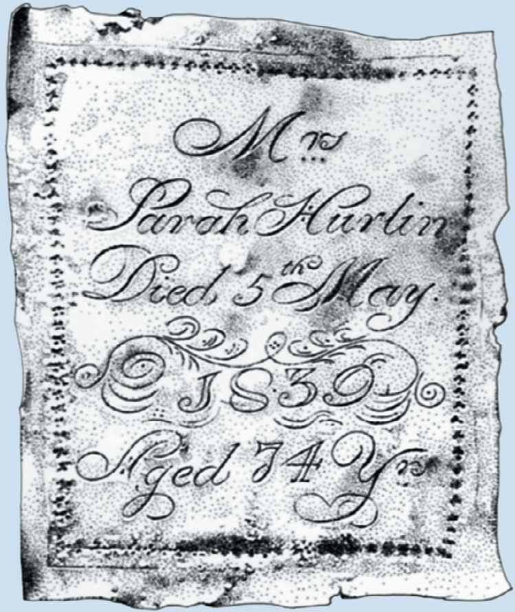
A rare opportunity to test the accuracy of different methods of ageing skeletal material came in 1984–86 with the clearance by archaeologists of almost a thousand inhumations in the crypt of Christ Church, Spitalfields, in east London. No fewer than 396 of the coffins had plates attached giving information on the name, age, and date of death of the occupants, who were all born between 1646 and 1852 and died between 1729 and 1852. Females and males were equally represented, and one-third were juveniles. The mean age at death of the adults was fifty-six for both sexes and the oldest was aged ninety-two.
11.5 Coffin plate of Sarah Hurlin, giving her name, age, and date of death.
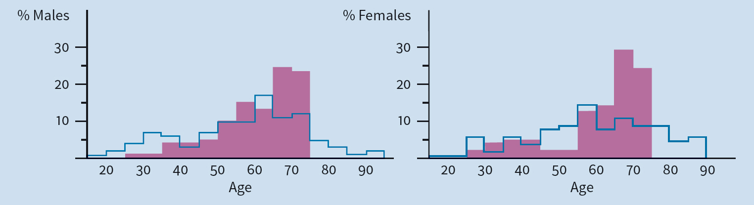
A range of techniques was used on the skeletons to evaluate apparent age at death, including the closure of cranial sutures, degeneration of the pubic symphysis (a joint between the pubic bones), the study of thin sections of bone tissue, and amino-acid racemization in teeth. The results were then compared with the true ages as documented on the coffin plates. It was found that traditional methods of determining age at death are inaccurate. All the methods applied to the Spitalfields skeletons tended to underestimate the age of the old, and overestimate the age of the young, a result that reflects the bias inherent in cemeteries composed primarily of individuals who died of natural causes. Those who die young have presumably failed to achieve their potential and already have “old bones,” while those who live to a great age are survivors and have “young” bones at death.
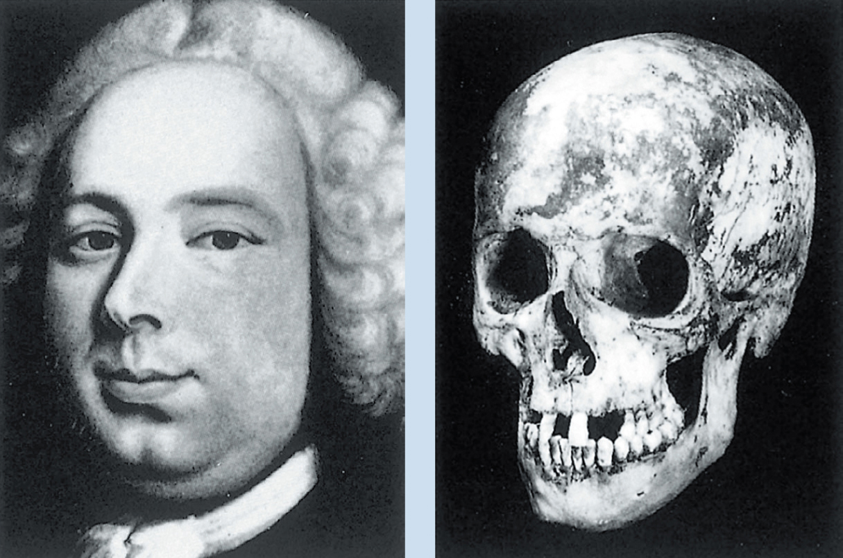
11.6–7 Peter Ogier (1711–1775), a master silk weaver, in life and death: a portrait compared with his actual skull.
In the Spitalfields population, children were small for their age compared to children today, but the material helped analysts develop and test methods that can give a fairly precise assessment of juvenile age. The Spitalfields adults began ageing later (after fifty) and at a slower rate than people today, which should make one cautious in applying data from modern reference samples to skeletal material from the past. As a result of the findings from Spitalfields, it would be rash, given existing methods, to try to age an adult more precisely than as biologically young, middle-aged, or old.
11.8 Comparison of the ages at death estimated from bone analysis (shaded) with real ages reveals that many mature adults had been given too high an age because they have “old bones.” The cut-off at seventy-five years old is due to the scale used for the reference population.
By measuring tooth growth ridges in fossil specimens, Tim Bromage and Christopher Dean concluded that previous studies overestimated the age at death of many early hominins. The famous 1–2-million-year-old australopithecine skull from Taung, South Africa, for example, belonged to a child who probably died at just over three years of age, not at five or six as had been believed. These conclusions have been confirmed by analyses of root growth patterns and by independent studies of dental development patterns in early hominins by Holly Smith, and by an investigation of the Taung skull’s dental development using computerized (or computed) axial tomography scans (CT or CAT scans). All this suggests that our earliest ancestors grew up more quickly than we do, and that their development into maturity was more like that of the modern great apes. This is supported by the biologically known fact that smaller creatures reach maturity sooner than larger ones (our earliest ancestors were considerably shorter than we are—see below).
Bromage and Dean, together with Chris Stringer, have also studied the Neanderthal child from Devil’s Tower Cave, Gibraltar, dating to perhaps 50,000 years ago, and changed its age at death from about five years to three years, a result confirmed by analysis of the temporal bone in the skull. An analysis of a Belgian Neanderthal child has likewise indicated that, at eight, its dental development was that of modern children several years older. There may, however, have been great variation in Neanderthal populations.
Other aspects of teeth can also provide clues to age. After a tooth’s crown has erupted fully, its root is still immature and takes months to become fully grown—its stage of development can be assessed by radiograph—and so, up to the age of about twenty, results can be obtained with some accuracy. The fully grown roots of a young adult’s teeth have sharp tips, but they gradually become rounded. Old teeth develop dentine in the pulp cavities, and the roots gradually become translucent from the tip upward. Measurement of the transparent root dentine of an 8,000-year-old skeleton from Bleivik, Norway, suggested an age at death of about sixty. Accumulated layers of cement around the roots can also be counted to estimate the years since a tooth erupted, although this procedure is not without problems.
Bones are also used in assessing age. The sequence in which the articulating ends (epiphyses) of bones become fused to the shafts gives a timescale that can be applied to the remains of young people. One of the last bones to fuse is the inner end of the clavicle (collarbone) at about eighteen to thirty years old; after that, different criteria are needed to age bones. Fusion, the joining of separate pieces of bone, can also indicate age: for instance, the five parts of the sacrum (the base of the spine) unify at between sixteen and twenty-three years old.
The degree of fusion of the sutures between the plates of the skull can be an indicator of age, but the presence of open sutures should not necessarily be taken as an indication of youth: open sutures can persist in old individuals. Skull thickness in immature individuals on the other hand does bear a rough relationship to age—the thicker the skull the older the person. But in old age all bones usually get thinner and lighter, although skull bones actually get thicker in about 10 percent of elderly people. Ribs can also be used to provide an age at death for adults, since their sternal end becomes increasingly irregular and ragged with age, as the bone thins and extends over the cartilage: this method was used on the man thought to be either Philip II of Macedon (Alexander the Great’s father) or Philip III (Alexander’s half-brother) found in a tomb at Vergina, northern Greece (see p. 547): it suggests he was closer to forty-five than thirty-five (historical evidence indicates that Philip II was forty-six when murdered). Other skeletal features used in age assessment include the pubic symphysis and the sacroiliac joint, which connects the lower spine to the pelvis.
11.9 Assessing age: the years at which bone epiphyses fuse.
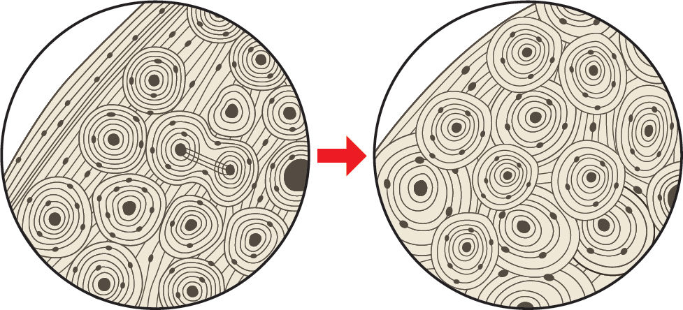
But what if the bone remains are small fragments? The answer lies under the microscope, in bone microstructure. As we age, the architecture of our bones changes in a distinct and measurable way. A young long bone, at about twenty years old, has rings around its circumference, and a relatively small number of circular structures called “osteons.” With age the rings disappear, and more and smaller osteons appear (see ill. 11.10). By this method, even a fragment can provide an age. Putting a thin section of a femur under the microscope and studying the stage of development is a technique that, in blind tests with documented known skeletons, has achieved accuracy to within five years. On material from Spitalfields, however, it proved no more accurate than other methods.
11.10 Assessing age: changes in bone structure are visible under the microscope as humans grow older. The circular osteons become more numerous and extend to the edge of the bone.
Akira Shimoyama and Kaoru Harada (1927–2010) applied a chemical method to a skeleton from a seventh-century CE burial mound in Narita, Japan. They measured the ratio of two sorts of aspartic acid in its dentine. This amino acid has two forms, or isomers, that are mirror images of each other. The L-isomer is used in building teeth, but converts slowly to the D-isomer during life. The D:L ratio increases steadily from the age of eight to eighty-three, and is therefore directly proportional to one’s age. In this case, it was shown that the skeleton was that of a fifty-year-old. Since the L-isomer continues to convert to the D-isomer after death, depending on temperature, the burial conditions have to be taken into account in the calculation.
Interpreting Age at Death.
It must be stressed that we can only calculate average age at death for the bodies and skeletons that have survived and been discovered. Many scholars used erroneously to believe that to dig up a cemetery, and assess the age and sex of its occupants, provided an accurate guide to the life expectancy and mortality pattern of a particular culture. This entails the considerable assumption that the cemetery contains all members of the community who died during the period of its use—that everyone was buried there regardless of age, sex, or status; that nobody died elsewhere; and that the cemetery was not reused at another time. This assumption cannot realistically be made. A cemetery provides a sample of the living population, but we do not know how representative that sample might be. Figures on life expectancy and average age in the literature should therefore be looked at critically before they are accepted and used by archaeologists.
But it is not sufficient to have a population broken down by age and sex. We also want to know something of their build and appearance.
What Was Their Height and Weight?
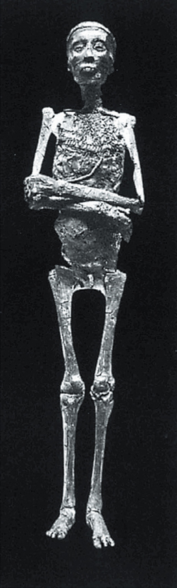
Height is easy to calculate if a body is preserved whole—as long as one allows for the shrinkage caused by mummification or desiccation. But it is also possible to assess stature from the lengths of some individual long bones, especially the leg bones. Tutankhamun’s height, for example, was estimated from the mummy and from his long bones as 1.69 m (5 ft. 6.5 in.), which corresponded to that of the two wooden guardian statues standing at his burial chamber door.
The formula for obtaining a rough indication of height from the length of long bones is called a “regression equation”—the metrical relationship of bone length to full body length. Different populations, however, require different equations because they have differing body proportions. Australian Aborigines and many Africans have very long legs that constitute 54 percent of their stature; but the legs of some Asian people may represent only 45 percent of their height. Consequently, people of the same height can have leg bones of very different lengths. The answer, in cases where the source population of the skeletal material is unknown, is to use a mean femoral stature (an average of the different equations), which will provide an adequate estimate of height, probably accurate to within 5 cm or a couple of inches, which is good enough for archaeological purposes. In Roman Cirencester, southern England, people seem to have been a little shorter than today: the average female height was 1.57 m (5 ft. 2 in.), and the tallest woman was equivalent in height to the average man (1.69 m or 5 ft. 6.5 in.).
Arm bones, where necessary, can also be used to estimate stature, as in the legless Lindow Man (c. 2 BCE–111 CE; hand stencils have also occasionally been used. And footprints also give a good indication, since foot length in adult males is reckoned to be equivalent to 15.5 percent of total height; in children under twelve it is thought to be 16 or 17 percent. The Laetoli footprints in Tanzania (see p. 443), which date to 3.75–3.60 million years ago, are 18.5 and 21.5 cm (7.3 and 8.5 in.) in length, and were therefore probably made by hominins of about 1.2 and 1.4 m (3 ft. 11 in. and 4 ft. 7 in.) in height, assuming that the same calculation is equally valid for premodern people.
Weight can also be calculated from intact bodies, since it is known that dry weight is about 25 to 30 percent of live weight. An Egyptian mummy of 835 BCE at Pennsylvania University Museum (designated “PUM III”) was therefore reckoned to have weighed between 37.8 and 45.4 kg (83–100 lb) when alive. Simply knowing the height can also be a guide, since from modern data we know the normal range of weight for people of either sex at given heights, who are neither obese nor unusually thin. Therefore, armed with the sex, stature, and age at death of human remains, we can make a reasonable estimate of weight. A single leg bone could indicate not only the height but also the sex, age, and bulk of its owner. Where early hominins are concerned, body size is more a matter of conjecture. Nevertheless, because the skeleton of the australopithecine nicknamed “Lucy” (see pp. 441 and 443) is 40 percent complete, it has been possible to reckon that this hominin was about 1.06 m (3 ft. 6 in.) tall, and weighed about 27 kg (60 lb).
So far, we have a sexed body of known age and size; but it is the human face that really serves to identify and differentiate individuals. How, therefore, can we pull faces out of the past?
What Did They Look Like?
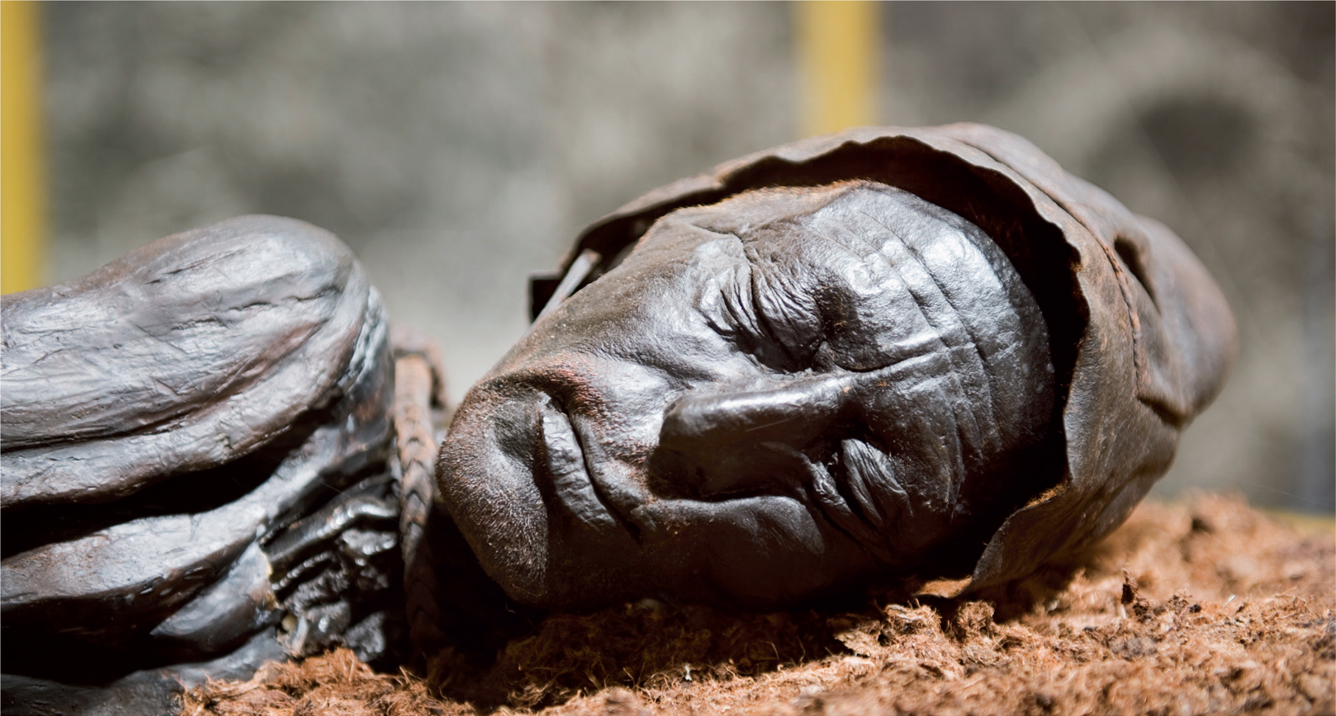
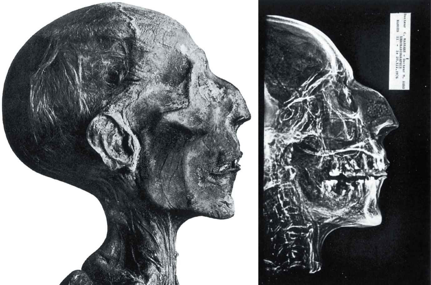
Once again, it is preserved bodies that provide us with our clearest glimpses of faces. Tollund Man, one of the remarkable Iron Age bog bodies from Denmark, is the best-known prehistoric example. Another finely preserved face belongs to the fifty-year-old man from Tomb 168 near Jinzhou in China, who was buried in the second century BCE and perfectly preserved by a mysterious dark red liquid. Discoveries at Thebes in Egypt in 1881 and 1898 of two royal burial caches have given us many mummified pharaohs, their faces still vivid, even if some shrinkage and distortion have taken place.
Thanks to artists from the Upper Paleolithic onward, we also have a huge array of portraits. Some of them, such as images painted on mummy cases, are directly associated with the remains of their subject. Others, such as Greek and Roman busts, are accurate likenesses of well-known figures whose remains may be lost forever. The extraordinary life-size terracotta army found near Xi’an, China, is made up of thousands of different models of soldiers of the third century BCE. Even though only the general features of each are represented, they constitute an unprecedented “library” of individuals, as well as providing invaluable information on hairstyles, armor, and weaponry (see ill. 5.6). From later periods, we have many life- or death-masks, sometimes used as the basis for life-size funerary effigies or tomb figures, such as those of European royalty and other notables from medieval times onward.
Identifying and Reconstructing Faces.
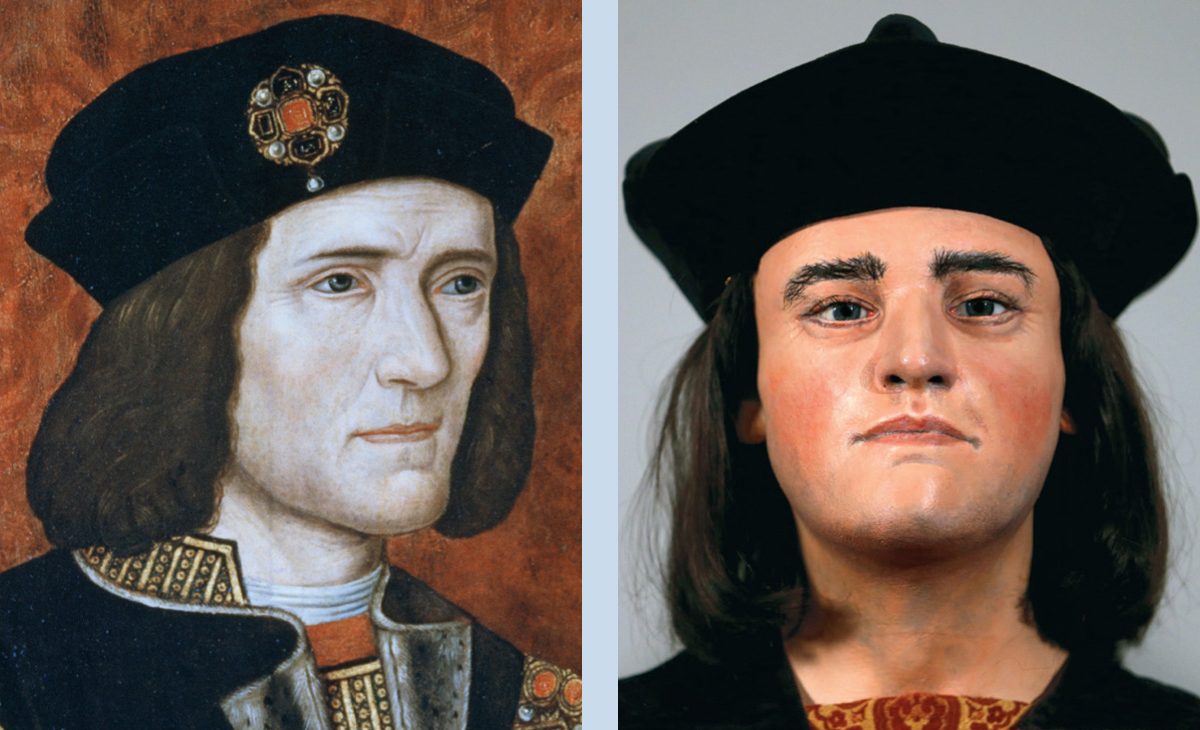
Occasionally, we can identify historical individuals by juxtaposing bones and portraits. Belgian scholar Paul Janssens developed a method of superimposing photographs of skulls and portraits. By this means one can confirm the identity of skeletons during the restoration of tombs. For instance, a photo of the skull thought to belong to Mary, Duchess of Burgundy (1457–1482), was superimposed on a picture of the head from her tomb’s sculpture and the match proved to be perfect. Superimposition of photos and skulls was also used to help identify the skulls of Tsar Nicholas II, his wife Alexandra, and their children, murdered by the Bolsheviks in 1918 and excavated some years ago from the pit in a Russian forest where they had been buried.
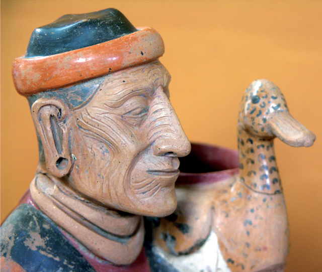
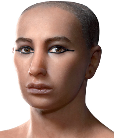
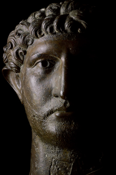
11.11 Faces from the past. Tollund Man, the Iron Age bog body from Denmark.11.12 Faces from the past. An old man with a wrinkled face is portrayed (with an accompanying duck) on this late Tiwanaku period (500–1100 CE) vase from the island of Pariti in Lake Titicaca, Bolivia.11.13 Faces from the past. Tutankhamun’s mummy was unwrapped in 1923, revealing within the bandages a shrunken body. The young king’s original height was estimated by measuring the long bones.11.14 Faces from the past. Tutankhamun’s facial features were reconstructed in 2014 using CT scans of his skull as a base—three teams separately produced very similar reconstructions, one of which is shown here.11.15 Faces from the past. Bronze head of the Roman emperor Hadrian (reigned 117–138 CE, from the River Thames.
Facial Reconstructions
Attempts to reconstruct faces were already being carried out in the nineteenth century by German anatomists in order to produce likenesses from the skulls of celebrities such as the poet Friedrich Schiller (1759–1805), the philosopher Immanuel Kant (1724–1804), and the composer Johann Sebastian Bach (1685–1750). But the best-known exponent of the technique in the twentieth century was the Russian Mikhail Gerasimov (1907–1970), who worked on remains ranging from fossil humans to those of Tsar Ivan the Terrible (reigned 1547–1584). It is now felt that much of his work represented “inspired interpretation,” rather than factual reconstruction. The process has now reached a higher degree of accuracy.
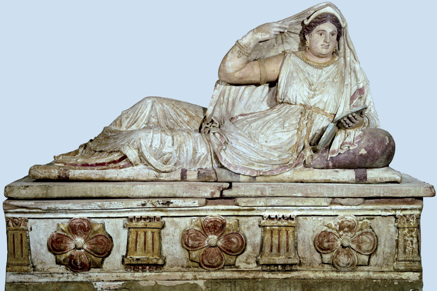
One of the most intriguing reconstructions has been of the best-preserved Etruscan skeleton known today, that of a noblewoman called Seianti Hanunia Tlesnasa, who died about 2,200 years ago in central Italy. Since 1887 her remains have been housed in the British Museum inside a splendid painted terracotta sarcophagus that bears her name engraved on it. The lid of this sarcophagus features a life-size image of the dead woman, reclining on a soft pillow, with a bronze mirror in her jeweled hand.
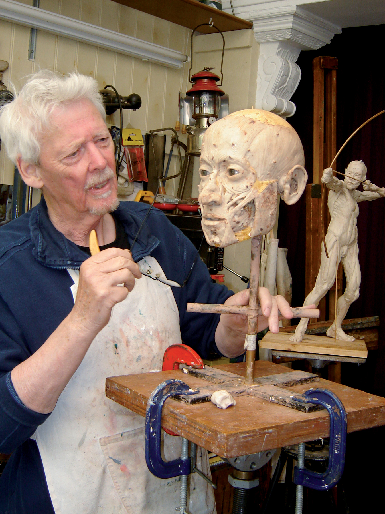
For years there had been doubts as to whether the bones in the casket were really hers. A team led by Judith Swaddling and John Prag set out to investigate the lady’s remains, and specialist Richard Neave was asked to reconstruct the dead woman’s face from her skull in order to compare it with her depiction.
11.16 Richard Neave reconstructs a face.
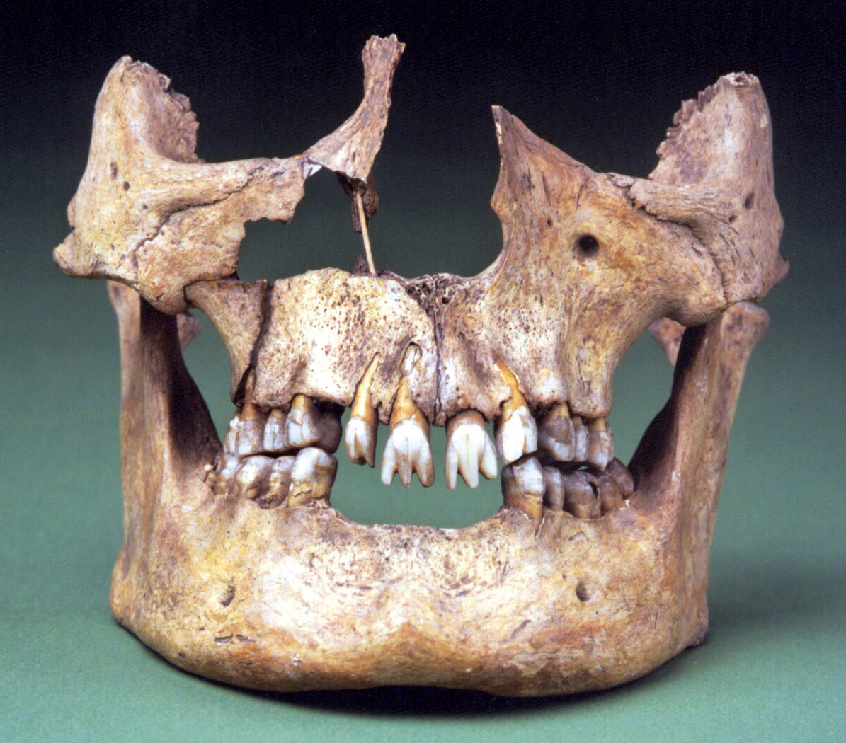
Anthropologists deduced from the skeleton that the woman was about 1.5 m (4 ft. 11 in.) tall, and middle-aged at death. Damage and wear on her bones, and the fact that she was almost toothless, had at first suggested old age, but in fact she had incurred severe injuries, most likely a riding accident, which had crushed her right hip and knocked out the teeth of her right lower jaw. The bone was damaged where the jaw joins the skull, and opening her mouth wide would have been painful. This prevented her from eating anything but soups and gruels, and from keeping her remaining teeth clean—most of them subsequently fell out. Seianti would also have had painful arthritis and increasing disabilities.
11.17 The sarcophagus of Seianti Hanunia Tlesnasa, which contained her bones; the lid takes the form of a life-size image of the dead woman—but how accurately did it represent her appearance?
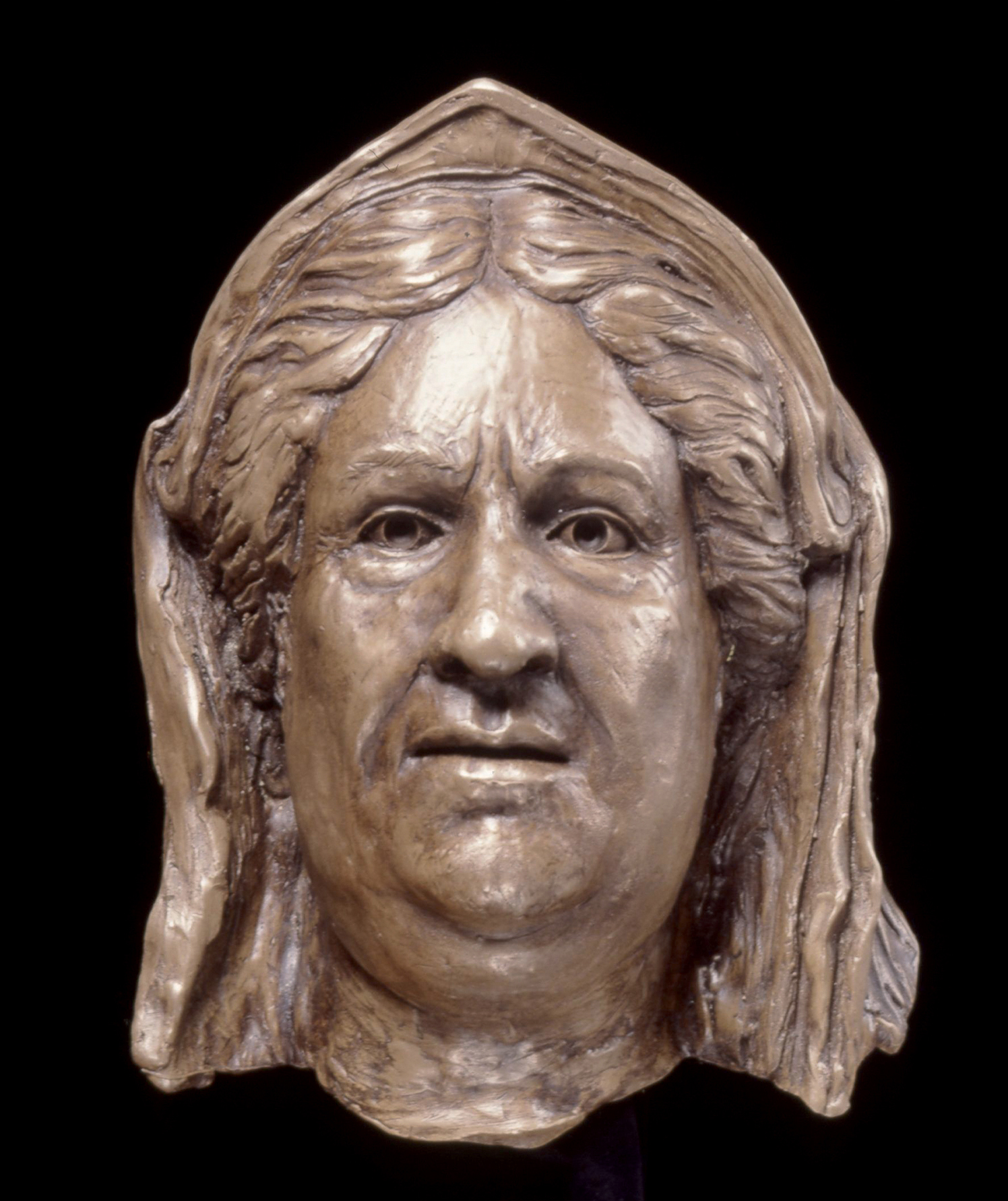
Two of the surviving teeth confirmed, from analysis of the dentine, that she died aged about fifty. And radiocarbon dating of the bones produced a result of 250–150 BCE, which proved that the skeleton was genuinely ancient and of the right period. The facial reconstruction showed a middle-aged woman who had grown rather obese. How did it compare with the coffin image?
From the side, there were differences, since the artist had given Seianti a prettier nose, but from the front the resemblances were clearer. The final confirmation came from a computerized technique for matching facial proportions and features—the computer photocomparison of the reconstruction and the portrait left no doubt that this was the same person. The sarcophagus image showed her as some years younger, with fewer chins, and a smaller, more girlish mouth. In other words, the sculptor had made flattering improvements to the portrait of this short, portly, middle-aged woman, but also captured Seianti’s likeness extremely well.
11.18 The reconstruction made from the skull found in the sarcophagus.
A case study of the facial reconstruction of an Etruscan woman, including computer photocomparison with a sarcophagus portrait, is discussed in the box on p. 439. Some facial reconstructions are now done with a laser-scanning camera connected to a computer containing information about the skull’s muscle-group thickness, and a computer-controlled machine then cuts a 3D model out of hard foam: this method has been used, for example, to recreate the face of a Viking fisherman at York. Such reconstructions are useful for museum display and TV programs, as well as to help identify an individual, but are not done routinely.
Any jewelry or clothing found associated with bodies or skeletons are also invaluable in assessing how these people looked during life. Footprints provide clues about footwear. Nearly all Ice Age prints are barefoot, but one of those in the French late Upper Paleolithic cave of Fontanet seems to have been made by a soft moccasin.
How Were They Related?
Increasingly, it is possible to assess the relationship between two individuals by looking at skull shape (which can be affected by many factors, such as diet), analyzing the hair, or studying ancient DNA. There are however, other methods of achieving the same result, such as by examining dental morphology. Some dental anomalies (such as enlarged or extra teeth, and especially missing wisdom teeth) run in families.
Blood groups have been determined from soft tissue, bone, and even from tooth dentine up to more than 30,000 years old, since the polysaccharides responsible for blood groups are found in all tissues, not just in red blood cells, and survive well. Indeed, protein analysis by radioimmunoassay (the detection of reaction to antibodies) can now identify protein molecules surviving in fossils that are thousands or even millions of years old, and can decipher taxonomic relationships of fossil, extinct, and living organisms. In the near future, we may obtain useful information on the genetic relationships of early hominins.
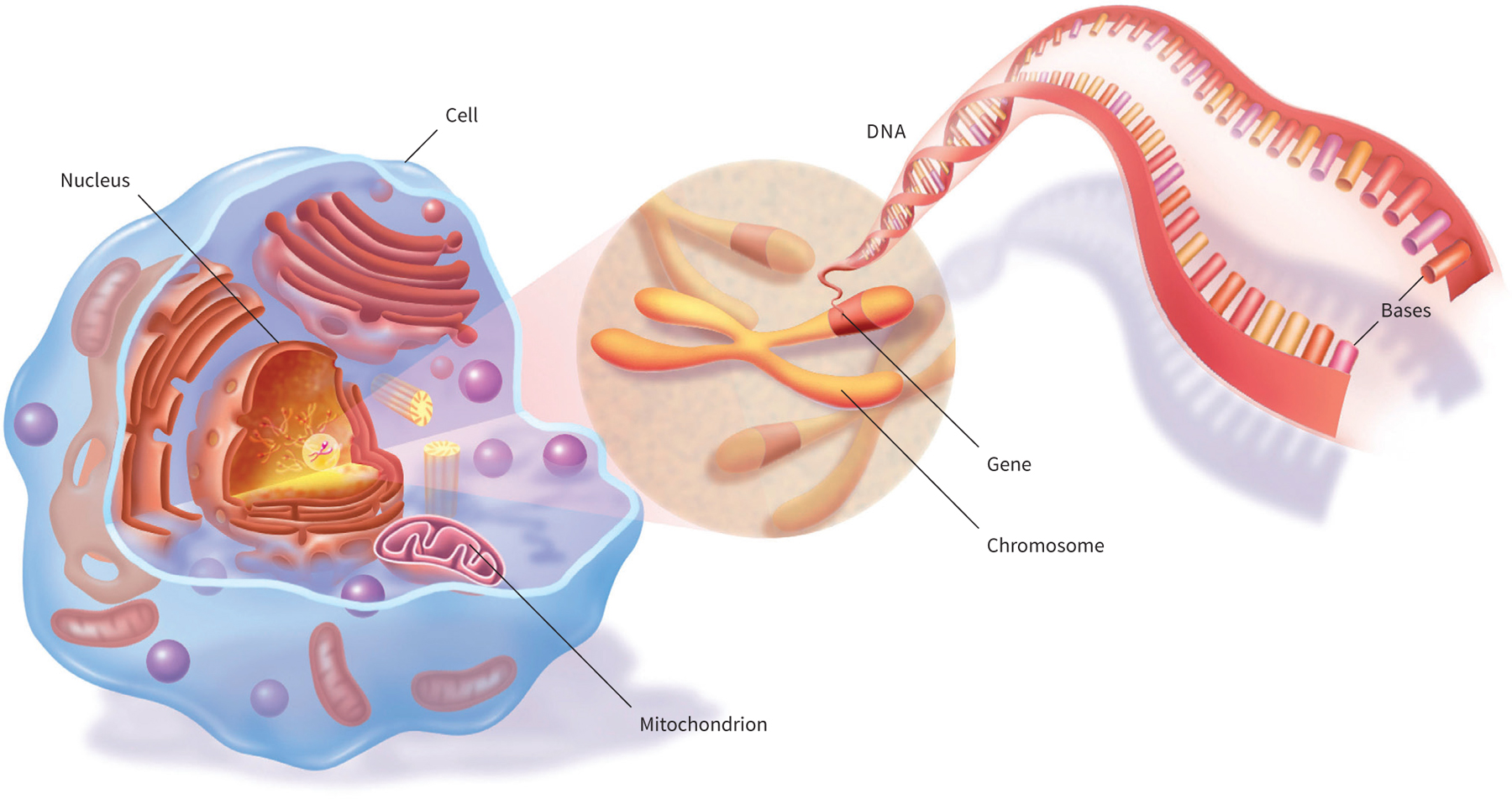
11.19 Genes, the organizers of inheritance, are composed of DNA (deoxyribonucleic acid), which carries the hereditary instructions needed to build a body and make it work. Genes are copied, or “replicated,” with every new generation of living cell. The majority of DNA is found in the nucleus of the cell and is called nuclear DNA. It is copied every time a new cell is produced. The mitochondria (small organelles) within the cell also contain DNA (mitochondrial DNA, or mtDNA) in relatively small loops that replicate separately from nuclear DNA. Both nuclear and mtDNA are important in archaeological genetic research.
Since blood groups are inherited in a simple fashion from parents, different systems—of which the best known is the A-B-O system, in which people are divided into those with blood types A, B, AB, and O—can sometimes help clarify physical relationships between different bodies. For example, it was suspected that Tutankhamun was somehow related to the unidentified body discovered in Tomb 55 at Thebes in 1907. The shape and diameter of the skulls were very similar, and when radiographs of the two crania were superimposed there was almost complete conformity. Robert Connolly and his colleagues therefore analyzed tissue from the two mummies, which showed that both had blood of group A, subgroup 2 with antigens M and N, a type relatively rare in ancient Egypt. This fact, together with the skeletal similarities, made it almost certain that the two were closely related. Subsequent DNA analysis has led to claims that the Tomb 55 body is Tutankhamun’s father, the pharaoh Akhenaten, and also to the presumed identification of Tutankhamun’s mother, grandparents, wife, and children—although not all specialists have accepted these conclusions.
Results from genetics, however, do show that family relationships can be worked out through DNA analysis (see ill. 11.19). In 1985 the Swedish scientist Svante Pääbo first succeeded in extracting and cloning mitochondrial DNA (mtDNA) from the 2,400-year-old mummy of an Egyptian boy. Over such a long time period, the DNA molecules are broken up by chemical action, so there is no question of reconstituting a functioning gene, far less a living body. But information on the DNA sequences of, for example, Egyptian mummies may determine whether members of a dynasty did indeed practice incest, as is commonly believed: an analysis of DNA from six mummies of 2200 BCE found at Hagasa, Egypt, has proved that they were a family group (for a recent study of the DNA of a family group in Neolithic Germany, see box overleaf). Currently, a databank in Manchester, England, of thousands of tissue samples is being compiled from mummies all over the world, for future research into everything from the spread of diseases to human migrations.
Genetic material has also been removed from ancient human brain cells in Florida by Glen Doran and his colleagues. Brain material has been recovered from ninety-one of 168 individuals buried in Windover Pond, a peat bog near Titusville, between 8,000 and 7,000 years ago. Some of the skulls, when placed in a scanner, proved to contain well-preserved and largely undamaged brains. DNA extracted from them may make it possible to discover whether there are any survivors from this particular Indian group.
It is now possible also to extract the tiny amounts of DNA left in bones and teeth. Researchers at Oxford, using the “polymerase chain reaction,” have been able to amplify minute amounts of DNA for study.
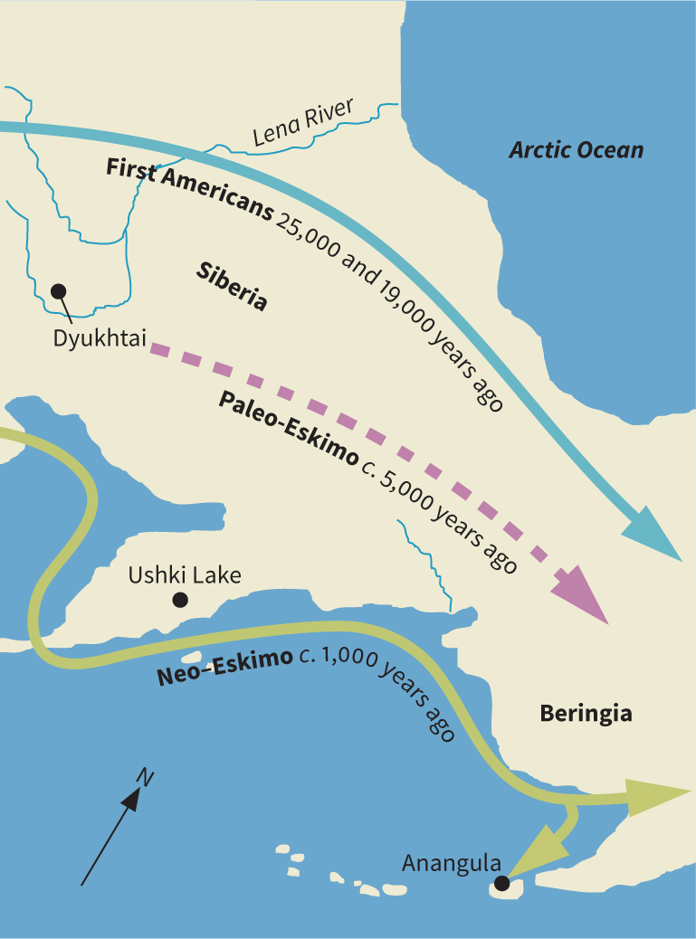
Pääbo has retrieved DNA molecules from the brains, bones, and teeth of Archaic-period Native American (more than 7,000 years old) found in 1988 in Little Salt Spring, Florida. The molecules contained a previously unknown mitochondrial DNA sequence, which suggests that America was entered by a group of humans entirely separate from the three lineages known to have migrated there (see box, p. 473), but that they died out some time after their arrival. This may represent the only demonstrated instance of the recent extinction of a group of Native Americans with no close surviving relatives.
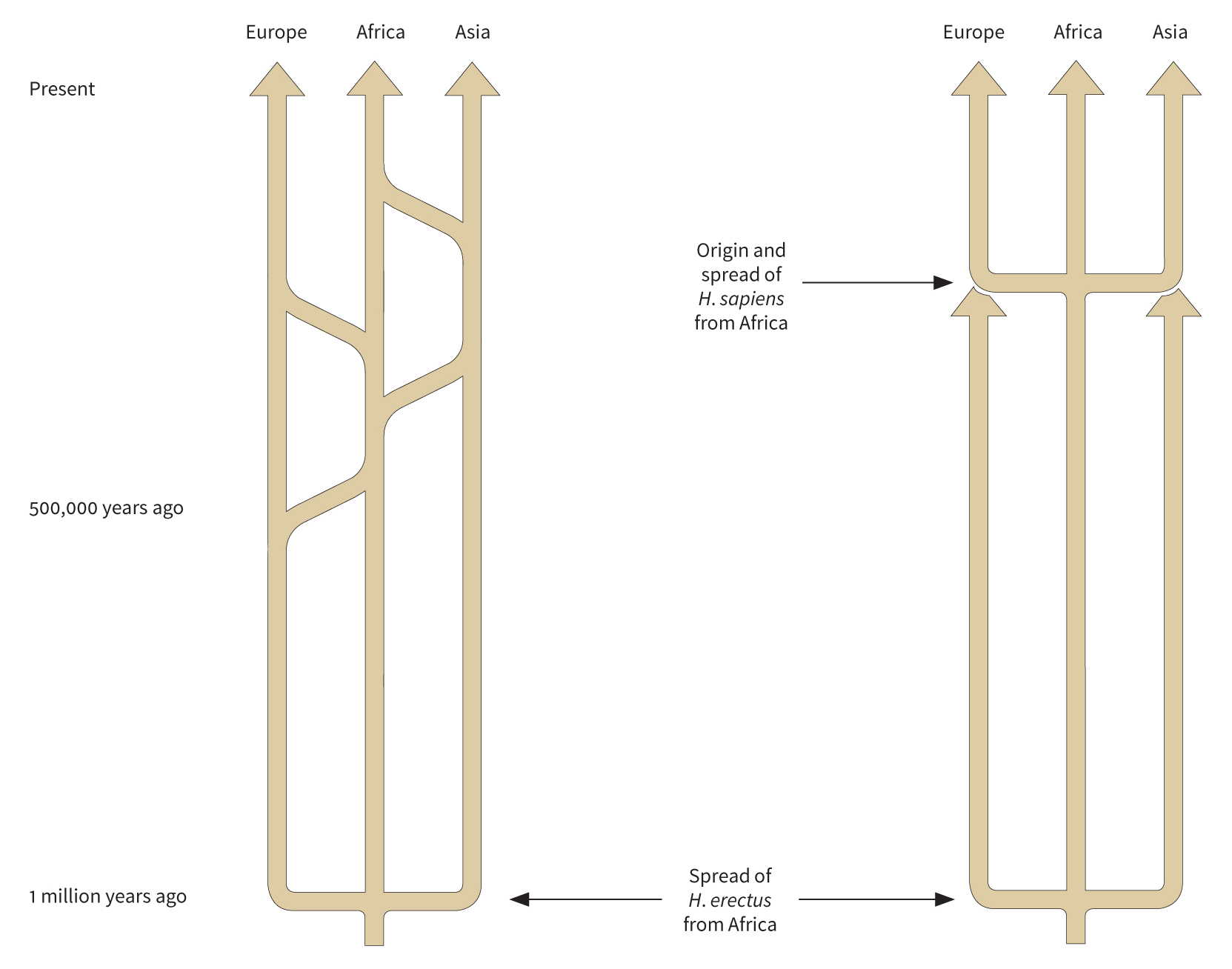
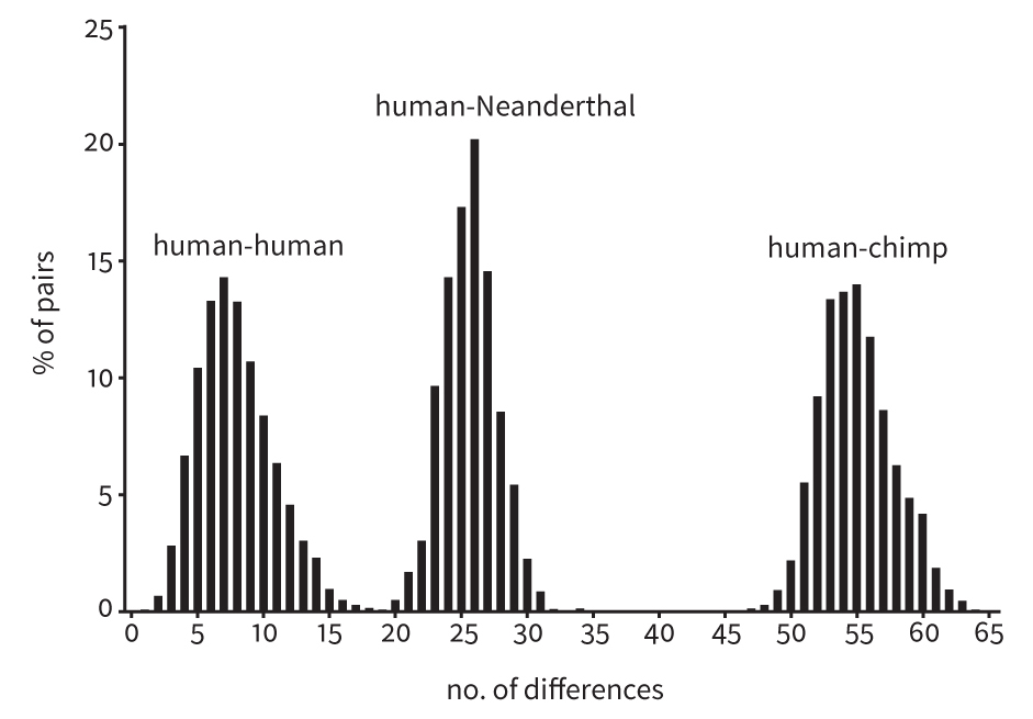
A highly significant breakthrough was achieved in 1997 by Matthias Krings, Svante Pääbo, and their colleagues with the extraction of mtDNA from 40,000-year-old hominin fossil remains. Even more remarkable was the analysis in 2010 of 4 million base pairs of Neanderthal nuclear DNA—effectively the entire Neanderthal genome. As discussed below (see p. 471), this has changed current thinking about the Neanderthals and has opened a new era in biological anthropology.
The recent advances in genetic engineering therefore open up fascinating possibilities for future work in human evolution, past human relationships, and the origin and evolution of disease.
So far in this chapter, we have learned how one can deduce a great deal about our ancestors’ physical characteristics; but the picture is still a static one. The next step is to learn how one reconstructs the way these bodies worked and what they could do.
Assessing Human Abilities
The human body is a superb machine, capable of performing a great variety of actions, some requiring strength and force and others involving fine control and specialized skills, but it has not always been able to perform these tasks. How, then, do we trace the development of human abilities?
Walking
One of the most basic uniquely human features is the ability to walk habitually on two legs—bipedalism. A number of methods provide insights into the evolution of this trait. Analysis of certain parts of the skeleton, and of body proportions, is the most straightforward method, but skulls are often the only parts of our early ancestors to have survived. One exception is the 40 percent complete australopithecine skeleton nicknamed “Lucy,” dating from around 3.18 million years ago and found at Hadar in the Afar region of Ethiopia—hence its scientific name, Australopithecus afarensis. Much attention has been focused on the lower half of Lucy’s skeleton. The American paleoanthropologists Jack Stern and Randall Susman believe that it could walk, but still needed trees for food and protection—their evidence consists of the long, curved, and very muscular hands and feet, features that suggest grasping.
Finding a Neolithic Family
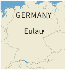
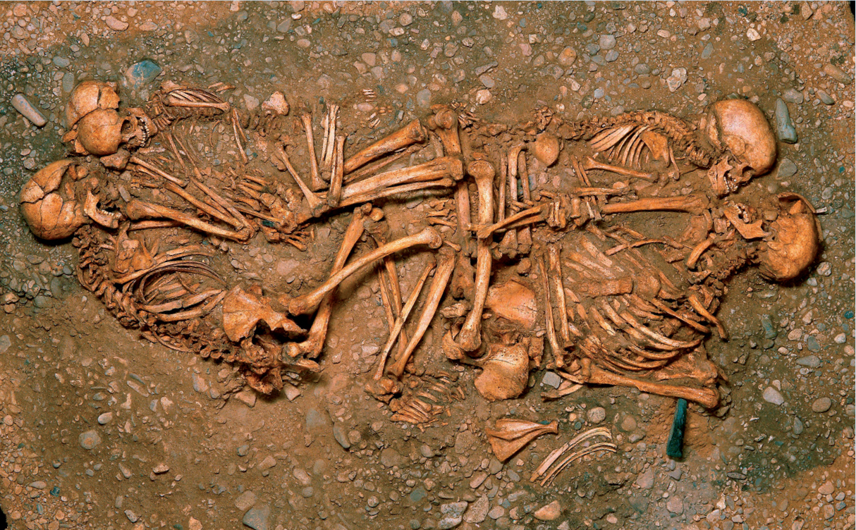
In 2005, at Eulau in Saxony (eastern Germany), archaeologists discovered four closely grouped and well-preserved multiple burials dating to the Neolithic Corded Ware culture, around 4600 years ago. Each contained a group of adults and children buried facing each other. Their simultaneous interment and signs of conflict showed that they must have been the victims of some kind of violent event. The team adopted a multidisciplinary approach, combining the methods of archaeology and anthropology with analyses of radiogenic isotopes, to determine the origins of the individuals, and examinations of ancient DNA, to investigate their relationships.
11.20 The skeletons in “Tomb 99” at Eulau.
Identifying a Family Group
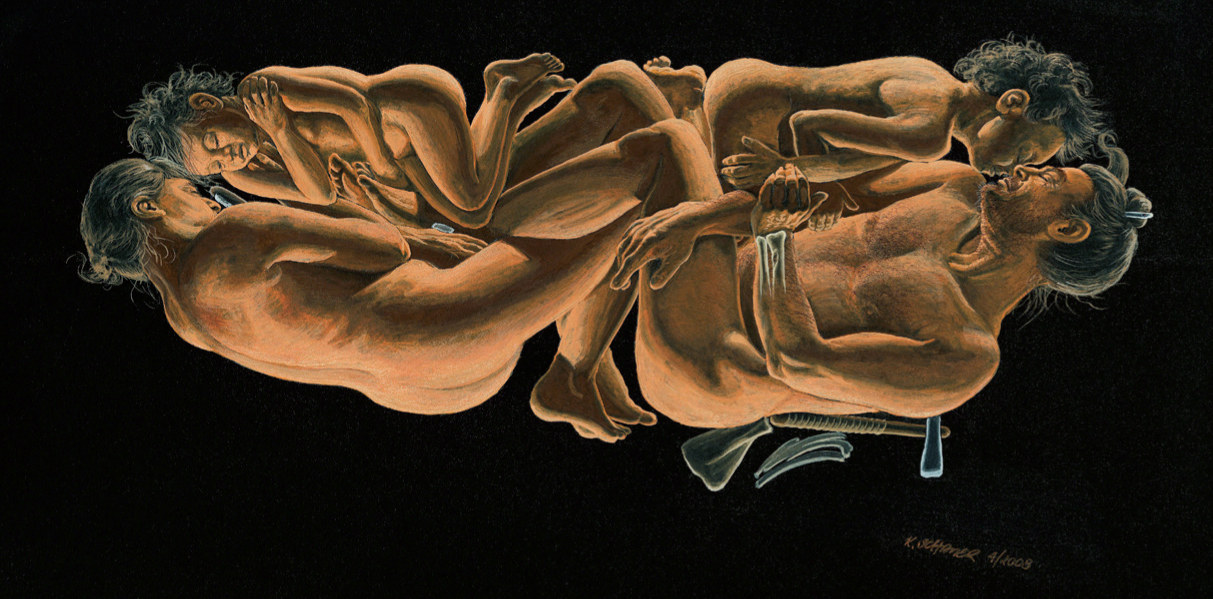
“Tomb 99” produced the clearest results. Anatomical analysis showed that it contained a man aged between forty and sixty, a woman aged between thirty-five and fifty, and two boys aged four to five and eight to nine. Each adult was buried facing one of the boys, their arms and hands linked. DNA analyses have proved that they were father, mother, and sons—the woman and boys had the same mitochondrial DNA, while the boys had the same Y-chromosome haplogroup as the man. This constitutes the earliest known genetic evidence for a nuclear family unit.
11.21 A reconstruction painting of how the bodies were arranged in “Tomb 99.”
Evidence for Violence and Social Origins
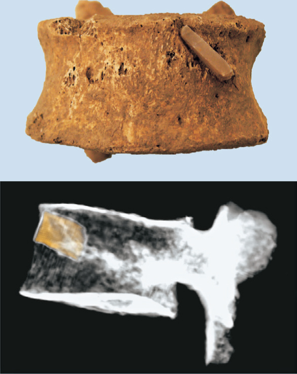
The other three graves contained a total of nine people, mostly women and children. Many bore signs of a violent end, such as a female with a flint projectile point embedded in a vertebra, two skulls with fractures, and some individuals with cutmarks on their hands and forearms suggesting an attempt at self-defense. Perhaps they were slaughtered in a raid and later buried by the survivors. There are no adolescents or young adults among the dead. There were few grave-goods—stone axes for the men and boys, flint tools or animal-tooth pendants for the women and girls. Butchered animal bones indicated at least one food offering per grave.
Isotope analyses of tooth enamel reflect the levels of dietary strontium derived from soils during childhood (see pp. 308–9), and vary between individuals from different regions. At Eulau, such analyses have shown that the men and children were local, while the women had a different origin, which suggests that this was an exogamous society (i.e., wives came from outside the area) and also patrilocal (i.e., females moved to the location of the males, where they had their offspring).
11.22–23 Photo (top) and radiograph (above) of a flint arrowhead embedded in a woman’s vertebra—the victim of a violent raid?
Another American researcher, Bruce Latimer, and his colleagues think that Lucy was a fully adapted biped. They doubt that curved finger and toe bones are proof of a life in trees, and find that the lower limbs were “totally reorganized for upright walking”: the orientation of the ankle is similar to that in a modern human, implying that the foot was less flexible in its sideways movements than an ape’s. More recent work has shown that Lucy and its relatives had rigid arches in their feet, and so could not use them for grasping. By contrast, foot bones of the earlier species Ardipithecus ramidus (4.4 million years ago) and of the later species Australopithecus sediba (2 million) suggest that they were both bipedal and tree-climbers.
Other remains of australopithecus exist that have been interpreted differently. In the mid 1990s in Sterkfontein, South Africa, four articulating foot bones were discovered, probably belonging to Australopithecus africanus. Analysis of the remains (called “Little Foot”) led some specialists to believe that, while clearly adapted for bipedalism, the foot showed apelike traits that would make it perfect for tree-life. Others insisted that these were simply relict anatomical traits, and that these australopithecines spent all their time on two legs on the ground.
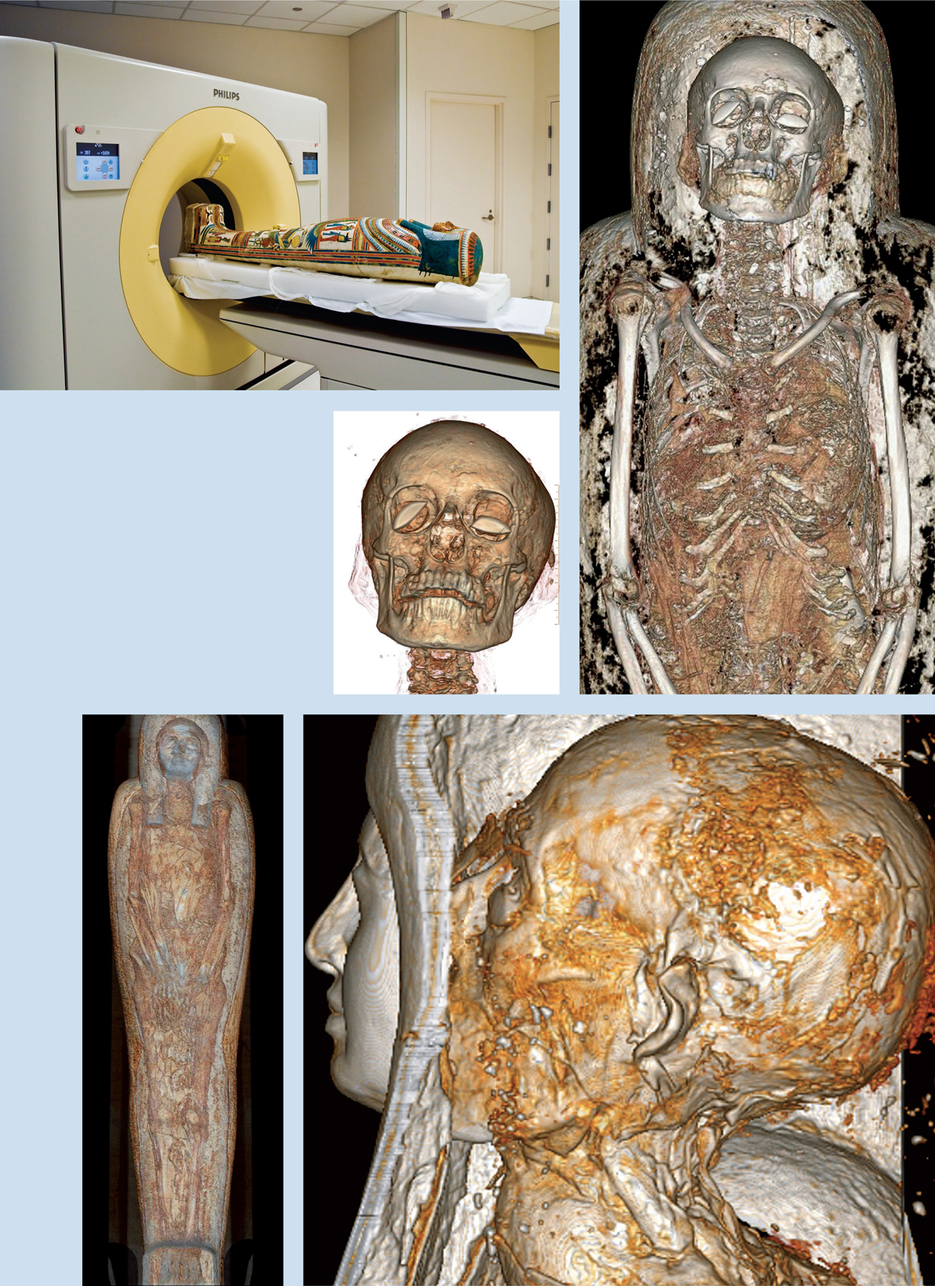
A different type of evidence for upright walking can be found in skulls. The position of the hole at their base, for example, where the spinal column enters, tells a great deal about the position of the body during locomotion. Even fossil skulls trapped inside a rock-hard matrix can now be examined through the technique of computerized (computed) axial tomography (CT), in which X-ray scans made at tiny intervals produce a series of cross sections that the computer can reformat to create vertical, oblique, or 3D images as required. A skull can therefore be seen from any angle. The technique is also useful for studying mummies without unwrapping them, and for revealing which organs still remain inside them (see box, pp. 452–53).
Dutch scientists Frans Zonneveld and Jan Wind have CT-scanned the very complete skull of Australopithecus africanus, 2–3 million years old, from Sterkfontein, South Africa, known as “Mrs. Ples.” The scans revealed the semi-circular canals of the inner ear entombed inside the solid fossil. This feature is of special interest because it helps with balance and provides an indication of the carriage of the head: the horizontal canal has a relationship with the angle of the head in upright-walking humans. The angle in “Mrs. Ples” suggested that she walked with her head at a greater forward-sloping angle than in modern humans.
Dutch anatomist Fred Spoor and his colleagues have studied the canals in a series of different hominins, and found that in australopithecines this feature is decidedly apelike—supporting the view that they mixed bipedalism with tree-climbing—while Homo erectus was similar to modern humans in this respect.
Footprints in Time.
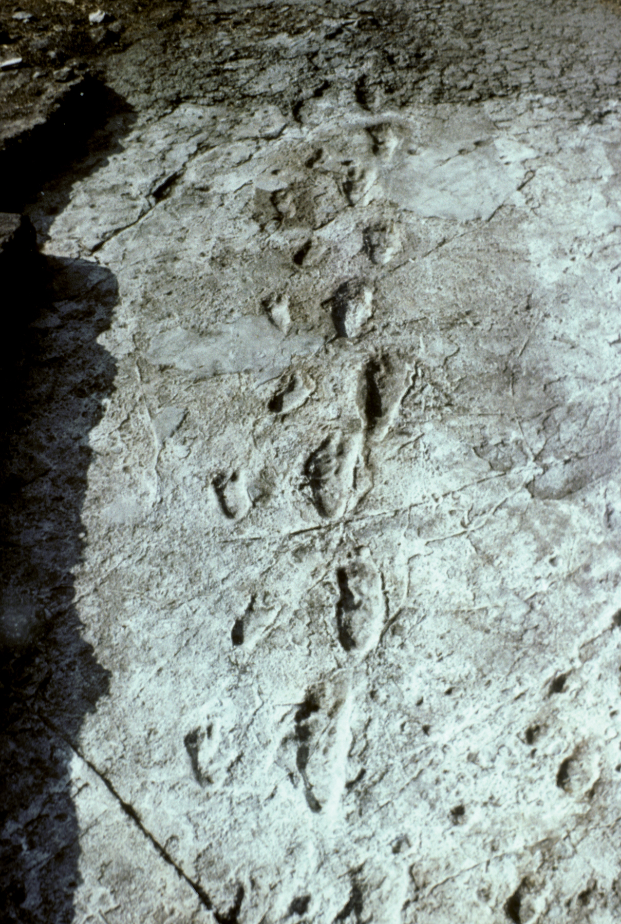
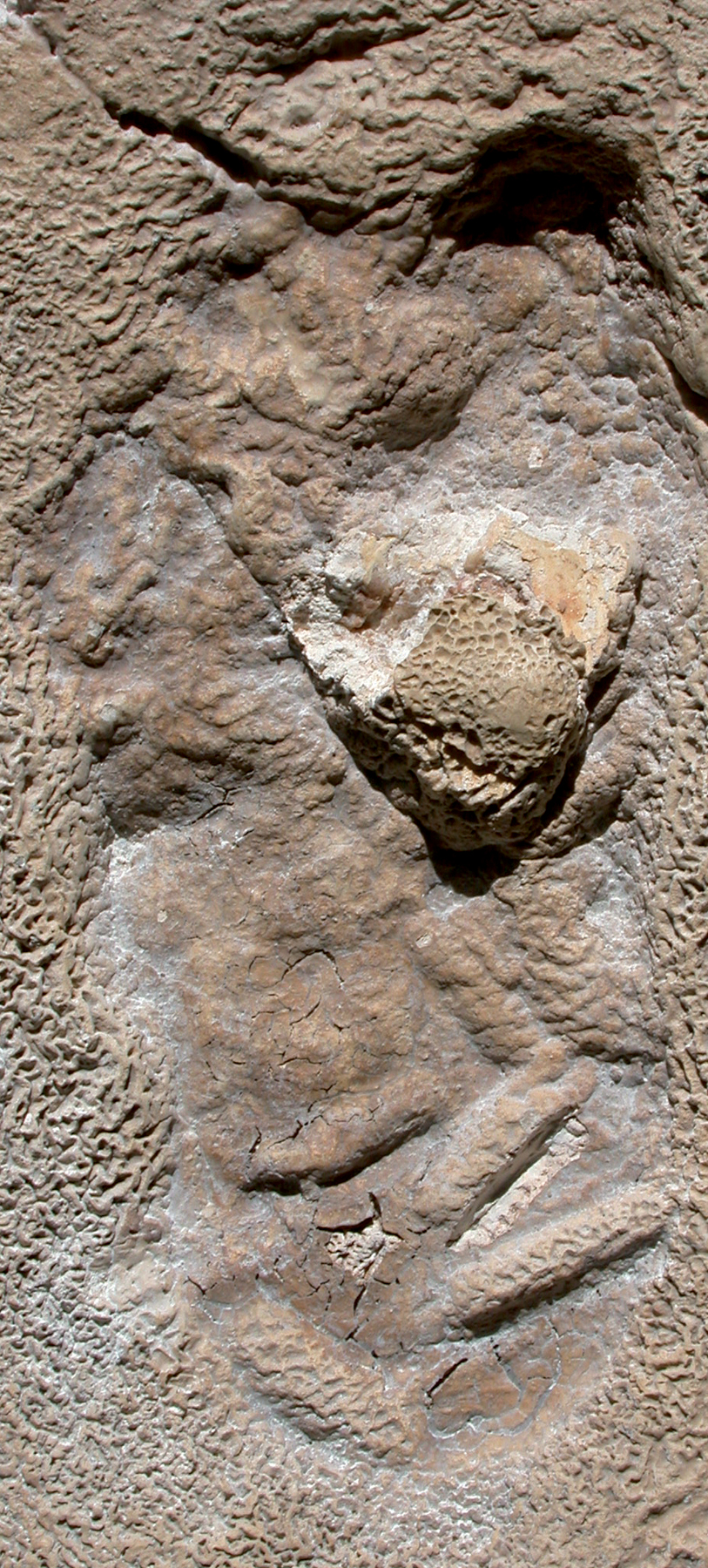
A great deal can be learned from the actual traces of human locomotion: the footprints of early hominins. The best-known examples are the remarkable trails discovered at Laetoli, Tanzania, by Mary Leakey. These were left by small hominins around 3.75–3.60 million years ago, according to potassium-argon dates of the volcanic tuffs above and below the footprints. They walked across a stretch of moist volcanic ash, which was subsequently turned to mud by rain, and then set like concrete.
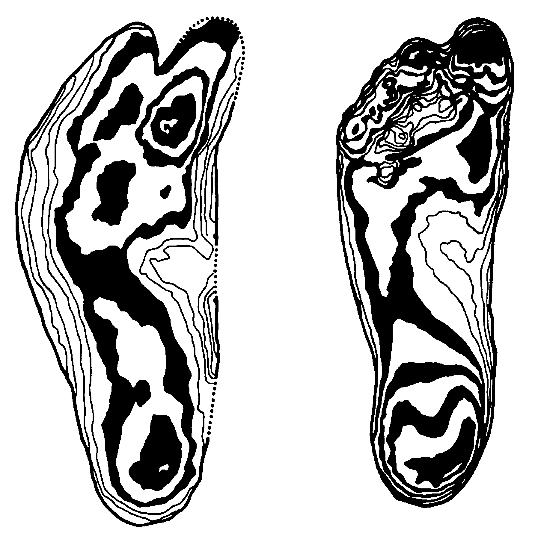
11.24 The Laetoli footprints. One of the remarkable footprint trails left by early hominins 3.75–3.60 million years ago at this East African site.11.25 The contour pattern of one of the Laetoli footprints (left) is strikingly similar to that of a modern male foot impression made in soft ground (right).
Analysis revealed to Mary Leakey and her colleagues that the feet had a raised arch, a rounded heel, a pronounced ball, and a big toe that pointed forward. These features, together with the weight-bearing pressure patterns, resemble the prints of upright-walking humans. The pressures exerted along the foot, together with the length of stride (average 87 cm, or 34 in.), indicate that the hominins (probably early australopithecines) had been walking slowly. In short, all the detectable morphological features imply that the feet that did the walking were very little different from our own.
A detailed study has been made of the prints using photogrammetry, which created a drawing showing all the curves and contours of the prints. The result emphasized that there were at least seven points of similarity with modern prints, such as the depth of the heel impression, and the deep imprint of the big toe. Michael Day and Ernest Wickens also took stereophotographs of the Laetoli prints and compared them with modern prints made by men and women in similar soil conditions. Once again, the results furnished possible evidence of bipedalism, a trait that is definite in the more recently discovered prints at Ileret, Kenya, dating to 1.5 million years ago. Footprints therefore provide us not merely with rare traces of the soft tissue of our remote ancestors, but evidence of upright walking that in many ways is clearer than can be obtained from analysis of bones.
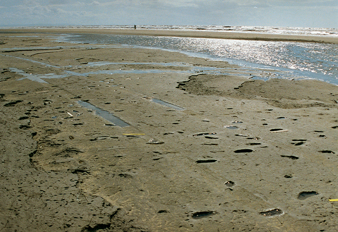
The earliest preserved footprints outside Africa are found in Happisburgh, England, dating to 800,000 years old. Discovered on the foreshore of an ancient estuary after a storm had removed the beach sand, the prints are thought to have belonged to Homo antecessor, a possible common ancestor of both Homo sapiens and Neanderthals. The study of fossil prints is by no means restricted to such remote periods. Hundreds of prints are known, for example, in French caves, dating from the end of the last Ice Age. Research by Léon Pales, using detailed silicone resin molds, has revealed details of behavior. In Fontanet Cave, one can follow the track of a barefoot child who was chasing a puppy or a fox. In Niaux Cave, the prints show that children’s feet were narrower and more arched than today.
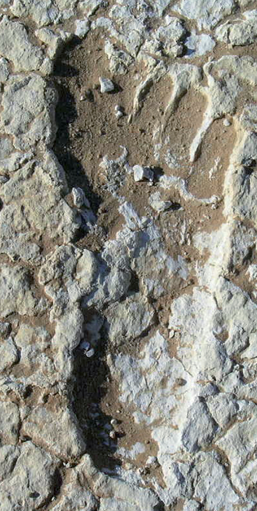
In 2003 the largest collection of Pleistocene footprints in the world was discovered in the Willandra Lakes of southeastern Australia. Optically dated (see p. 159) to between 23,000 and 19,000 years ago, they comprise more than 450 prints in trackways, and were made by a dozen individuals—adults, adolescents, and children—crossing what was then a moist clay surface. One man, probably 2 m (6 ft. 7 in.) tall, was sprinting at about 20 km/h (12 mph), while the smallest prints were from a child 1 m (3 ft. 3 in.) tall.
More recent prints are known from the surface of ancient Japanese paddy fields, from early Holocene surfaces on the Argentine seashore, and especially from 3,600-year-old mudflats in northwest England’s Mersey estuary where 145 footprint trails show a mean adult male height of 1.66 m (5 ft. 5 in.) and a female height of 1.45 m (4 ft. 9 in.). Many children are present, moving slowly like the women (perhaps gathering shellfish), while the men moved rapidly. Some of the prints show abnormalities such as toes missing or fused, providing potential information on medical conditions.
11.26 The Happisburgh footprints in Norfolk, eastern England, discovered in 2013. These human footprints date to perhaps as early as 800,000 years ago, and were likely made by the human ancestor Homo antecessor.11.27 Neanderthal footprint from Vârtop Cave, Romania. More than 62,000 years old, it is 22 cm (8.5 in.) long, suggesting a body height of 1.46 m (4 ft. 9.5 in.).11.28 An early Homo sapiens footprint, dating to around 20,000 years ago, one of more than 450 discovered in 2003 in the Willandra Lakes area of southeastern Australia. Males and females are both represented, as are a variety of ages and speeds of walking and running.
Which Hand Did They Use?
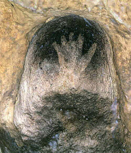
Many more people today are right-handed than left-handed, but can we trace this same pattern far back in prehistory? Much of the evidence comes from stencils and prints found in Australia and in many Ice Age caves in France, Spain, and Tasmania. Where a left hand has been stenciled, this implies that the artist was right-handed, and vice versa (assuming the hand was stenciled palm-downward). Even though the paint was often sprayed on by mouth, one can assume that the dominant hand assisted in the operation. Of 158 stencils in the French cave of Gargas, to which we shall return below (ills. 11.35–36), 136 have been identified as left, and only twenty-two as right: right-handedness was therefore predominant. In the few cases where an Ice Age figure is depicted holding something, it is mostly, though not always, in the right hand.
Clues to right-handedness can also be found by other methods. Right-handers tend to have longer, stronger, and more muscular bones on the right side; as long ago as 1911, the French paleontologist Marcellin Boule (1861–1942) noted that the Neanderthal skeleton from La Chapelle aux Saints had a right upper arm bone that was more robust than the left. Similar observations have been made on other Neanderthal skeletons, such as La Ferrassie I and Neanderthal itself, while skeletons of the eleventh to sixteenth centuries CE from the English village of Wharram Percy, Yorkshire, have been found to have right arms longer than the left in 81 percent of skeletons, and the left longer in 16 percent.
Fractures and cutmarks are other sources of evidence. Right-handed soldiers tend to be wounded on the left. The skeleton of a forty- or fifty-year-old Nabataean warrior, buried 2,000 years ago in the Negev Desert, Israel, had multiple healed fractures to the skull, the left arm, and ribs. Pierre-François Puech, in his study of scratches on the teeth of fossil humans (Chapter 7), noted that the Mauer (Heidelberg) jaw of around 500,000 years ago has marks on six front teeth; these were made by a stone tool, and their direction indicates that the jaw’s owner was right-handed.
Tools themselves can be revealing. Long-handled Neolithic spoons of yew wood, preserved in Alpine lake villages dating to 3000 BCE, have survived; the signs of rubbing on their left side indicate that their users were right-handed. The late Ice Age rope found in the French cave of Lascaux consisted of fibers spiraling to the right, and was therefore tressed by a right-hander.
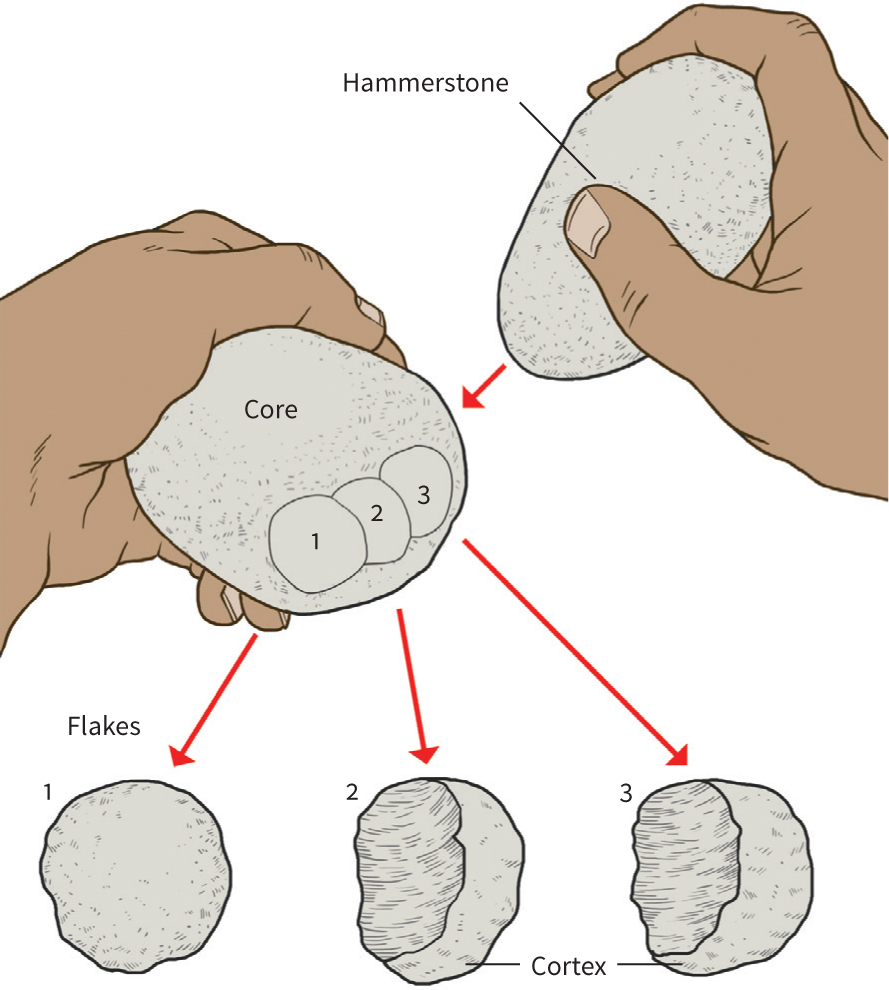
11.29 Nick Toth’s experiments showed that, typically, 56 per cent of the flakes produced by a right-handed stone toolmaker will have the cortex on the right, as shown here. Tools more than 1.5 million years old from Koobi Fora, Kenya, display an almost identical ratio.
Occasionally we can determine whether stone tools were used in the right hand or the left. In stone toolmaking experiments, Nick Toth, a right-hander, held the core in his left hand and the hammerstone in his right. As the tool was made, the core was rotated clockwise, and the flakes, removed in sequence, had a little crescent of cortex (the core’s outer surface) on the side; 56 percent of flakes had the cortex on the right, 44 percent on the left. A left-handed toolmaker would produce the opposite pattern. Toth has applied these criteria to the similarly made pebble tools from a number of early sites (before 1.5 million years) at Koobi Fora, Kenya, probably made by Homo habilis. At seven sites, he found that 57 percent of the flakes were right-oriented, and 43 percent left, a pattern almost identical to that produced today.
About 90 percent of modern humans are right-handed: we are the only mammal with a preferential use of one hand. The part of the brain responsible for fine control and movement is located in the left cerebral hemisphere, and the above findings suggest that the hominin brain was already asymmetrical in its structure and function not long after 2 million years ago. Among Neanderthals of 70,000–35,000 years ago, Marcellin Boule noted that the La Chapelle aux Saints individual had a left hemisphere slightly bigger than the right, and the same was found for brains of specimens from Neanderthal, Gibraltar, and La Quina.
When Did Speech Develop?
Like fine control and movement, speech is also controlled in the left part of the brain. Some scholars believe we can learn something about early language abilities from brain endocasts. These are made by pouring latex rubber into a skull; when set, the latex forms an accurate image of the inner surface of the cranium, on which the outer shape of the brain leaves faint impressions. The method gives an estimate of cranial capacity—in this way, American anthropologist Ralph Holloway examined two reconstructed skulls from Koobi Fora (KNM-ER 1470 and 1805), and calculated their brain volumes. Skull 1470, dating to about 1.89 million years and usually attributed to Homo habilis, had a capacity of either 752 cc or about 775 cc (46–47 cu. in.), while 1805, dating to about 1.65 million years and belonging to either Homo or Australopithecus, had a brain of australopithecine size (582 cc, or 35.5 cu. in.). According to American scholar Dean Falk, 1470’s endocast shows clearly human features, while 1805 was more gorilla- or chimpanzee-like.
The speech center of the brain is a bump protruding on the surface of the left hemisphere, which an endocast should theoretically record. Certainly Dean Falk, following on from analyses done by South African paleoanthropologist Phillip Tobias (1925–2012), argues that this area of 1470’s brain is already specialized for language and that this hominin was perhaps capable of articulate speech. Others, however, are unconvinced that features of this type in fossils are ever sufficiently clear for reliable interpretation.
Since fine control, movement, and speech are all located in the same part of the brain, some scholars go on to argue that these may be interconnected. Symmetry in tools could therefore be a sign of the sort of intellectual skill needed to understand language. The increasing abundance and perfection of the Acheulian hand-ax, or an increase in the number of tool categories, might imply an elevation in intellectual—and therefore language—capacity. Others, however, deny any correlation between spatial (technological) abilities and linguistic behavior, arguing that toolmaking and language are not conceived or learned in the same way. Much of the apparent standardization of tools, they say, is probably the result of material and manufacturing constraints, as well as in our archaeological classifications. Stone tools alone, it is concluded, cannot tell us much about language.
It is encouraging, however, that research in molecular genetics is making progress with the issue. A serious speech defect in three generations of a family resident in London (referred to for purposes of anonymity as “KE”) has been linked to a mutation in a specific gene designated “FOXP2.” Molecular genetic studies of this gene suggest that the specific (and, for humans, normal) version is common to all humans but not found in other primates, and that it may be a preferential mutation that took place about 100,000 years ago. This positive mutation is related to the capacity to control fine movement of the mouth and face. Molecular genetics is already illuminating the evolutionary history of the articulatory skills involved in developed language—but not yet that of the underlying symbolic skills. Some geneticists argue that FOXP2 is involved only in the mechanical aspects, while others claim the gene plays a role in both the mechanical and cognitive sides. As with many studies in this field, research continues.
Reconstructing the Vocal Tract.
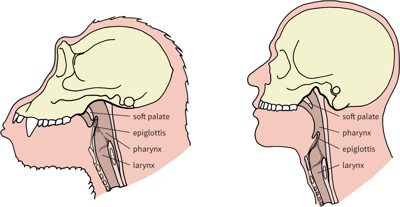
Another approach to assessing speech ability is to try to reconstruct the vocal tract in the throat. Philip Lieberman and Edmund Crelin (1923–2004) compared the vocal tract of Neanderthals, chimpanzees, and modern newborn and adult humans, and claimed that the adult Neanderthal upper throat most closely resembles that of modern infants. Neanderthals, they argued, lacked a modern pharynx (the cavity above the larynx or voice box) and therefore could make only a narrow range of vowel sounds, not fully articulated speech. This claim rests on fragile evidence and has not been widely accepted.
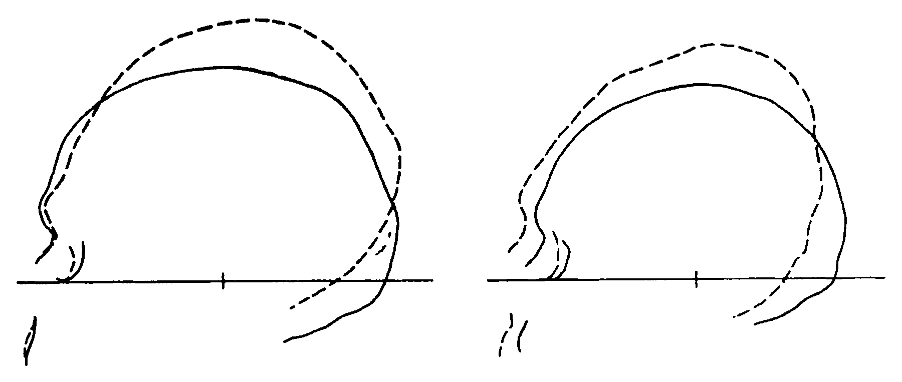
11.30(Left) Vocal tracts of a chimpanzee and (right) a modern human. The human larynx is lower, and the base of the skull is also more arched—the origins of this trait can be studied in the fossil record.
The vocal-tract work, however, has received support from Jeffrey Laitman using a different method. He noted that the shape of the base of the skull, which forms a “ceiling” to the throat, is linked to the position of the larynx. In mammals and human infants, the base is flat, and the larynx high, below a small pharynx; but in adult humans, the base is curved and the larynx low, with a large pharynx allowing greater modulation of vocal sounds.
Turning to fossil hominins, Laitman found that in australopithecines the base of the skull was flat, and the pharynx therefore small—albeit slightly bigger than in apes. Australopithecines could vocalize more than apes, but probably could not manage vowels. Moreover, like apes and unlike humans, they could still breathe and swallow liquids at the same time. In skulls of Homo erectus (1.6 million to 300,000 years ago), the skull-base is becoming curved, indicating that the larynx was probably descending. According to Laitman, full curvature of modern type probably coincides with the appearance of Homo sapiens, though he agrees that Neanderthals very likely had a more restricted vocal range than modern humans.
Debate about Neanderthal speech abilities was rekindled by the find, at Kebara Cave, Israel, of a 60,000-year-old human hyoid; the movement of this small U-shaped bone affects the position and movement of the larynx to which it is attached. The size, shape, and muscle-attachment marks put the find within the range of modern humans, therefore casting more doubt on Lieberman’s view and suggesting that Neanderthals were indeed capable of speaking a language. Several scholars, however, have pointed out that language is a function of the brain and of mental capacity.
Analysis of the hypoglossal canal, a perforation at the bottom of the skull adjacent to where the spinal cord links to the brain, has shown that as much as 400,000 years ago these canals were comparable in size to those of modern humans. This suggests that they contained a similar complement of nerves leading to the tongue, and that humanlike speech capabilities may have evolved far earlier than had previously been thought, and certainly long before the Neanderthals.
Identifying Other Kinds of Behavior
Use of Teeth.
As we saw in Chapter 7, marks on the teeth of our early ancestors can sometimes suggest that they often used their mouths as a sort of third hand to grip and cut things. In Neanderthals this is indicated by the extreme wear on the teeth even of fairly young adults and by the very high incidence of enamel chipping and microfractures.
The history of dental hygiene may seem of remote interest to archaeologists, but it is certainly intriguing to know that science can now indicate use of toothpicks of some kind by our early ancestors. David Frayer and Mary Russell found grooves and striations on the cheek teeth of Neanderthals from Krapina, Croatia, consistent with regular probing by a small, sharp-pointed instrument. Such marks have also been observed on the teeth of Homo erectus and Homo habilis. For a much more recent period, the sixteenth century CE, analysis in the scanning electron microscope of the front teeth of King Christian III of Denmark (reigned 1534–1559) revealed striations which indicated by their form and direction that the king had cleaned his teeth with a damp cloth impregnated with abrasive powder.
Ancient Cannibals?
The traditional urge to uncover cannibalism suffered a massive jolt with the appearance forty years ago of a groundbreaking work by anthropologist William Arens that, for the first time, showed that the vast majority of claims for cannibalism in the ethnographic or ethnohistorical record were untrustworthy. In recent decades, a better understanding of taphonomy, greater familiarity with the huge variety of funerary rituals around the world, and a more objective assessment of the facts, have helped to weed out many claims for prehistoric cannibalism. Meanwhile, new claims have been put forward that rely on more plausible evidence than before.
The Earliest Evidence
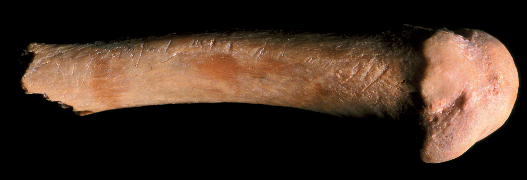
At Atapuerca, near Burgos in northern Spain (see box, pp. 156–57), the bones of a human ancestor called Homo antecessor, dating to perhaps 1 million years ago and found in the Gran Dolina site, bear abundant cutmarks that have been interpreted as evidence for cannibalism, and it is difficult to disagree with this inference. It is known that cannibalism can occur in other species, including the chimpanzee, and it can happen today in cases of starvation or mental health problems, so there is no reason to deny its possible existence at times in prehistory. In the case of Gran Dolina, at such a remote point in prehistory, when we have little idea what our ancestors were like or how they lived, there is no reason to doubt the presence of cannibalism, and there is absolutely no evidence for any kind of funerary rituals or other secondary treatment of the dead. No other explanation for the cutmarks is conceivable in the present state of our knowledge. They are most probably butchery marks, and therefore are an indication of consumption of human flesh by other humans.
11.31 Cutmarks on this human bone from Gran Dolina were almost certainly caused by butchering.
A later site at Atapuerca, the Sima de los Huesos, also presents the earliest evidence in the world for some kind of funerary ritual, perhaps some 600,000 years ago (see box, pp. 390–91). Ethnographic and ethnohistorical records all over the globe show clearly that a huge variety of often bizarre funerary practices exists, some involving cutting, smashing, and burning of bones, either shortly after death or long afterward when bodies are exhumed. The archaeological record contains many instances from different periods, stretching back into prehistory, that can plausibly be attributed to such practices. Atapuerca demonstrates, therefore, that all human remains from 600,000 years ago onward need to be interpreted with great circumspection, since funerary rituals thenceforth are an ever-present possibility, and indeed are one of the distinctive marks of humanity.
Categories of Evidence
In order to decide whether human remains were produced by cannibalism or by funerary activities (or warfare, etc.), there are two main categories of evidence. The first is the presence of human bones with marks of cutting, smashing, or burning. Fruitless attempts have been made to isolate specific criteria by which one might recognize cannibalism, but none of them is truly diagnostic, and alternative explanations are always available. The second is the presence of human bones mixed with animal bones, with similar marks and treatment; since the animal bones are obviously the remains of food, the same must apply to the human bones. Things may not be so simple, however, since the people who left the archaeological record were humans, capable of all kinds of complex and odd behavioral patterns. The human and animal bones are not necessarily the results of the same phenomenon, so one must avoid jumping to simplistic and “obvious” conclusions.
The data are always ambiguous, as can be seen clearly in one of the many Neanderthal examples that have been advanced as evidence for cannibalism. The hundreds of fragments of Neanderthal bones unearthed in 1899 in the cave at Krapina, in Croatia, were first attributed to a cannibal feast; they were badly broken and scratched and mixed with animal remains, and it was assumed the flesh had been cut off the human bones for food. But a re-examination by Mary Russell showed that the marks are quite different from those on defleshed meatbones but very similar to those found on Native North American skeletons that have been given secondary burial. In other words, the Krapina bodies were not eaten, but the bones were probably scraped clean for reburial. Moreover, her reanalysis showed that most of the damage to them could better be explained by roof falls, crushing by sediments, and the use of dynamite in the excavations.
At Fontbrégoua, a Neolithic cave in southeast France dating to 4000 BCE, Paola Villa and her colleagues found animal and human bones that shared the same types of cutmarks. Six people had been stripped of their flesh with stone tools shortly after death, and their limb bones cracked open. Although there was no direct evidence of consumption of flesh or marrow, this was presented as the most plausible case of prehistoric cannibalism yet discovered. Ethnographic evidence from Australia, on the other hand, suggests that it could well be a mortuary practice. Similarly, a reassessment by Heidi Peter-Röcher of numerous claims for cannibalism in the prehistory of central and eastern Europe found absolutely no evidence for the practice, with secondary funerary rituals able to account for all the finds.
Dramatic claims have also been made for cannibalism among the Ancestral Puebloan people of the American Southwest, around 1100 CE, including supposed human fecal material containing human tissue; but once again alternative explanations are available, involving not only funerary practices but also the extreme violence and mutilation inflicted on enemy corpses in warfare. In addition, the fecal material may actually be from a scavenging coyote.
Although many early claims for cannibalism have been debunked, the possibility remains that it may have existed occasionally, not merely in the remote times of Homo antecessor but also much later among Neanderthals and even modern humans. But the evidence is always ambiguous, and must be assessed carefully and objectively, rather than with wishful, melodramatic thinking, as has so often been the case in the past. The practice must certainly have occurred from time to time in cases of starvation; its existence as “custom cannibalism,” however, is far harder to prove. In any case, even if cannibalism existed occasionally, the contribution of human flesh to diet must have been minimal and sporadic, paling into insignificance beside that of other creatures, especially the big herbivores.
Use of Hands and Fingers.
We can study surviving hands and fingers to assess manual dexterity and labor. Randall Susman has shown that the first (thumb) metacarpal bone has a broad head in relation to its length in humans but not in chimpanzees, and since this bone has a similar configuration in Homo erectus, it follows that this hominin must have had a well-muscled thumb capable of generating the force needed for tool use and manufacture; conversely, the thumb of Australopithecus afarensis did not have this potential—it could not have grasped a hammerstone with all five fingers, but its hands were still better adapted to tool use than those of apes. It has been claimed that Australopithecus africanus was capable of the forceful opposition of thumb and fingers—a power and precision grip—required for tool use. Casts of Neanderthal thumb and index-finger bones have been scanned and used in 3D simulations, which revealed that their manual dexterity was not significantly different from that of modern humans. The manicured fingernails of Lindow Man suggested that he did not undertake any heavy or rough work.
Stresses on the Skeleton.
Human beings repeat many actions and tasks endlessly through their lives, and these often have effects on the skeleton that biological anthropologists can analyze and try to interpret.
Squatting has been suggested by Erik Trinkaus as a habitual trait among Neanderthals, on the basis of a high frequency of slight flattening of the ends of the thigh bone and other evidence. Female prehistoric Chinchorro mummies from Arica, on the Chilean coast, demonstrate bone structures on the feet and ankle joints, often called squatting facets, which are thought to have been caused by working crouched, perhaps opening shellfish on the beach.
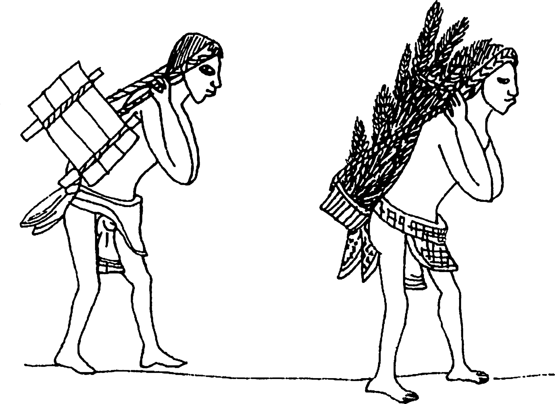
Load-carrying can lead to degenerative changes in the lower spine, though not all such changes can be assumed to be the result of this activity. In New Zealand such changes have been found in both sexes, but in other regions of the world they are predominantly associated with men. On the other hand, females seem to have done most of the carrying in Neolithic Orkney. In his analysis of the skeletons from the Orkney chambered tomb of Isbister, Judson Chesterman (1903–1987) noted that several skulls had a visible depression running across the top of the cranium; it was associated with a markedly increased attachment of neck muscles to the back of the skull. These features are known from the Congo region of central Africa, where women get them from carrying loads on their back, held by a strap or rope over the head. In parts of Central and South America, northern Japan, and other regions, the strap goes across the forehead, and can leave a similar depression there. Numerous Aztec codices depict porters carrying goods in this way in pre-Columbian times.
11.32 In Mesoamerica, without beasts of burden, porters, such as these Aztecs, carried loads using straps around the forehead.
Sexual Behavior and Childbirth.
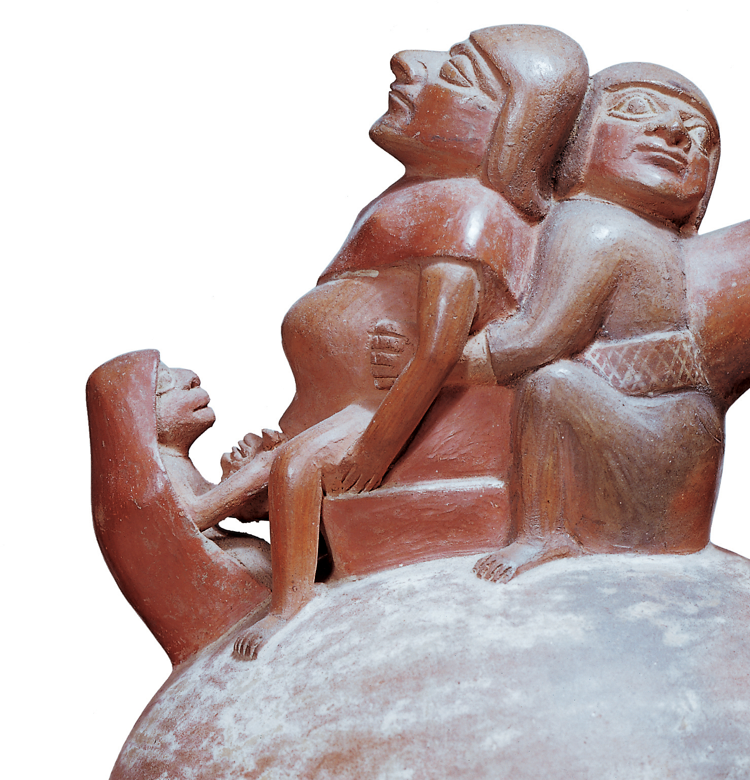
Art and literature provide evidence for innumerable human activities in the past, some of which, such as having sex, may not be detectable from any other source. The abundant and finely modeled Moche pottery of Peru gives us a vivid and detailed display of sexual behavior in the period between 200 and 700 CE. If it can be taken as an accurate record, it appears that there was a strong predominance of anal and oral sex (with rare homosexuality and bestiality)—were these methods perhaps adopted as a means of contraception rather than out of preference? We also learn from pottery representations the position that Moche women adopted for childbirth.
11.33 A depiction of childbirth: a scene from a Peruvian vase produced during the Moche period.
Cannibalism.
The eating of human flesh by other humans has often been claimed to exist in different periods of the past, usually on the flimsiest of evidence. Ever since the nineteenth century, numerous archaeologists have been prone to interpreting some human skeletal remains which they encountered in caves or elsewhere as the remains of cannibalistic feasts. In most cases, the reasons for choosing this interpretation were slight or, apparently, simply the whim of the excavator—the taphonomy of human bones was not yet understood, and it was simply assumed that cannibalism was a “primitive” trait and must therefore have existed in prehistory. Such claims still occur regularly, and of course the media adore cannibalism stories and always give them great prominence (see box on previous pages).
Disease, Deformity, and Death
So far, we have reconstructed human bodies and assessed human abilities. But it is necessary to look at the other, often more negative aspect of the picture: What was people’s quality of life? What was their state of health? Did they have any inherited variations? We may know how long they lived, but how did they die?
Where we have intact bodies, the precise cause of death can sometimes be deduced—indeed, in some cases, such as the asphyxiated people of Pompeii and Herculaneum, it is obvious from the circumstances (the effect of the eruption of the volcano Vesuvius). For the more numerous skeletal remains that come down to us, however, cause of death can be ascertained only rarely, since most afflictions leading to death leave no trace on bone. Paleopathology (the study of ancient disease) tells us far more about life than about death, a fact of great benefit to the archaeologist.
In parallel, anthropologists are increasingly using techniques developed within archaeology to assist them with the recovery and study of human remains. Indeed, a new subdiscipline has now developed—forensic archaeology (see box, p. 126)—which helps in the recovery and interpretation of murder victims, as well as trying to identify individuals within mass burials, as encountered in Rwanda and the former Yugoslavia as a result of the genocides committed there in the 1990s.
Evidence in Soft Tissue
Since most diseases rarely leave detectable traces in bones, the most commonly examined source of information on ancient diseases is surviving soft tissue. Soft tissue rarely survives except in specific environments. The surface tissue sometimes reveals evidence of illness, such as eczema. It can also reveal some causes of violent death, such as the slit throats of several bog bodies.
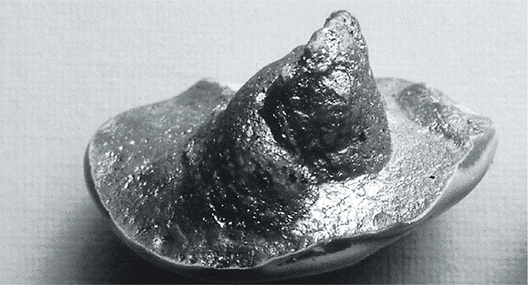
11.34 Cast of a finger-end produced by the City of London Police from a hole in a 5,000-year-old pot from the River Thames.
Where inner tissue is involved, a number of methods are at the analyst’s disposal. Radiographs can provide much information, and have been used on Egyptian mummies, but more powerful methods are now available (see box overleaf). Occasionally, one can study soft tissue that is no longer there: the footprints, handprints, and hand stencils mentioned in an earlier section. Fingerprints have survived on dozens of pieces of fired loess from around 26,000 years ago at the sites of Pavlov and Dolní Vĕstonice in the Czech Republic and on artifacts from many other periods, such as Babylonian clay disks and cuneiform tablets from Nineveh (3000 BCE), and ancient Greek vases, where they help to identify different potters.
Some handprints and stencils may supply intriguing pathological evidence. In three or four caves, most notably that of Gargas, France, there are hundreds of late Ice Age stencils of hands with apparently severe damage. Some have all four fingers missing. Debate still continues as to whether the stencils were made with the fingers folded, as a kind of sign language, or whether the damage is real but caused by mutilation or disease.
11.35 Hand stencils from the late Ice Age cave of Gargas, France. Photograph of one of the stencils.
Other forms of art from all periods yield evidence for illnesses. The small figures carved in medieval churches and cathedrals in western Europe illustrate various maladies and ills. The Mexican Monte Albán danzante figures carved on stone slabs have sometimes been interpreted as a kind of early medical dictionary, with internal organs displayed, although the current view is that these figures represent slain or sacrificed captives (Chapters 10 and 13).
11.36 Chart showing the numbers of hands found with particular types of “mutilation.” Debate still continues as to whether the hands were indeed mutilated or simply had folded fingers.
Bacteria, Parasites, and Viruses
Particularly where soft tissue survives, one can usually find parasites of some sort. The first place to look is in the bodies themselves, and principally in the guts, although body and head lice can also be detected (lice have also been found in combs in Israel dating from the first century BCE to the eighth century CE). Parasites can be identified from their morphology by a specialist. A huge diversity of such infestations has been found in Egyptian mummies—indeed, almost all have them, no doubt because of inadequate sanitation and an ignorance of the causes and means of transmission of diseases. The Egyptians had parasites that caused amoebic dysentery and bilharzia, and they had many intestinal occupants. Pre-Columbian mummies in the New World have eggs of the whipworm and the roundworm. Grauballe Man in Denmark (see box pp. 454–55) must have had more or less continuous stomach ache through the activities of the whipworm Trichuris, since he had millions of its eggs inside him.
Another important source of information about parasites is human feces (Chapter 7). The parasite eggs are passed out of the host in the feces; encased in hard shells, they survive very successfully. Parasites are known in prehistoric dung from Israel, Colorado, and coastal Peru—but it is worth noting that fifty feces from Lovelock Cave, Nevada, proved to have none at all. It is not uncommon for hunter-gatherers in temperate latitudes and open country to be parasite-free. On the other hand, some 6,000-year-old samples from Los Gavilanes, Peru, analyzed by Raul Patrucco and his colleagues, had eggs from the tapeworm Diphyllobothrium, with which one becomes infested from eating raw or partially cooked sea-fish. Feces in other parts of the New World have yielded eggs of the tapeworm, pinworm, and thorny-headed worm, as well as traces of ticks, mites, and lice. Parasites can also be detected in medieval cesspits, while sediments from a French Upper Paleolithic cave at Arcy-sur-Cure, dating to between 30,000 and 25,000 years ago, have been found to contain concentrations of the eggs of the parasitic intestinal roundworm Ascaris which are almost certainly from human excrement.
Examining Bodies
When examining human remains, it is essential to extract the maximum information while causing minimum damage to the remains themselves. In some cases, such as the mummies ofthe Egyptian pharaohs, the authorities permit examination only under exceptional circumstances. But considerable information can be gained by “seeing” into a body, and modern technology has placed several effective methods at scientists’ disposal.
Non-Destructive Techniques
Archaeologists are often surprised by what X-rays (or, more properly, radiographs) of coffins and wrapped mummies reveal: animal bodies where human remains were anticipated, additional bodies in one coffin, or a mass of jewelry. Xeroradiography goes a step further. This technique is rather like a cross between X-rays and a photocopy, in that it produces electrostatic images through colored powder being blown onto a selenium plate. The result is a much sharper definition than that produced by normal X-rays, and the wide exposure latitude allows both soft and hard tissue to show clearly on the same image. With edge enhancement, features are outlined like a pencil drawing. The technique can be used on mummies, either wrapped orin their coffins. When used on the head of the pharaoh Ramesses II (reigned c. 1279–1213 BCE), xeroradiography revealed a tiny animal bone inserted by the embalmer to support the nose; and in cavities behind the nose, a cluster of tiny beads became apparent.
Computerized (computed) axial tomography (CT) using a scanner is an important method that also allows wrapped mummies and other bodies to be examined in some detail non-destructively. The body is passed into the machine and images produced of cross-sectional “slices” through the body. CT scanners are more effective at dealing with tissues of different density, enabling soft organs to be viewed as well. Helical scanners move spirally around the body and produce continuous images rather than slices, a much quicker method.
Another technique for looking at internal organs is Magnetic Resonance Imaging (MRI), which lines up the body’s hydrogen atoms in a strong magnetic field and causes them to resonate by radio waves. The resulting measurements are fed into a computer, which produces a cross-sectional image of the body. This method is only suitable for objects containing water, and is therefore of limited use in studying desiccated mummies.
11.37–38 When the mummy of Ramesses II was taken to Paris for specialized medical treatment in the 1970s, it was subjected to xeroradiography.
By using a fiber-optic endoscope—a narrow, flexible tube with a light source—analysts can look inside a body, see what has survived, and assess its condition. Endoscopy occasionally reveals details of the mummification process as well as disease. When inserted into the head of the pharaoh Ramesses V (reigned 1149–1145 BCE), the fiberscope showed an unexpected hole at the base of the skull through which the brain had been removed (the brain was often broken up and removed through the nose during mummification); a cloth had later been put inside the empty skull.
Destructive Techniques
In cases where it is acceptable for the body to have samples taken from it for microscopic analysis, there are several techniques at the disposal of the scientist. (Fiber-optic endoscopy is also used in some cases for removing tissue.)
When tissue samples are removed, they are rehydrated in a solution of bicarbonate of soda (becoming very fragile in the process). They are then dehydrated, placed in paraffin wax, and sliced into thin sections, which are stained for greater clarity under a microscope. Using this technique on Egyptian mummies, analysts have detected both red and white blood cells, and have even been able to diagnose arterial disease.
Finally, analytical electron microscopy (similar to scanning electron microscopy) permits elements in tissue to be analyzed and quantified. When Rosalie David’s Manchester mummy team applied it to one Egyptian specimen, they found that particles in the lung contained a high proportion of silica and were probably sand—evidence of pneumoconiosis in ancient Egypt, where this lung disease was evidently quite a common hazard.
11.39–43 The coffin of Meresamun, an ancient Egyptian singer-priestess of c. 800 BCE in the temple at Karnak, was acquired by the Oriental Institute in Chicago in 1920 and has remained unopened. It has been CT-scanned three times as technology improved—most recently in 2008, when a state-of-the-art 256-slice scanner was used. The data can be rendered in 3D and manipulated in different ways, effectively allowing one to strip away successive layers, and to isolate particular bones or features of interest for analysis; movie sequences can also be created. Many details missed in the previous scans were uncovered, from items of jewelry and dental features to degenerative spinal changes and minor post-mortem fractures.
Certain parasites cause medical conditions that can be recognized if soft tissue survives. Some prehistoric mummies from the Chilean desert, dating from 7050 BCE to 1500 CE, had clinical traces or DNA of Chagas disease—notably an inflamed and enlarged heart and gut. The muscles of these organs are invaded by the causative parasites left on the skin in the feces of bloodsucking bugs.
Scabs and viruses can also survive in recognizable form in soft tissue, and may possibly even pose problems for the unwary archaeologist. We do not know for certain how long microbes can lie dormant in the ground. Most experts doubt that they pose any danger after a century or two, but there is a claim that anthrax spores survived in an Egyptian pyramid, and infectious microorganisms may also persist in bodies buried in the Arctic, preserved by the permafrost. The dangers in decaying bone and tissue may be very real—especially as our immunity to vanished or currently rare diseases has now declined.
A safer approach is provided by genetics, since some diseases leave traces in DNA. Smallpox and polio, for example, are caused by viruses, and a virus is simply DNA, or closely related RNA (ribonucleic acid), in a “protective overcoat” of protein. A virus infects by releasing its DNA into the unfortunate host, and some of the host’s cells are then converted to the production of viruses. In this way, viral infections can leave traces of the DNA of the virus. Analysis of ancient genetic material may therefore help to trace the history of certain diseases. For example, American pathologist Arthur Aufderheide (1922–2013) and his colleagues isolated fragments of DNA of the tuberculosis bacterium from lesions in the lungs of a 900-year-old Peruvian mummy, thus proving that this microbe was not brought to the Americas by European colonists. Ancient DNA studies have succeeded in identifying an extinct strain of the human hepatitis B virus in 4,500-year-old human remains from burials found in Europe and Asia.
Grauballe Man: The Body in the Bog
In 1952 peatcutters at Grauballe, Denmark, encountered a beautifully preserved bog body. It was lying in a prone position, with the left leg extended and the right leg and arm flexed. The various studies made of the body by multidisciplinary teams both in 1952 and, with new techniques, more recently have yielded remarkable insights into the life and death of this individual, now radiocarbon dated to 400–200 BCE, most likely around 290 BCE.
11.44 The body of Grauballe Man during excavation in 1952.11.45 Grauballe Man’s beautifully preserved feet, shortly after excavation.
Age and Sex
The body is that of a man aged about thirty. When found, he had a 1-cm (0.4-in.) long beard and moustache, but these fell off during conservation. As facial hair grows about 2.5–3.5 mm (0.10–0.14 in.) per week, and post-mortem skin shrinkage causes beard hair to stick out by 4–5 mm (0.16–0.20 in.), his beard was therefore about two weeks old when he died.
Physique
Grauballe Man appears to have been of average build for the period. His height has been estimated at between 1.65 and 1.70 m (5 ft. 5 in. and 5 ft. 7 in.), but this is uncertain due to shrinkage of the remains.
Appearance
There were no traces of clothing or artifacts with the body. His hair is 15 cm (6 in.) long. Its relatively straight-cut ends suggest it was cut by scissors. It is now reddish-brown, but this may be due to its immersion in the bog, so one cannot be sure what color it was originally. The hands have well-preserved fingerprints, and the rounded-off nails, like the clear lines on the palms, indicate that he had not been involved in heavy manual work—the same seems to be true of many other male bog bodies in northwest Europe.
State of Health
Grauballe Man was apparently fit and healthy—his body displays no sign of illness or disease—but he did have incipient arthritis in the thoracic vertebrae, which rarely occurs before the age of thirty. He still had twenty-one teeth, though several had fallen out since his death. They were worn, due to a coarse diet, and dental analysis revealed periods of starvation or poor health during his early childhood. The existence of periodontitis and cavities here and there showed that he had endured terrible toothache at times. Analysis of his hair shows that in his final months he had a terrestrial-based diet, with most protein coming from animal sources. His last meal was a gruel, a kind of poor muesli—it was dominated (80 percent) by seeds from a very small number of weed species, but there was also cereal bran. Small fragments of bone, including some from a pig, showed the presence of meat. The food would have been nutritious but not palatable—it is not known if it was typical of the everyday diet or not. The absence of fruit and greens suggests that he died in the winter.
11.46 Grauballe Man’s body, today displayed in the Moesgaard Museum, Denmark.
How Did He Die?
The cause of his death was a deep cut to the throat from ear to ear, made with a large sharp blade, while his head was bent sharply backward—it was so deep that the jugular vein and carotid arteries were severed. It was originally thought that a blow to the head in the temple area had been inflicted by a blunt instrument, but a recent CT scan has proved it to be post-mortem damage. An oblique fracture on the left tibia, however, was clearly caused by a heavy blow—perhaps to bring him to his knees so that his throat could be cut more easily. We do not know why he, or any other bog body, died—perhaps as a sacrifice, or as an executed criminal—but we have been able to learn a great deal about the life and death of Grauballe Man thanks to the very wide variety of tests and analyses that have been applied to him.
11.47 This deep throat wound, severing the jugular vein and carotid arteries, was the cause of Grauballe Man’s death.11.48 CT scan of Grauballe Man’s fractured left tibia. The most likely cause was a direct blow from a heavy object, and while it is thought that this occurred in life or around the point of death, it is impossible to be certain.11.49 Lumps visible on the lung of a 900-year-old Peruvian mummy were caused by tuberculosis, ascertained by isolating DNA of the disease in the lesion. This is proof that TB was not brought to the Americas by the European colonists.
The nature of plague, carried by the bacterium Yersinia pestis, is becoming much clearer through ancient DNA analysis. The first historically attested epidemic attributed to Yersinia pestis dates to 547–787 CE, the initial years of which are known as “the Plague of Justinian.” The second historic epidemic began around 1350 CE, commonly known as “the Black Death.” Y. pestis has three different pathogenetic forms. The bubonic form (“buboes” are swollen lymph nodes) is generally transmitted by the bite of an infected flea, whereas the septicemic and pneumonic forms are transmitted by contacts between humans (for instance, by cough droplets). Ancient DNA analysis of dental plaque from human teeth has now shown that pneumonic (but not bubonic) plague was prevalent in Europe and Central Asia from as early as 3500 BCE. The study of the prehistory of disease using ancient DNA is an important and developing field.
Skeletal Evidence for Deformity and Disease
Skeletal material, as we have seen, is far more abundant than preserved soft tissue, and can reveal a great deal of paleopathological information. Effects apparent on bone can be divided into those caused by violence or accident and those caused by disease or congenital deformity.
Violent Damage.
Where violence or accidents resulting in skeletal trauma are concerned, observations by experts can often reveal how the damage was caused and how serious its consequences were for the victim. For example, one of the Upper Paleolithic skeletons of children from Grimaldi, Italy, had an arrowhead buried in its backbone, a wound that was very probably mortal—as was the Roman ballista bolt famously found by Mortimer Wheeler (see p. 31) in the spine of an ancient Briton at the Iron Age hillfort of Maiden Castle, southern England.
Microbial Archaeology
Microbial archaeology investigates the relationships in the past between people and microorganisms, or microbes—a diverse range of single-celled organisms that include bacteria (single cell organisms without an enclosed nucleus), such as E. coli, eukaryotes (cells with an enclosed nucleus), such as yeasts, and archaea (cells without a nucleus but distinctive from bacteria and usually living in extreme conditions). Being microscopic, microbes are easy to overlook, but they greatly influence our daily lives. Microbes are estimated to make up half of the earth’s total biomass, and slightly more than half of the cells in the human body are in fact bacterial. Although many people equate microbes with disease-causing pathogens, the vast majority of microbes we encounter on a daily basis are either neutral or beneficial. Soil positively teems with millions of species of microbes—and yet despite coming into contact with us every day, they rarely make us sick. Other microbes, such as yeasts and lactic acid bacteria, are key components of our foods, converting grapes to wine, milk to yoghurt, and cabbage to sauerkraut. Additionally, the native microbial inhabitants of our colon, our gut microbiome, help us digest our food and are important producers of B and K vitamins. Today, we interact with microbes in myriad ways, and so did our ancestors in the past.
Food and Fermentation
Recent DNA and protein analyses of food containers and vessels have identified traces of lactic acid bacteria (LAB) and yeasts that were used in prehistory to intentionally ferment wine, make bread, and sour milk. Preserved organic remains from the ancient cultures of Xinjiang, for example (see p. 302), have revealed detailed information about the daily foods consumed during the Bronze Age in northwestern China, many of which were fermented. At the sites of Xiaohe, Gumugou, and Subeixi (c. 1500–300 BCE), preserved remains of sourdough bread, sour milk, and kefir have been identified, as well as the microbes that were used to produce them, including bread yeast (Saccharomyces cerevisiae), kefir yeast (Kluyveromyces) and a diverse range of LAB (Lactobacillus, Weissella, and Leuconostoc).
Bones and Disease
Bones yield important microbial information. For example, diseases such as tuberculosis can infect the skeleton, producing characteristic lesions in the spine, ribs, and other bones. Genetic testing of these infected bones has led not only to the positive diagnosis of tuberculosis in these individuals but also to the identification of the specific Mycobacterium tuberculosis complex (MTBC) strains that caused the infection. One of the most surprising findings of this research has been the discovery that pre-Columbian tuberculosis in the Americas (see above) is more closely related to zoonotic (animal) forms of tuberculosis than to Old World human strains, and thus may have been spread to the New World by migrating animals, such as seals, rather than humans.
11.50 Simplified evolutionary tree showing the genetic relatedness of tuberculosis-causing bacterial strains. The tree shows that pre-Columbian tuberculosis in the Americas was more closely related to animal than human forms of the disease, suggesting a prehistoric introduction by migrating animals.
Teeth and Dental Calculus
Teeth are essential for understanding the evolution of the human microbiome, the history of past epidemics, and the evolution of disease. A single tooth can simultaneously reveal information about the oral microbiome present in dental calculus, traces of pathogens trapped in the pulp chamber, and the communities of bacteria decomposing the body. Dental calculus is a mineralized form of dental plaque that builds up on the surfaces of teeth. Forming incrementally over time, like tree-rings, dental calculus is the only part of the human body that fossilizes during life, and it is also the richest known source of ancient DNA in the archaeological record. Composed mostly of oral bacteria, dental calculus preserves detailed information about oral health, disease, and immune function. Recent studies of dental calculus, such as research done by Christina Warriner at Harvard University, have shown that the oral bacteria Tannerella forsythia, Porphyromonas gingivalis, and Treponema denticola have been associated with periodontal disease for more than a thousand years. In addition to providing information about the beneficial and pathogenic bacteria in the mouth, teeth are also excellent sources of DNA from bloodborne pathogens. During life, dental pulp is rich in blood and other tissues, and bloodborne pathogens can become trapped in the pulp chamber after death. Studies of pathogen DNA retrieved from teeth have revealed a Bronze Age origin for the plague bacterium Yersinia pestis, and identified a rare strain of Salmonella enterica as a cause of the 1545 Mexican cocoliztli epidemic where millions died from enteric fever.
11.51 Teeth reveal information about the (a) oral microbiome in dental calculus, (b) traces of pathogens adhering to the pulp chamber walls (arrow), and (c) the diverse bacteria that decompose the body.
Paleofeces
Feces are highly informative about the health and well-being of past societies, and animal dung can be instructive about how ancient peoples managed their livestock and organized their household space. Genetic analysis has been used to investigate bacteria preserved in human paleofeces and gastrointestinal tissues found in dry caves and Alpine glaciers. It has revealed a highly diverse prehistoric human gut microbiome containing Prevotella and Treponema, as well as the presence of Helicobacter pylori—a species of bacteria associated with ulcers and gastric cancer—in the stomach of the Alpine Iceman (see box, pp. 70–71). Chemical analysis of fecal bacterial by-products, such as coprostanol, can also be used to validate paleofeces and to detect the location of latrines and fecal accumulation in occupational sediments. During digestion, bacteria in the gut irreversibly convert a portion of cholesterol present in bile into coprostanol, which is then excreted in feces. Microscopic traces of coprostanol were recently used to identify activity at Neanderthal sites.
Studies of ancient microbes have revealed important new insights about the human past. Whether by causing disease, fermenting our foods, or supporting our health, microbes have profoundly shaped the course of human history, and they are important subjects of archaeological study.
A study by Douglas Scott and Melissa Connor of the skeletal remains at the site of the famous Battle of the Little Bighorn in Montana—where General Custer and his entire force of 267 men were wiped out by mostly Sioux and Cheyenne warriors in 1876—showed the extensive use of clubs and hatchets to deliver a coup de grâce. One poor soldier, aged about twenty-five years old, had been wounded in the chest by a .44 bullet, then shot in the head with a Colt revolver, and finally had his skull crushed with a war club. In cases where the bones are masked by soft tissue, X-ray analysis is necessary (see box, pp. 452–53).
Individual wounds and fractures, however compelling the personal stories they reveal, are nevertheless of limited interest to medical history. Instead, the frequency and type of injuries on a population level are more useful to the archaeologist. Hunter-gatherers must have encountered different dangers from those faced by farmers, so their injuries would therefore be different as well. The aim should be to study traumas, along with other pathological conditions, as they occur in entire groups and communities.
Survival with major injuries also tells us about the capacity and willingness of the group to help those in need. That occurred far back in time. For example, one of the Neanderthals found in Shanidar Cave, northern Iraq, a man aged about forty, had suffered a blow to the left eye, making him partially blind. He also had a useless, withered right arm, caused by a childhood injury, a fracture in one foot bone, and arthritis in the knee and ankle. He may only have survived through the help of his community, although humans are good at adapting.
Intentional Alterations to Bone.
Skeletons can also be altered in other ways while someone was living or after death. Some human communities, such as the Maya, shaped skulls deliberately by binding the brow or back of the head of growing infants, with or without a board, to produce an unusually shaped head that was an irreversible and life-long mark of social status or group affiliation. Analysis of two of the Neanderthals found in Shanidar Cave has led Erik Trinkaus to claim that deliberate skull shaping was already practiced at this early date.
11.52 Skull deformation. (Right) Skull outlines of an artificially deformed Melanesian male—dashed line—and a normal male. (Left) A 13,000-year-old skull from Kow Swamp, Australia—dashed line—compared with that of a modern male Aborigine, suggesting that the Kow skull was deformed deliberately.
Life and Death among the Inuit
In 1972 two collective burials were discovered under an overhanging rock at Qilakitsoq, a small Inuit settlement on the west coast of Greenland dating to about 1475 CE. The eight bodies had all been mummified naturally by a combination of low temperature and lack of moisture. In one grave were three women and a four-year-old boy, and a six-month-old infant. In the second grave were three female bodies. The over- and under-clothing (a total of seventy-eight items including trousers, anoraks, boots) had also survived in perfect condition.
The bodies were sexed by the genitalia of those unwrapped and from X-ray examination of the intact mummies; in addition, facial tattoos were usually restricted to adult women in this society.
Ageing was done from dental development and other physical features. Three of the women died in their late teens to early twenties, but the other three had reached about fifty—a good age, since even at the turn of the twentieth century the average age of death for women in Greenland was only twenty-nine.
11.53 The eight bodies, layers of animal skins between them, lay protected by overhanging rock. Their frozen, moistureless grave resulted in natural mummification.11.54 Cold, dry conditions resulted in remarkable finds at Qilakitsoq. This six-month-old child was the best preserved of all the mummies.
The young boy and one woman may have been in much pain. X-rays of the boy showed that he had a misshapen pelvis of a kind often associated with Down’s syndrome. A disorder known as “Calvé–Perthes disease” was also destroying the head of a thigh bone, and he may have had to move around on all fours.
11.55 Drawing of a woman’s garment made from feathers carefully chosen from different birds, and worn next to the skin for extra warmth.
The woman, one of those who was aged around fifty, had broken her left collarbone at some stage; it had never knitted, perhaps impairing the function of her left arm. In addition, she had naso-pharyngeal cancer (at the back of her nasal passage), which had spread to surrounding areas causing blindness in the left eye, and also some deafness.
11.56 Infrared photography has made the tattoo design on this woman’s face clear.
Some of her features could be attributed to particular activities: her left thumbnail had fresh grooves on it, caused by cutting sinew against it with a knife (and, incidentally, showing that she was right-handed). She had also lost her lower front teeth, no doubt from chewing skins and using her teeth as a vice. The youngest woman’s lungs contained high levels of soot, probably from seal-blubber lamps. On the other hand, hair samples from the mummies showed low levels of mercury and lead, far lower than in the region today.
11.57 Leather clothing also survived well in the cold. These short trousers are made of reindeer skin.
How these people met their deaths remains a mystery. At any rate, they did not die of starvation. The woman with cancer had Harris lines (see p. 462) showing arrested bone growth as a child caused by illness or malnutrition, but she was well nourished when she died. The youngest woman had a sizable quantity of digested food in her lower intestine. Isotopic analysis of the boy’s skin collagen (p. 308) revealed that 75 percent of his diet came from marine products (seals, whales, fish) and only 25 percent from the land (reindeer, hare, plants).
Finally, analysis was carried out to ascertain the possible relationships among these individuals. Tissue typing established that some were not related at all, while others might have been. Either of two of the younger women could have been the mother of the four-year-old boy buried above them; while two of the women aged about fifty (including the one with cancer) may have been sisters. They also had identical facial tattoos, perhaps by the same artist, which were just like those on the earliest known portrait from this area (c. 1654 CE). Another woman had a tattoo so different in style and workmanship that she probably came from a different region and married into the group.
The practice also seems to have existed in Pleistocene or early Holocene Australia. Peter Brown compared deliberately shaped Melanesian skulls with normal ones in order to identify the changes caused by cranial shaping. He then applied his results to skulls from early Australian sites in Victoria, including Kow Swamp, and established beyond doubt that they had been artificially shaped. The oldest specimen, Kow Swamp 5, is 13,000 years old.
Other practices besides the skull modeling of infants are detectable. Tim White used a scanning electron microscope to analyze the skull of “Bodo,” a large male Homo erectus or archaic Homo sapiens from Ethiopia, about 300,000 years old, and came to the conclusion that it had been scalped. Analysis revealed two series of cutmarks, one on the left cheek under the eye socket, and the other across the forehead. These were made before the bone had hardened and fossilized, and therefore just before or just after death. Pre-Columbian Native American skulls that were scalped have similar marks in the same positions.
Identifying Disease from Human Bone.
The small number of diseases that affect bone do so in three basic ways—they cause bone formation, bone destruction, or both. It is their characteristic distribution pattern that is key to the diagnosis of the disease. Furthermore, the bone lesions associated with various illnesses can differ in terms of their number and location in the skeleton. Some afflictions leave quite clear signs, whereas others do not. The former include several infections, nutritional deficiencies, and cancers. It is also possible to detect growth disorders by the overall size and shape of bones.
Leprosy, for example, which is a bacterial infection, destroys the front part of the upper jawbone and the extremities in a distinctive manner, and there is clear evidence of skeletons from medieval Denmark with leprosy, as elsewhere in Europe, though none from the pre-Columbian New World. DNA from the leprosy bacterium has been isolated from a number of skeletons. Certain cancers also have a noticeable effect on bone (see box, pp. 458–59), such as the pathological changes to the leg bones of the elderly Neanderthal man of La Ferrassie 1, France, which are likely to have been caused by lung cancer.
Australian archaeologist Dan Potts and his colleagues have discovered the world’s earliest known polio victim in a 4,000-year-old grave in the United Arab Emirates; the skeleton of young woman, aged between eighteen and twenty years old, showed classic signs of the condition, such as the small size and inflammation of muscle attachments, thinness of all long bones, one leg 4 cm (1.6 in.) shorter than the other, a curved sacrum, and asymmetrical pelvis.
Richard III
In 2012 the world was startled to learn that a skeleton believed to belong to King Richard III of England (reigned 1483–1485 CE) had been uncovered in Leicester. Immortalized by Shakespeare as a villainous hunchback, the last of the Plantagenet kings was killed in battle at Bosworth Field in 1485, and it was known that he was buried in Leicester’s Greyfriars church. The church was demolished in the sixteenth century—along with all its burials, it was assumed—although there was also a story that Richard’s body had earlier been exhumed and thrown into the nearby river.
The project was an unusual collaboration between professional and academic archaeologists, an amateur group (the Richard III Society), and the City of Leicester. Excavation began in August 2012, in what had become a parking lot, and rapidly revealed the foundations of part of the church. The skeleton in question was found on day one, in a location that turned out to be a high-status position beneath the church’s choir. The body was in an irregularly cut grave, with a concave base and sloping sides, which was too short for it, whereas other graves in the choir were neatly rectangular and of the correct length. It seems the deceased was treated with little reverence. There was no evidence of a coffin or shroud; the lower limbs were fully extended, but the torso was twisted sideways to the north, and the head was propped up against a corner of the hole. In other words, it seemed the body had been crammed feet first into this small hole, implying great haste and/or a lack of respect.
11.58 Excavations in the Leicester parking lot where the burial believed to be of Richard III was found. The grave cut can be made out at the bottom of the photo. Richard’s remains were reinterred inside Leicester Cathedral in a formal ceremony in March 2015.
If this is indeed Richard, the haste may have been caused by the fact that his body had been on public display for several days, in the height of summer. The hands were crossed at the wrist, which may indicate that they were bound. The feet had been lost to some digging by nineteenth-century workmen, but otherwise the skeleton is in good condition—with 135 bones and twenty-nine teeth. It is that of an adult male of gracile build, aged, according to bone growth and tooth development, in his late twenties to late thirties (Richard was thirty-two when he died). The most remarkable feature is a severe scoliosis, i.e. a spine badly curved to the side (as opposed to the hunched back of legend).
11.59 The skeleton believed to be of Richard III, showing pronounced curvature of the spine.
The scoliosis developed around the age of ten to thirteen. As the man aged, some back ligaments would have turned to bone, stiffening the curve, and he also developed some osteoarthritis. The progressive scoliosis would have put a strain on his heart and lungs, possibly causing shortness of breath and pain. Had he not had this condition, his thigh bones indicate that he would have stood around 1.73 m (5ft. 8 in.) tall, above average for the period, but the disability would have reduced this substantially, to perhaps 1.42 m (4ft. 8 in.), and his right shoulder would have stood higher than the left. In other words, this man had a squat torso and uneven shoulders, which fits the few contemporary accounts of Richard’s appearance. Radiocarbon dating of the remains produced a result of 1456–1530 CE, which is consistent with the Battle of Bosworth in 1485.
The teeth of the man were a little worn, but had no cavities, and he was infected with roundworms. Analysis of nitrogen and carbon in the ribs by different laboratories revealed a high protein diet, including about 25 percent seafood, which implies high status. Oxygen and strontium isotopes from fluids he ingested as a child reflect the geology of the water source, and are consistent with Richard’s origins in Northamptonshire. It then seems the person moved further west by the age of seven, possibly to the Welsh border (Richard resided at Ludlow Castle, Shropshire, in 1459). On the other hand, there is a significant increase in the oxygen isotope composition in the last few years of his life, which analysts claim cannot be accounted for by beer and food, so they have attributed it to grape juice, in the form of wine—the equivalent of a modern bottle a day would lead to such a raised signature!
11.60 The facial reconstruction of the Leicester skeleton bears a remarkable likeness to the few portraits of Richard III, but this was not a blind test as the sculptor had been told to whom the skull had likely belonged.
The skeleton displays at least eleven wounds, all of them perimortem (i.e., inflicted around the time of death), since none shows signs of healing. Two large wounds under the back of the skull are consistent with blows from a halberd and sword, and would probably have caused almost instant death. There is a third, smaller penetrating wound to the top of the skull, possibly from a dagger. None of the skull wounds could have been inflicted on someone wearing a fifteenth-century helmet. Two other wounds—a cut on a right rib, and one on the right pelvis (probably caused by a thrust through the right buttock)—are also unlikely on someone wearing armor. So these may be “humiliation injuries,” delivered after death. In short, this person almost certainly died in battle, and the nearest battle in this time-range was that of Bosworth, 24 km (15 miles) west of Leicester. Finally, genetic analysis has been carried out on mitochondrial DNA, and a link has been claimed with two modern-day descendants of Richard’s elder sister, Anne of York (1439–1476). The researchers therefore believe that they have proven the identity of this skeleton “beyond reasonable doubt,” as it was expressed by Richard Buckley in a press conference in 2013. Whether or not they are correct, the project has displayed archaeology’s occasional and unique potential for mass appeal to the world’s media.
Radiographic analysis of bone may reveal evidence of arrested growth known as “Harris lines” (see box, pp. 458–59). These are narrow radio-opaque deposits of bone at the ends of long bones. They are laid down when growth resumes after being interrupted in childhood or adolescence as a result of illness or malnutrition. They are usually clearest in the lower tibia (shinbone). The number of lines can provide a rough guide to the frequency of difficult periods during growth. If the lines are found in whole groups of skeletons, they can indicate frequent subsistence crises or, perhaps, the consequences of social inequality sufficient to have had an effect on health. Similarly, Beau’s lines on finger- and toenails are shallow grooves indicating slowed growth caused by disease or malnourishment. The one surviving fingernail of the Alpine Iceman of 3300 BCE has three such grooves, suggesting that he had been subject to bouts of crippling disease four, three and two months before he died (see box, pp. 70–71).
Deformity in bone may be congenital, i.e. a person can be born with the deformity or develop it in childhood (such as the curved spine of King Richard III, see box on pp. 460–61). The tiny mummified fetus of a female, one of two found in the tomb of Tutankhamun, was shown by radiographic analysis to have Sprengel’s deformity—where the left shoulder blade is congenitally high and spina bifida is present—which probably explains why the infant, perhaps Tutankhamun’s own child, was stillborn (see ill. 11.62). Generally speaking, the Pharaonic practice of marrying one’s own sister might be expected to produce offspring with a high incidence of congenital abnormality.
Egypt also provides skeletal evidence of dwarfism, another congenital condition, and the same has been found among Paleo-Indians in Alabama. The earliest known example of a dwarf is a male from the tenth millennium BCE, no more than 1.1–1.2 m (3 ft. 7 in.–3 ft. 11 in.) tall, who died at the age of about seventeen and was buried in the decorated shelter of Riparo del Romito, Calabria, Italy.
11.62 A tiny mummified fetus from Tutankhamun’s tomb was shown by X-ray analysis to have Sprengel’s deformity, probably explaining why the child, a female, was stillborn.
Art may also provide evidence of congenital deformities. The most common motif in the Olmec art of Mexico is an anthropomorphic figure, a child with feline facial features known as the “were-jaguar motif.” Such figures often display a cleft forehead, and a downturned, open mouth, with canine teeth protruding; the body is usually obese and sexless. Carson Murdy (1946–2018) suggested that the motif represents congenital deformities, and Michael Coe has further argued that the cleft forehead represents spina bifida, which is associated with a number of cranial deformities. Such conditions usually occur only about once in every thousand live births and may therefore have been restricted to a certain social group, or even to a single extended family. Murdy also hypothesizes that a chief’s family may have used the phenomenon to reinforce their status, identifying their children’s deformities with the characteristics of the supernatural jaguar. If “jaguar blood” ran in the family, it would be only natural to produce “were-jaguar” offspring.
For adults, perhaps the most common ailment in prehistoric and early historic societies was arthritis, which could affect any joint in the body. For example, at Mesa Verde, Colorado, in the period 550 to 1300 CE, everyone more than thirty-five years old suffered from osteoarthritis, some more so than others. (See also Grauballe Man above, and the Iceman in Chapter 2.)
Sometimes the body produces hard structures distinct from bone, such as stones in the gallbladder or kidney, and they occasionally survive to be excavated along with the skeleton. Straightforward observation (or X-ray analysis of mummies) is sufficient to identify most of these unusual structures.
Lead Poisoning.
Analysis of bone—including X-rays revealing lead lines in long bones—can show that the danger of poisoning from toxic substances is by no means confined to our own times. Some Roman inhabitants of Poundbury in Dorset, southern England (see p. 465), had a remarkably high concentration of lead in their bones, probably thanks to the regular use of lead cooking vessels and lead water pipes. Lead has also been found in face-powder from a 3,000-year-old Mycenaean tomb in Greece, probably used as a cosmetic.
Three British sailors who died and were buried over 170 years ago, in 1846, on Canada’s Beechey Island, Northwest Territories (see Chapter 2), had been crew members of the Franklin expedition attempting to find a navigable Northwest Passage. Their bodies, well preserved in permafrost, were exhumed by the Canadian anthropologist Owen Beattie and his colleagues. Analysis of bone samples revealed an enormously high lead content, enough to have caused poisoning if ingested during the expedition. The poisoning probably came from the lead-soldered tins of food, lead-glazed pottery, and containers lined with lead foil. Combined with other conditions, such as scurvy, this poisoning could have been lethal.
Lead in skeletons has also provided insights into the lives of colonial Americans. Arthur Aufderheide analyzed bones from burial grounds in Maryland, Virginia, and Georgia, dating from the seventeenth to nineteenth centuries. He found that the people there had been exposed to lead from the glaze in their ceramics, and also from pewter containers, which they used for storing, preparing, and serving food and drink. Only the affluent, though, could afford to poison themselves in this way, and this is the key to obtaining social data from the lead content. In two populations from plantations in Georgia and Virginia, white tenant farmers tended to have more lead than free blacks or slaves, but less than the wealthier plantation owners. On the other hand, white servants usually had low levels, especially those working for white tenant farmers. This suggests sharp segregation from their employers.
Teeth
Food not only affects the integrity of bones but also has a direct impact on the teeth, so that study of the condition of the dentition can provide much varied information. Analysis of the teeth of ancient Egyptians such as Ramesses II (see p. 452), for example, shows that the frequently heavy wear and appalling decay was caused not just by grains of sand entering the food but also by the consistency of the food and the presence of hard material in plants. X-ray analysis can, in addition, reveal dental caries and abscesses. Like the human remains from ancient Egypt, the skeletons from Roman Herculaneum had a low incidence of tooth decay, which indicates a low sugar intake compared with today and was probably helped by a water supply with lots of fluoride.
11.63 Part of an adult female skull from a Jomon-period site in Fujiidera City, Osaka, Japan, with teeth extracted and some decorated—presumably for ceremonial or decorative reasons.
When analyzing dentition one needs to remember that healthy teeth were sometimes extracted for ceremonial or aesthetic reasons. This practice was very common in the Jomon period in Japan (especially around 4,000 years ago) and was applied to both sexes over the age of fourteen or fifteen. Certain incisors, and occasionally premolars, were removed. Indeed, in the later Jomon (3,000–2,200 years ago), three different regional styles developed.
In Australia the Aboriginal custom of tooth avulsion—the knocking out of one or two upper incisors as part of a male initiation ceremony—has been found in a burial at Nitchie, New South Wales, dating to around 7,000 years ago, while the skull from Cossack, Western Australia, some 6,500 years old, also seems to have had a tooth removed long before death. Of course, it may be difficult to distinguish between extracted teeth and those lost naturally.
Finally, there is early evidence of dentistry. A Neanderthal from El Sidrón Cave, Spain, had a dental abscess and seems to have treated it with poplar (which contains a natural painkiller), as traces of this have been found in the tooth’s plaque. The oldest fillings known at present date to 14,000–13,000 years ago at Upper Paleolithic sites in Italy: for example, at Riparo Fredian, two front teeth had holes drilled into them which were filled with tar in which hairs and plant fibers were embedded. At Mehrgarh, in Pakistan, round holes likewise seem to have been made in teeth with flint drills about 9,000 years ago. Another early filling has been found in a Neolithic tooth from Lonche Cave, Slovenia, dating to 6,500 years ago; this cracked canine from a man aged between twenty-four and thirty had been packed with beeswax, presumably to alleviate pain and sensitivity when chewing. Another early filling was found in Israel, in the tooth of the Nabataean warrior buried 2,000 years ago in the Negev Desert (see p. 445). Investigation by Joe Zias found that one of his teeth was green because it had been filled with a wire that had oxidized. It is likely that the dentist had cheated him. Instead of inserting a gold wire, he had installed one made of bronze, which is corrosive and poisonous. The oldest known example of false teeth is Phoenician, dating to the sixth–fourth century BCE, made of gold wire with two ivory teeth. About twenty examples are known among the Etruscans of Italy in the same period—their false teeth may have been made from gold or from human or animal teeth—while an iron specimen was precisely fitted to the jaw of a 1,900-year-old Gaul from Chantambre, near Paris.
The examination of the skull of Isabella of Aragon (1470–1524), an Italian noblewoman and possible inspiration for Leonardo da Vinci’s Mona Lisa, revealed that her teeth were coated with a black layer which she had tried so desperately to remove that the enamel on her incisors was rubbed away. Analysis of the black layer showed that it was caused by mercury intoxication: inhalation of mercury fumes was common in that period as a treatment for syphilis and other complaints, especially skin conditions. The result of the protracted treatment was a serious inflammation of the teeth, and it is probable that Isabella’s death was caused by the mercury treatment rather than the syphilis.
Medical Knowledge
Documentary sources are important to our understanding of early medicine. Egyptian literature mentions the use of wire to prevent loss of teeth by holding them together. Roman texts also tell us something about dental treatment. Where general medicine is concerned, there are medical papyri from Egypt, and ample documentary and artistic evidence from Greece and Rome, as well as from later cultures.
The most common and impressive archaeological evidence for medical skill is the phenomenon of trepanation, or trephination, the cutting out of a piece of bone from the skull, probably to alleviate pressure on the brain caused by skull fracture, or to combat headaches or epilepsy. Far more than 1,000 cases are known, especially in the Andean region, and more than half had healed completely—indeed, some skulls have up to seven pieces cut out. Amazingly, this practice dates back at least 7,000 or 8,000 years. In France there is evidence of an Early Neolithic forearm amputation about 6,900 years ago.
11.64 Roman surgical instruments from the “House of the Surgeon” in Pompeii.11.65 In the wreck of the English warship the Mary Rose, which foundered and sank during a battle with the French fleet off the coast of southern England in 1545, the surgeon’s chest was found unopened in his cabin. It contained the full range of his equipment, although only the wooden handles of the steel-bladed instruments were preserved.
Other evidence for early medical expertise includes bark splints found with broken forearms dating to the third millennium BCE in Egypt. The ancient Egyptians also fitted artificial toes made of wood or cartonnage (stiffened cloth). The dismembered skeleton of a fetus from the fourth-century CE Romano-British cemetery at Poundbury Camp, Dorset, has cutmarks that correspond precisely to the operation described by Soranus, a Roman doctor, for removing a dead infant from the womb to save the mother; while a second-century thigh bone from a cemetery near Rome still shows the serrated marks of the surgeon’s saw that amputated the leg.
Examples of surgeons’ equipment include sets of instruments unearthed at Pompeii and a full Roman medical chest with contents (including wooden lidded cylinders of medicines) recovered from a shipwreck off Tuscany, central Italy. A similar kit was discovered in the wreck of the Mary Rose, the sixteenth-century English warship raised from the seabed in 1982; it originally included flasks, jars, razors, a urethral syringe, knives, and saws.
In Asia the remains of an eleventh-century CE hospital attached to a Buddhist monastery outside the city of Polonnaruva, Sri Lanka, contained medical and surgical instruments, and glazed storage vessels, suggesting a sophisticated level of medical care.
In the pre-Columbian New World, a set of surgical instruments has been found in Peru, dating to the Chimú period, 450–750 CE. It consists of scalpels, forceps, bandages of wool and cotton, and, most interestingly of all, some metal implements closely resembling modern instruments that are used to scrape a uterus in order to induce an abortion. It comes as no surprise that the ancient Peruvians had achieved this level of skill—we know from other evidence that they routinely did trepanation, and added artificial parts to support faulty limbs. Their pottery displays detailed medical knowledge, including the different stages of pregnancy and labor (see ill. 11.33). It is also clear from Maya codices and Spanish records of the Aztecs that other peoples of the New World had sophisticated medical know-how, including the use of hallucinogenic fungi.
Archaeologists and paleopathologists use a wide variety of methods to provide fascinating insights into the health of people living in the past. By combining these approaches with data on subsistence (as discussed in Chapter 7), we can now go on to examine the quality of diet of our ancestors and the likely character and size of their populations.
Assessing Nutrition
“Nutrition” can be described as the measure of a diet’s ability to maintain the human body in its physical and social environment. We are of course interested to be able to learn that a particular group of people in the past enjoyed good nutrition. In his investigations in northeast Thailand, the archaeologist Charles Higham found that the prehistoric people of 1500–100 BCE had abundant food at their disposal, and displayed no signs of ill health or malnutrition; some of them lived to be more than fifty years old. But, in many ways, what is more informative is to discover that the diet was deficient in some respect, which may have noticeably affected bone thickness and skeletal growth. Furthermore, comparison of nutrition at different periods may significantly add to our understanding of fundamental changes to the pattern of life, as in the transition from hunting and gathering to farming.
Malnutrition
What are the skeletal signs of malnutrition? In the previous section, we mentioned the Harris lines that indicate periods of arrested growth during development and that are caused by malnutrition. A similar phenomenon occurs in teeth, where patches of poorly mineralized enamel reflect growth disturbance brought about by a diet deficient in milk, fish, oil, or animal fats (or sometimes by childhood diseases such as measles). A lack of vitamin C produces scurvy, an affliction that causes changes in the gums and underlying jawbones in particular, and it has been found in human remains from many parts of the world. Scurvy was also common among sailors until the nineteenth century because of their poor diet.
11.66 Evidence for malnutrition: detail of a wall relief from the complex surrounding the Pyramid of Unas, at Saqqara in Egypt, depicting famine victims, c. 2350 BCE.
The general size and condition of a skeleton’s bones and teeth can provide an indication of aspects of diet. As mentioned earlier, sand in food, or the grit from grindstones, can have drastic effects on teeth. The excessive abrasion of teeth among certain California Native Americans can be linked to their habit of leaching the tannins out of acorns (their staple food) through a bed of sand, leaving a residue in the food.
Additional evidence for malnutrition can be obtained from art and literature. Vitamin B deficiency (beriberi) is mentioned in the Su Wen, a Chinese text of the third century BCE, and the Greek geographer Strabo also refers to a case among Roman troops in the late first century BCE. Egyptian art provides scenes such as the well-known “famine” depicted at Saqqara, dating to around 2350 BCE.
Comparing Diets: the Rise of Agriculture
Chemical analysis of bone allows further insights. Much has been done with the stable isotopes of carbon and nitrogen (see Chapter 7), which vary among individuals according to what they ate. The carbon isotopes incorporated in bone—the stable ones, not the carbon-14 that is used for dating purposes—can be used to detect a diet high in certain plants or in marine resources. The consumption of maize, in particular, can be detected, so it has been used to detect a shift in subsistence strategies in parts of the prehistoric New World. In eastern North America, for example, a shift in the stable carbon isotope signature of human bones about a millennium ago corresponds nicely to a marked change in the representation of maize in the plant remains from habitation sites. This is one example where independent lines of evidence—the composition of bones and the kinds of carbonized plant remains—complement one another, increasing confidence in the inferences one makes about the past.
Clark Larsen compared some 269 hunter-gatherer skeletons (2200 BCE–1150 CE) and 342 agricultural community skeletons (1150–1550 CE) from thirty-three sites on the Georgia coast (see also Chapter 7). He discovered that, through time, there was a decline in dental health attributable to an increase in maize consumption. On the other hand, the sort of joint disease related to the mechanical stress of being a hunter decreased (men of both periods suffered from this osteoarthritis much more than women).
There was also a reduction in the size of the face and jaws—but only females had a decrease in tooth size, and it was females who had the greater increase in dental decay and the most marked decrease in cranial and overall skeletal size (probably related to a reduction in protein intake and an increase in carbohydrates). These results suggest that the shift to agriculture affected women more than men, who perhaps carried on hunting and fishing while the women did the field preparation, planting, harvesting, and cooking. Taken together, therefore, the eastern North American data are quite consistent in highlighting the differential effects of maize agriculture on males and females.
At a broader level of analysis, it is difficult to distinguish the effects of different aspects of the adoption of agriculture—not merely a changed diet, but a settled way of life, greater concentrations of population, differential access to resources, and so on. Nevertheless, studies of skeletal lesions in many areas are beginning to form a pattern, suggesting that the adoption of agriculture (and its accompanying effects on group size and permanence of settlement) commonly led to increased rates of chronic stress, including infection and malnutrition. As in the case of Georgia, a decrease in mechanical stress was replaced by an increase in nutritional stress.
Population Studies
In the preceding sections of this chapter, we have looked at individuals or at small groups of people. The time has now come to extend the discussion to larger groups and to entire populations, a field of research known as demographic archaeology, which is concerned with estimates from archaeological data of various aspects of populations, such as size, density, and growth rates. It is also concerned with the role of population in culture change. Simulation models based on archaeological and demographic data can be used to gain an understanding of the link between population, resources, technology, and society, and have helped clarify the first peopling of North America and Australia and the spread of agriculture into Europe.
An allied field is paleodemography, which is primarily concerned with the study of skeletal remains to estimate population parameters such as fertility rates and mortality rates, population structure, and life expectancy. All the techniques mentioned so far can be of assistance here, by helping us to investigate the lifespan of both sexes in different periods. Study of disease or malnutrition can be combined with sex and age data to cast light on differential quality of life. But there remains one fundamental question: How can one estimate the size of population, and hence population densities, from archaeological evidence?
There are two basic approaches. The first is to derive figures from settlement data, based on the relationship between group size and total site area, roofed area, site length, site volume, or number of dwellings. The second is to try to assess the richness of a particular environment in terms of its animal and plant resources for each season, and therefore how many people that environment might have supported at a certain level of technology (the environment’s “carrying capacity”). For our purposes, the first approach is the most fruitful. In a single site, it is necessary to establish, as best we can, how many dwellings were occupied at a particular time, and then we can proceed to the calculation. (On waterlogged, or very dry sites as in the American Southwest, remains of timber dwellings can often be tree-ring dated to the exact years when they were built, occupied, and then abandoned. Usually such results indicate that fewer buildings were lived in during a particular phase than archaeologists had previously imagined.) Assessments of occupied floor areas are potentially the most accurate means of achieving population figures. The most famous equation is that proposed by the demographer Raoul Naroll (1920–1985). Using data derived from an examination of eighteen modern cultures, he suggested that the population of a prehistoric site is equal to one-tenth of the total floor area in square meters.
This claim was later refined and modified by a number of archaeologists, who found that it was necessary to take into account the variation in dwelling environments. But just as Naroll’s original formula was overgeneralized, some more recent equations have perhaps been too narrowly focused on a particular area—for example, “Pueblo population = one-third of total floor area in square meters.” One useful rule of thumb developed by Sherburne Cook and Robert Heizer, if one is starting with non-metric data, is to allow 25 sq. ft. (2.32 sq. m) for each of the first six people, and then 100 sq. ft. (9.29 sq. m) for every other person.
In the case of longhouses of the Neolithic Linearbandkeramik (LBK) culture in Poland, Sarunas Milisauskas first applied Naroll’s formula and obtained a figure of 117 people for a total of ten houses. He then tried using a colleague’s ethnographic evidence, which assumes one family for every hearth in a longhouse, and thus one family for every 4–5 m (13–16 ft.) of house length, and he obtained a figure of 200 people for the same houses.
Samuel Casselberry further refined the procedure for multi-family dwellings of this sort. Using data from ethnography, he established a formula for New World multi-family houses, claiming that “population = one-sixth of the floor area in square meters.” Applying this to the Polish LBK houses, he reached a figure of 192 people for the ten dwellings, which is close enough to Milisauskas’s second result to suggest that methods of this type are steadily achieving greater reliability. The important factor is that the ethnographic data used are from types of dwelling similar to those under investigation in the archaeological record.
Other techniques are possible. In her attempt to assess the population of a pa (hillfort) in Auckland, New Zealand, Aileen Fox (1907–2005) used ethnographic data, which showed that Maori nuclear families were relatively small in the late eighteenth and early nineteenth centuries CE. Archaeological evidence indicated an average of one household utilizing two storage pits on the pa terraces. A combination of both sets of data led to a formula of six adults to every two storage pits; the site’s thirty-six pits indicated eighteen households and 108 people—a far smaller figure than had previously been believed. Population estimates may also be made from the frequency of artifacts or the amount of food remains, though these calculations depend on even more assumptions
In some situations it is possible to estimate the size of a community from the number of people buried in a cemetery. To do so, however, we must be able to demonstrate that all members of the community were buried in the cemetery and ensure that all their skeletons are excavated and correctly identified. Individuals may have been excluded from the cemetery for some reason, perhaps because they were newborns, or conditions in the soil may not have favored the preservation of the small skeletons of children. One would also have to estimate the duration of cemetery use as well as the overall mortality rate. With care, however, cemetery information can be used to check estimates generated from the number of structures or other archaeological information (which are also subject to error).
It is also ethnography (primarily through studying the !Kung San of the Kalahari Desert and the Australian Aborigines) that has given us the generalized totals of about twenty-five people in a hunter-gatherer local group or band, and about 500 people in a tribe. Since bands in Australia and elsewhere vary considerably in size through time and with the seasons, often numbering fewer than twenty-five members, it follows that such figures provide only a rough guide. Nevertheless, given that we can never establish exact population numbers for prehistoric peoples, figures of this sort do provide useful estimates that are certainly of the right order of magnitude. Even crude estimates give one some idea of the potential human impact on the environment or the manpower available for building projects and suchlike.
But what of the population of large areas? Where archaeological evidence is concerned, we can only count the number of sites for each region, assume how many in each cultural phase were occupied at the same time, estimate the population of each relevant site, and then arrive at a rough figure for population density. For historical periods, it is sometimes possible to use written evidence. On the basis of censuses and grain imports and other data, for example, it has been estimated that the population of Classical Attica, Greece, was 315,000 in 431 BCE and 258,000 in 323 BCE. In another Classical example, this time of a city rather than a region, the population of ancient Rome has been estimated to be about 450,000 on the basis of the population densities of Pompeii and Ostia, as well as of hundreds of pre-industrial and modern cities. In general, the best way to tackle demographic estimates is to use two or three independent methods and see if they agree.
11.67 Trends in world population: the rate of growth increased considerably after the farming revolution, and has accelerated dramatically in the last two centuries.
Population estimates for wide areas during prehistory however, are no more than guesses. Estimates for world population in the Paleolithic and Mesolithic vary from 5 million to more than 20 million. Perhaps in the future, with improved knowledge of the population densities of different economic groups and the “carrying capacities” of past environments, we may be able to achieve a more informed guess for the tantalizing question of world population.
Diversity and Evolution
Finally, we come to the question of identifying the origins and distribution of human populations from human remains. Modern techniques have ensured that such studies are on a sounder and more objective footing than they were before World War II.
Genes: Our Past within Ourselves
Much the best information on early population movements is now being obtained from the “archaeology of the living body,” the clues to be found in the genetic material we all carry ourselves. For example, light has recently been cast on the old problem of when people first entered the Americas, and it has come not from archaeological or fossil evidence but from the distribution of genetic markers in modern Native Americans (see box, p. 473).
It is proving possible to compare ancient DNA, such as that extracted from ancient brains in Florida (see p. 441), with that of modern Native Americans. If the ancient DNA has patterns that no longer exist, this might indicate that the ancient group in question had disappeared or greatly changed. In the case of “Kennewick Man,” dated to around 8,500 years ago, DNA results have differed from cranial analyses in this respect (see pp. 556–57).
In 1987 Rebecca Cann, Mark Stoneking, and Allan Wilson (1934–1991) wrote an influential paper. It focused upon mitochondrial DNA (mtDNA), which is contained not in the cell nucleus but in other bodies in our cells (the mitochondria), and is passed on only by females. Since mtDNA is inherited only through the mother, unlike nuclear DNA, which is a mixture of both parents’ genes, it preserves a family record that is altered over the generations only by mutations. Cann and her colleagues analyzed mtDNA from 147 present-day women from Africa, Asia, Europe, Australia, and New Guinea and concluded that the people of sub-Saharan African descent showed the most differences among themselves, which implied that their mtDNA had had the most time to mutate, and hence that their ancestors must be the earliest. This would imply that our species, Homo sapiens, originated in sub-Saharan Africa.
Genetics and Language Histories
Genetic methods are increasingly being used in conjunction with linguistics to investigate population history. In many parts of the world, the language spoken by a human community is the best predictor of the genetic characteristics (as seen, for example, in blood groups) that community will have.
Laurent Excoffier and his colleagues have studied African populations, measuring the frequencies of the varieties of gamma globulin in the blood of different populations. The frequencies were used to compute similarities and differences between the various populations, which were then plotted in tree form.
It was found that this classification, based on genetic evidence (gamma globulin frequencies), actually arranges the populations of Africa into their language families. The Bantu-speaking populations, for example, are classed together. The Afroasiatic speakers of North Africa form another group, and the pygmies, with languages of the Khoisan family, another group again. So striking a correlation between genetic composition and language is impressive.
Luca Cavalli-Sforza (1922–2018) and his colleagues suggested a very widespread correlation between genetic and linguistic classifications, arguing that both are the products of similar evolutionary processes. But language change takes place much more quickly than genetic change, which is governed by the mutation rate for individual genes. Instead, the correlation is partly explained by the processes underlying language replacement (see box, pp. 484–85).
If a farming dispersal introduces large numbers of a new human population speaking a language new to the territory, language replacement may be accompanied by genetic replacement too. Later migrations can also result in population replacement, which can be accompanied by language replacement (see Chapter 12).
DNA and Languages
Increasingly, mtDNA, Y-chromosome, and whole genome studies are being used to investigate the affinity of populations defined by the languages they speak. The situation becomes more complex, yet more reliable, when ancient DNA, taken from human remains relevant to specific times and places, is available, as the “Clovis boy” from the Anzick site in Montana has shown (see p. 472).
The application of molecular genetics to population studies and to historical linguistics is still in its early stages, but the information potentially available is vast in quantity, and this is certain to be an expanding field.
There is some evidence from mtDNA studies in the Americas that the speakers of a particular language may have different haplogroup frequencies from those of their neighbors, and indeed that specific haplotypes may be seen to be characteristic of the speakers of a particular language. This phenomenon of “population-specific polymorphism” and its relation to specific languages remains to be explored further (see p. 196), but, as we have seen, it seems clear for African populations (where it is language families rather than specific languages that are being contrasted).
Molecular geneticists have also now studied the relationships between African language groups (including the !Kung and the Hadza) speaking the so-called “click languages,” which are often assigned to the Khoisan language family. They have shown, using mtDNA, that these different groups are only very distantly related genetically, with an estimated date for the common ancestor as far back as 27,000 years ago. If the specific linguistic characteristics that they share were indeed inherited from a common ancestor, they have been conserved for a remarkably long period.
Macrofamilies
Russian and Israeli linguists have made the controversial proposal that a number of major language families in the western part of the Old World (namely the Indo-European, Afroasiatic, Uralic, Altaic, Dravidian, and Kartvelian families) can be classified in a single, more embracing (and more ancient) macrofamily, to which the term “Nostratic” has been given. The American linguist Joseph Greenberg (1915–2001) proposed an analogous “Eurasiatic” macrofamily, although he drew the boundaries differently. In 1963 he classified the various languages of Africa into just four macrofamilies, a proposal that has been widely accepted, but his similar proposal for just three macrofamilies among the native languages of the Americas (named at the time Eskimo-Aleut, NaDene, and “Amerind”) has been widely criticized by historical linguists.
Despite this, there is some evidence from molecular genetics that has been taken as support of the Greenberg view, and (as we have seen) there is a correlation in Africa between his classification and the molecular genetic data there. The whole question is also caught up with that of the peopling of the Americas and Australia (see box, p. 473) and other continents. At present, it is probably wise for the archaeologist to treat concepts such as “Amerind” or “Nostratic” with considerable caution, in view of the reservations of many linguists. Even if the genetic data favor a classification that might correlate well with the linguistic “lumpers” (who favor long-range linguistic connections and macrofamilies, as against the “splitters” who are skeptical of both), there might be other explanations. Caution is in order until the linguistic picture is clearer.
Using an estimate for the mutation rate of mtDNA (of about 2–4 percent per million years) they could estimate the date, about 200,000 years ago, of the ancestral woman from whom we are all descended, whom they gave the nickname “Eve.” It was, however, stressed that she had a mother herself, and lived at the same time as other people. Indeed, many other males and females must have contributed to her or her children’s offspring in order to account for the genetic variability which we possess in nuclear DNA. The important point is that she was not the first woman, but the ancestor of everyone on earth today. Other females alive at the same time also had descendants, but Eve was the only one who still appears in everyone’s genealogy.
11.68 Two views of the origins of modern humans. (Left) The “multiregional hypothesis”: according to this view, after the migration of Homo erectus out of Africa around 1 million years ago, modern humans developed independently in different parts of the world. (Right) “Out of Africa”: the weight of genetic evidence now indicates that modern humans evolved first in Africa, migrating from there into other continents around 60,000 years ago and replacing earlier Homo erectus populations.
The conclusion seemed clear that the distribution of our species was the result of an expansion out of Africa, a process that was estimated to begin some 60,000 years ago. This important result, which is now widely accepted, argued against the alternative view, the “multiregional hypothesis,” in which there would have been an evolutionary process in different parts of the world involving the transition from our ancestor Homo erectus to Homo sapiens. It seems instead that the lineages derived from Homo erectus and living outside Africa became extinct, being replaced by the new sapiens humans some 60,000 years ago. This view has been supported by the study of Y-chromosome DNA, which is inherited in the male line (and which likewise does not recombine as the genetic material is passed on to the next generation).
The weight of evidence from mtDNA and Y-chromosome studies not only indicates an “Out of Africa” origin for our species but also offers an increasingly refined and well-dated picture for the first human movements from Africa and the various patterns of dispersal around the globe that followed. The discipline of archaeogenetics is currently being combined with the study of languages to produce interesting results. The conclusions currently being reached are tentative. The archaeology of our own cells has started to tell us much about ourselves and our past. It must be noted, however, that genetics based upon living populations can only tell us about past populations that left descendants; it can tell us nothing about people who died out. For that we have to turn to ancient DNA.
Ancient Neanderthal DNA
Most of the running in the application of molecular genetics has come from the study of samples taken from living populations. But the contribution of ancient DNA, from the remains of ancient burials and other human remains, will soon prove highly important. A significant advance came from the study of Neanderthal DNA from one of the original fossils found in the Neander Valley in western Germany in 1856, which gave its name to “Neanderthal Man.” Mathias Krings and Svante Pääbo in Munich, with Anne Stone and Mark Stoneking at Pennsylvania State University, were able to extract genetic material and then amplify segments of mtDNA. By using overlapping amplifications, they recovered mitochondrial DNA sequences more than 360 base pairs in length.
When these were compared with the comparable sequences in humans, twenty-seven differences were found, and this could be used to estimate the date at which the human and Neanderthal populations split (often called divergence). In any population, DNA tends to mutate at a constant rate, so comparing the degree of mutation between populations can show whether and at what point they diverged from a single ancestor species. The researchers first used the divergence of humans and chimpanzees as a starting point, estimating that their last shared ancestor was 4 to 5 million years ago, and measured the degree of genetic difference. This provided a rate of DNA mutation over time. Knowing this rate of mutation, and knowing also the degree of genetic difference between modern humans and Neanderthals, it was possible to estimate that Neanderthal and modern humans diverged around 690,000–550,000 years ago. Some more recent estimates would put the date for when the human and Neanderthal ancestral populations split rather later, some 370,000 years ago.
These divergence dates for Neanderthals and humans correspond reasonably well with current thinking and the “Out of Africa” hypothesis for human origins. The surprise is that the human–Neanderthal divergence date is so much earlier than had been thought. The Neanderthals may still just be considered our “cousins,” but according to the mtDNA evidence they are much more remote cousins than had previously been thought.
More recently, in 2010, the Neanderthal genome project based at the Max Planck Institute for Evolutionary Anthropology in Leipzig, led by Svante Pääbo, published the entire draft sequence of the Neanderthal genome, using Neanderthal bones from the Vindija Cave in Croatia, between 44,000 and 38,000 years old. This was one of the most ambitious projects at the time, based on ancient DNA, and indicated that, at roughly 4 billion base pairs, the Neanderthal genome is about the same size as the modern human genome. The project team estimated the date of divergence between modern humans and Neanderthals lies between 440,000 and 270,000 years ago, a rather more recent estimate than the one based upon mtDNA reported above. They also observed that the Neanderthals are significantly closer to living Europeans and Asians than to modern Africans. This they explained by concluding that there had been significant gene flow from Neanderthals into modern humans, estimated at between 1 percent and 4 percent of the genome. They argued that this took place in the Middle East before the expansion of modern humans into Eurasia. This conclusion remains hotly debated, but the work of the Neanderthal genome project represents a significant step forward in our understanding of the human past.
The picture has been complicated by the DNA analysis of a finger bone from Denisova in Siberia, revealing that the individual in question there is neither human nor Neanderthal but a hominin of a species which split from the human and Neanderthal lineage some 1 million years ago. The whole genome sequencing of the fossil fragment suggests that the Denisovans have an evolutionary history distinct both from western Eurasian Neanderthals and from modern humans, although the claim for a new species would traditionally be supported by some more impressive parts of the cranium and skeleton. Further Denisovan fossils have been found since 2010, such as a tooth fossil found also at Denisova Cave in 2015 with the same genetic distinctions. As yet, no evidence for Denisovans has been found outside this region.
11.69 Comparison of the degree of genetic difference between humans, humans and Neanderthals, and humans and chimpanzees, calculated by comparing the genomes of two individuals at a time. The x-axis shows the number of differences in the genetic sequence between the two individuals (for example, difference between a human individual and a Neanderthal individual). The y-axis shows the percentage of compared individuals within the given sample that demonstrated that degree of genetic difference. The graphs show that human–Neanderthal genetic differences are much more numerous than had been imagined and hence the Neanderthals to be much more remote cousins of humans.
Another significant landmark was reached in 2013 with the successful recovery of a mitochondrial genome sequence from a hominin of Middle Pleistocene date (more than 300,000 years old) by Matthias Meyer and his colleagues at the Max Planck Institute for Evolutionary Anthropology. The sample was from a femur recovered from the Sima de los Huesos in the Sierra de Atapuerca in Spain (see box, pp. 390–91). This is the oldest hominin DNA sequence yet recovered, and therefore breaks new ground, paving the way toward further research through ancient DNA on hominin evolution in the Pleistocene. The skeletal remains show features related to Homo heidelbergensis. Interestingly and unexpectedly, it is closely related to the lineage leading to the mitochondrial genomes of the Denisovans more than 200,000 years later. Many anthropologists would have expected a closer relationship with the Neanderthals than with the Denisovans, their Siberian contemporaries.
Ancient DNA of Homo sapiens
The first important applications of molecular genetics applied to human origins, including the formulation of the “Out of Africa” hypothesis for the origins of our own species, Homo sapiens, were based upon samples drawn from a wide range of living populations, from which inferences about their relationships and descent (i.e. phylogenetic) histories could be drawn. Working with ancient DNA (aDNA), where the samples are taken from preserved ancient remains—bone, teeth, hair, and even human feces—presents the problem of contamination from the DNA of the technicians working with the specimens. These were much easier to recognize when the ancient DNA came from Neanderthal skeletal remains, and it was partly for that reason that much of the most successful early work on ancient DNA was undertaken on Neanderthal remains. It is only more recently that work on anatomically modern humans of our own species has been undertaken on human remains from the Upper Paleolithic period, and indeed also on prehistoric human remains from the Holocene period, over the past 12,000 years.
Cold conditions are favorable to the preservation of ancient DNA, and so most of the positive results so far come from samples found in northern Europe, Siberia, or North America. A fragmentary femur recovered from a river bank at Ust’-Ishim in western Siberia, dated by radiocarbon to around 45,000 years ago, gave particularly interesting results, yielding autosomal data as well as Y-chromosome and mitochondrial DNA information. This is the oldest anatomically modern human yet to have yielded a high-quality genome sequence. With respect to genetic diversity, the population to which the Ust’-Ishim individual belonged was more similar to present-day Eurasians than to present-day Africans. Qiaomei Fu and his colleagues concluded it is possible that the Ust’-Ishim individual was associated with the Asian variant of the Initial Upper Paleolithic tool industry documented in the Altai Mountains at about 47,000 years ago. This individual would then represent an early modern human radiation into Europe and Central Asia that may have failed to leave any descendants among present-day populations. The population to which the Ust’-Ishim individual belonged diverged from the ancestors of present-day West Eurasian and East Eurasian populations before (or simultaneously with) their divergence from each other. The publication by Fu and his colleagues (in 2014) of these ancient DNA results seems an indication of how much we can learn from such studies about the details of early human dispersals and demographic processes.
The value of research in the ancient DNA field is illustrated also by the 2014 whole genome analysis by Morten Rasmussen and his colleagues in Copenhagen of a male infant from the Anzick burial site in western Montana, radiocarbon dated to about 12,500 years ago. Found associated with tools of the Clovis industry, it is the only human burial directly linked with the Clovis culture. The infant’s mtDNA belongs to what is thought to be one of the founder lineages carried by the “First Americans,” and his Y-chromosome lineage is also common to Native Americans. Comparison of his nuclear genome with that of Eurasians and Native Americans shows that he is more genetically similar to Siberians than to other Eurasians. These results lend weight to the recent reconstruction of Native American population history by David Reich and colleagues where three “streams” of Asian gene flow are envisaged. The Anzick boy would be part of the first stream, followed later by two further ancestor migrations conforming to the model illustrated in the box on p. 473.
In Europe ancient DNA has shown that the position is more complicated than one might appreciate if relying simply on DNA from living populations, as earlier studies had done. An early European farmer cluster, with origins in Anatolia or the Near East, can now be recognized in samples taken from Early Neolithic burials in Germany and Sweden (as well as the Alpine Iceman: see box, pp. 70–71). A west European hunter-gatherer cluster can be recognized from samples from Mesolithic contexts in Spain and Luxembourg, and an ancient North Eurasian cluster from sites in Siberia, including Mal’ta. It is now clear from the study of ancient DNA that the coming of farming to Europe from Anatolia was accompanied by a substantial input of a new population, the changes varying in their details from area to area. Then, at the beginning of the Bronze Age, a substantial migration associated with the presence of kurgan burial mounds brought a new pastoralist population from the steppe lands north of the Black Sea into central Europe (see Chapter 12), with an ensuing migration of Beaker Folk across the Channel to the British Isles a few centuries later.
Studying the Origins of Populations in the Americas and Australia
Northeast Asia and Siberia have long been accepted as the launching ground for the migration of the first humans into the New World. But was there one major wave of migration across the Bering Strait into the Americas, or several? And when did this event, or events, take place? Clues have come from both research linguistics and genetics research.
Evidence from Linguistics
The linguist Joseph Greenberg, since the 1950s, argued that all native American languages belong to just three major macrofamilies, called at the time “Amerind,” NaDene, and Eskimo-Aleut (see box, p. 469)—a view that gave rise to the idea of three main migrations into the Americas. Greenberg was in a minority among fellow linguists, most of whom criticized his three macrofamilies as oversimplistic, especially the Amerind linguistic group. More than 1,000 Native American languages have been documented—which are so divergent from each other that their relationships are impossible to reconstruct—which has led other linguists to argue that it is impossible to tie language groups in the Americas to migrations from Asia.
11.70 Research by Bastien Llamas and colleagues pointed to a single wave of migration from Siberia to North America taking place between 25,000 and 19,000 years ago. A second, later migration dated to some 5,000 years ago was proposed by David Reich.
Evidence from Genetics
Molecular genetic evidence, first from mtDNA, then from Y-chromosome studies, and finally from analysis of whole genomes, has provided additional insights. In 1994 Andrew Merriwether and colleagues suggested that all Native Americans are descended from a single incoming population wave because four main genetic markers in the mtDNA (A, B, C, D) are found nearly everywhere in the Americas. Merriwether later explained that the most straightforward theory to account for the DNA data is that one single wave of migration occurred, in which people possessed all four genetic markers. This would then have been followed by linguistic and cultural diversification after or during migration (Merriwether 1999, 126). In 2016 Bastien Llamas and colleagues proposed a date of 25,000–19,000 years ago for the wave of migration into Beringia (the area of dry land connecting present-day Alaska and Siberia). Y-chromosome evidence of Native American communities demonstrates that one specific mutation is carried by 85 percent of Native American males (sometimes ascribed to the “Native American Adam”), which supports this implication of a single migration into the continent.
The work of David Reich and colleagues supported the theory of a single population for the peopling of the Americas, following a genome-scale study of Native American populations in 2012. By examining the genetic markers of fifty-two Native American populations, Reich’s team found that forty-seven of these populations showed no difference in their relatedness to Eurasian populations. This led the team to the conclusion that the vast majority of Native Americans today descend from a single population, rather than from different sources in Eurasia. In 2015 genetic work by Eske Willerslev and colleagues estimated that the main lineage ancestral to Native Americans separated from the ancestors of present-day Siberian groups by 23,000 years ago, and this is in agreement with the findings from mtDNA made by Llamas’s team.
The genetic data have also confirmed later important movements of people into the Americas. In 2012 Reich proposed the immigration of a second population around 5,000 years ago that can be associated with material culture of the “Paleo-Eskimos” as well as with present-day speakers of NaDene languages; a scenario that he and colleagues validated in 2017. A further third migration included the arrival around 1,000 years ago of “Neo-Eskimo” speakers, including the Thule and ancestors of the Inuit. Originally it was thought these newcomers replaced Paleo-Eskimo lineages entirely, but later work by Reich has shown genetic mixing between the two populations, as well as with the First Americans.
Australia
Molecular genetic studies of contemporary indigenous Australian populations are now beginning to throw light on the very early settlement of Australia. The deep mtDNA and Y-chromosomal branching patterns between Australia and most other populations around the Indian Ocean point to a considerable isolation after the initial settlement some 50,000 years ago. Only minor secondary gene flow into Australia was detected, which could have taken place before the land bridge between Australia and New Guinea was submerged, some 8,000 years ago. This would call into question whether significant developments in later Australian prehistory, such as the emergence of the Pama-Nyungan language family or the evolution of a backed-blade lithic industry, could have been externally motivated.
Identity and Personhood
In this chapter, dealing with the archaeology of people, the topic of “What were they like?” has been covered from a number of perspectives, using many of the techniques of biological anthropology. The question naturally covers differences between individuals and groups, and embraces various issues of biological diversity. The question “Who were they?” is, however, more complex, depending upon how they constructed their own identity, or how they were perceived by others, both individually and collectively.
It is perhaps a paradox that, while the techniques of molecular genetics are currently proving enormously effective at tracing human lineages (lines of descent), and in doing so outlining the history of the peopling of the world, the significance of the various classificatory categories employed—the haplogroups—is less and less clear. As indicated at the beginning of this chapter, the notion of “race” as a supposedly objective concept seems increasingly imprecise and problematic. What is evident is that humans do form themselves into social groups, often based largely upon descent, and that these groups frequently hold considerable significance for those who belong to them. The diversity of human languages is moreover such that groups speaking the same language often regard themselves as natural social groups: many ethnic groups are of this kind. In this sense, ethnicity is a social phenomenon: it is discussed in the chapter on Social Archaeology, Chapter 5 (see pp. 193 and 195). The archaeology of the individual and of personhood of course goes beyond questions of ethnicity, involving issues of gender, age, kinship, class, religion, and other classificatory dimensions. These issues are dealt with further in Chapters 5 and 10.
Summary
The physical remains of past peoples provide direct evidence about their lives. Bioarchaeology is the study of human remains from archaeological sites. Though whole human bodies can be preserved in a variety of ways, including mummification and freezing, the vast majority of human remains recovered by archaeologists are in the form of skeletons and bone fragments.
An important part of the analysis of human remains is the identification of physical attributes. The sex of adult skeletal remains, for example, can be determined through observing the shape of the pelvis as well as other bones. Teeth can help establish an individual’s relative age at death, namely whether they were young, adult or old. It is even possible to reconstruct what an individual looked like through careful analysis of skull features.
When intact bodies such as mummies are found, the precise cause of death can sometimes be deduced. For skeletal remains, the cause of death can only rarely be determined as most afflictions leave no trace on bone. Only the effects of violence, accident, congenital deformity, and a handful of diseases can be seen on bones.
Evidence for early medicine is found through both written and physical sources. Those cultures that developed writing recorded a number of maladies and their respective cures. Physically, archaeological remains can, at times, show the marks of surgery. Surgical equipment has been recovered from contexts all over the world.
Demographic archaeology utilizes archaeological information to make estimates about the size, density, and growth rate of populations. This can be done through analysis of settlement data as well as the richness of a particular environment in terms of its animal and plant resources.
Much of the best evidence for early population movements comes from the analysis of modern genetic material. But the genetic analysis of living populations can only tell us about past cultures that have living descendants.
Further Reading
The following provide good general introductions to the study of human remains:
Aufderheide, A. C. 2003. The Scientific Study of Mummies. Cambridge University Press: Cambridge & New York.
Blau, S. & Ubelaker, D. H. 2008. Handbook of Forensic Archaeology and Anthropology. Left Coast Press: Walnut Creek, CA.
Brothwell, D. R. 1986. The Bog Man and the Archaeology of People. British Museum Publications: London; Harvard University Press: Cambridge, MA.
Chamberlain, A. T. & Parker Pearson, M. 2004. Earthly Remains: The History and Science of Preserved Human Bodies. Oxford University Press: New York.
Hassett, B. 2018. Built on Bones: 15,000 Years of Urban Life and Dead. Bloomsbury: London.
Larsen, C. S. 2002. Skeletons in Our Closet: Revealing Our Past through Bioarchaeology. Princeton University Press: Princeton.
Mays, S. 2010. The Archaeology of Human Bones. (2nd edn) Routledge: London.
Roberts, C. A. 2012. Human Remains in Archaeology: A Handbook. (Rev. edn) Council for British Archaeology: York.
Waldron, T. 2001. Shadows in the Soil: Human Bones and Archaeology. Tempus: Stroud; Charleston, SC.
White, T., Black, M. & Folkens, P. 2011. Human Osteology. (3rd edn) Academic Press: London & New York.
For the study of disease and deformity, one can begin with:
Ortner, D. J. 2003. Identification of Pathological Conditions in Human Skeletal Remains. (2nd edn) Academic Press: London.
Roberts, C. A. & Manchester, K. 2010. The Archaeology of Disease. (3rd edn) The History Press: Stroud (also Cornell University Press: Ithaca, NY, 2007).
For population studies see:
Chamberlain, A. T. 2006. Demography in Archaeology. Cambridge University Press: Cambridge & New York.
For the evolution of Neanderthals and modern humans see:
Johanson, D. & Edgar, B. 2006. From Lucy to Language. (2nd edn) Simon & Schuster: New York.
Stringer, C. & Andrews, P. 2011. The Complete World of Human Evolution. (2nd edn) Thames & Hudson: London & New York.
For the application of molecular genetics and stable isotope studies see:
Brown, T. A. & Brown, K. 2011. Biomolecular Archaeology: An Introduction. Wiley-Blackwell: Oxford.
Cavalli-Sforza, L. L., Menozzi, P. & Piazza, A. 1994. The History and Geography of Human Genes. Princeton University Press: Princeton.
Jobling, M. A., Hurles, M. E. & Tyler-Smith, C. 2004. Human Evolutionary Genetics: Origins, Peoples and Disease. Garland Science: New York.
Jones, M. 2001. The Molecule Hunt: Archaeology and the Search for Ancient DNA. Allen Lane: London & New York.
Matisoo-Smith, E. & Horsburgh, K. A. 2012. DNA for Archaeologists. Left Coast Press: Walnut Creek, CA.
Olson, S. 2002. Mapping Human History: Discovering the Past through Our Genes. Bloomsbury: London; Houghton Mifflin: Boston.
Reich, D. 2018. Who We Are and How We Got Here: Ancient DNA and the New Science of the Human Past. Oxford University Press: Oxford.
Renfrew, C. 2002. Genetics and language in contemporary archaeology, in Archaeology: The Widening Debate (B. Cunliffe, W. Davies & C. Renfrew eds.), 43–72. British Academy: London.
Renfrew, C. & Boyle, K. (eds.). 2000. Archaeogenetics: DNA and the Population Prehistory of Europe. McDonald Institute: Cambridge.
Wells, S. 2002. The Journey of Man: A Genetic Odyssey. Princeton University Press: Princeton.
A method of protein analysis whereby it is possible to identify protein molecules surviving in fossils which are thousands, and even millions, of years old.
An accurate image of the inner surface of the cranium made by pouring latex rubber into a skull. This method gives an estimate of cranial capacity and has been used on early hominin skulls.
Classificatory term in linguistics, referring to a group of language families showing sufficient similarities to suggest that they are genetically related (e.g. the “Nostratic” macrofamily, seen by some linguists as a unit embracing the Indo-European, Afroasiatic, Uralic, Altaic, and Kartvelian language families).