Describe how the structure of DNA was discovered, and explain the significance of DNA for determining the traits of life.
Explain how the use of DNA information has transformed the practice of medicine.
Describe how microbial molecular biology led to editing human DNA for therapy.
How did science change medicine during the twentieth century? Amid world wars and societal transformations, the field of microbiology exploded with new knowledge (see Table 1.1). Most of what we know about microbes today has been discovered since 1900 by scientists too numerous to cite in this book. Advances in biochemistry and microscopy revealed the fundamental structure and function of cell membranes and proteins. Researchers made key discoveries using the method of X-ray diffraction crystallography, observing the pattern of X-rays as they pass through a crystal formed by repeating units of a complex molecule. For example, in 1948 the alpha helix structure of protein (Figure 1.23) was first shown by Herman Branson (1914–1995, later a professor at Howard University) and Linus Pauling (1901–1994) at Caltech. Further molecular advances in microbiology in the twentieth century offered unprecedented advances in human medicine and industry, such as the tools of genetic engineering.
Figure 1.23X-ray Crystallography Reveals Protein Structure
More information
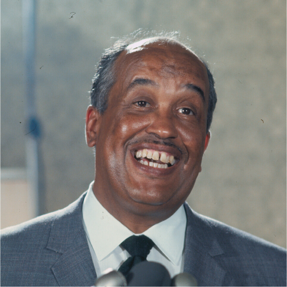
A photo of Herman Branson. Branson is smiling or laughing at something just off to the side of the camera. He is dressed in a grey suit with a white collared shirt and a black tie.
A. Herman Branson, professor of physics at Howard University and later president of Lincoln University.More information
A space filling model of the alpha helix structure of a protein. Carbons, represented by grey spheres, are arranged in a spiral. Other atoms, represented by spheres of other colors, are bound to the carbons. Blue spheres represent nitrogen. Red spheres represent oxygen. White spheres represent hydrogen. The majority of the atoms bound to the carbons are hydrogens.
B. An alpha helix structure of protein.
The Discovery of the Structure of DNA
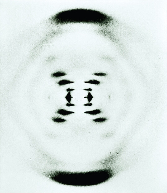
Scientists learned to read the cell’s central information molecule, deoxyribonucleic acid, better known as DNA (Figure 1.24). The famous double helix of DNA was determined by British researchers using X-ray crystallography. In 1953 at King’s College London, crystallographer Rosalind Franklin (1920–1958; Figure 1.25A) and her graduate student Raymond Gosling (1926–2015) obtained the most famous X-ray image ever taken, which revealed the structure of DNA. As a Jewish woman who supported relief work in Palestine, Franklin felt socially isolated at the male-dominated Protestant university. Nevertheless, the exceptional quality of her X-ray micrographs enabled her to propose that the DNA molecule is a double helix. The X-shaped pattern in her micrograph (Figure 1.25B) arises from the double-helical form of the DNA molecule.
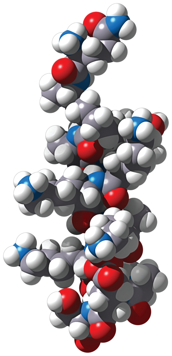
Figure 1.24 DNA: The Central Information Molecule
More information
A space filling model of the helical structure of double stranded D N A. The D N A is shown as a double helix comprised of several different atoms of various sizes. The atoms are represented by colored spheres. The atoms on the outsides of each helix are sulfur, oxygen, and carbon. The atoms between both helixes are nitrogen, oxygen, and carbon. The atoms are arranged such that the D N A has the appearance of a twisted ladder. Grey spheres represent carbon. Blue spheres represent nitrogen. Red spheres represent oxygen. Yellow spheres represent sulfur.
The sequence of base pairs in DNA encodes all the genetic information of an organism.
Figure 1.25Discovering the DNA Double Helix
More information
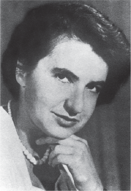
A historic portrait photo of Rosalind Franklin. She is smiling at the camera and resting her head on her hand. Her dark hair is styled into a neat updo.
A. Rosalind Franklin discovered that DNA forms a double helix.More information
An X ray crystallography pattern of D N A. The image appears as a circle. There is a thick, arch shaped shaded region at the top and bottom of the circle. In the center there are several black blobs oriented in an X shape. Three very faint, partial cocentric circles can be seen in the space from the sides of the x to right within the thick arch shapes and the outside of the circle.
B. “Photograph 51,” the famous X-ray crystallography pattern of DNA obtained by Rosalind Franklin and her graduate student Raymond Gosling.
More information
A historic photo of James Watson and Francis Crick standing with a floor to ceiling model of D N A. Watson and Crick are both wearing dark suits over a white collared shirt and black tie. Both are focused on the model between them. The model is an enlarged ball and stick model of the helical structure of D N A. They appear to be standing in a classroom.
C. James Watson and Francis Crick proposed a structure for the complementary pairing between bases of DNA.
Without Franklin’s knowledge, her colleague Maurice Wilkins (1916–2004) showed her data to a competitor, James Watson (born 1928). The X-shaped pattern led Watson and Francis Crick (1916–2004) to guess that the four bases of the DNA “alphabet” are paired in the interior of Franklin’s double helix (Figure 1.25C). They published their model in the journal Nature, without acknowledging their use of Franklin’s data.
The discovery of the double helix earned Watson, Crick, and Wilkins the 1962 Nobel Prize in Physiology or Medicine. Franklin died of ovarian cancer before the Nobel Prize was awarded. Before her death, however, she turned her efforts to the structure of ribonucleic acid (RNA). She determined the form of the RNA chromosome within tobacco mosaic virus, the first viral RNA to be characterized.
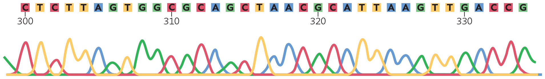
The structure of DNA base pairs led to the development of techniques for DNA sequencing, the reading of the sequence of DNA base pairs (the DNA sequencing technique is outlined in Section 8.6). Figure 1.26 shows an example of DNA sequence data, in which each color represents one of the four bases and each peak represents a DNA fragment terminating in that particular base. The order of fragment lengths yields the sequence of bases in one strand. Reading the sequence enabled microbiologists to determine the beginning and endpoint of genes and ultimately entire genomes.
Figure 1.26 DNA Sequence Data
More information
An illustration of sequenced D N A data. There is a genomic sequence of about 35 base pairs in length. The top part of the model is a line of colored boxes with letters inside of each. Each of the four letters has its own color. ‘C’ is in red, ‘T’ is in yellow, ‘A’ is in blue, and ‘G’ is in green. Below this line of colored letters is a graph like diagram. Four different colored lines run parallel to the letters above, each with several peaks. There are four line colors, each matching with a corresponding letter. The peaks of the colored lines appear when the corresponding base is listed directly above in the list of letters. The sequence reads C T C T T A G T G G C G C A G C T A A C T T A A G T T G A C C G.
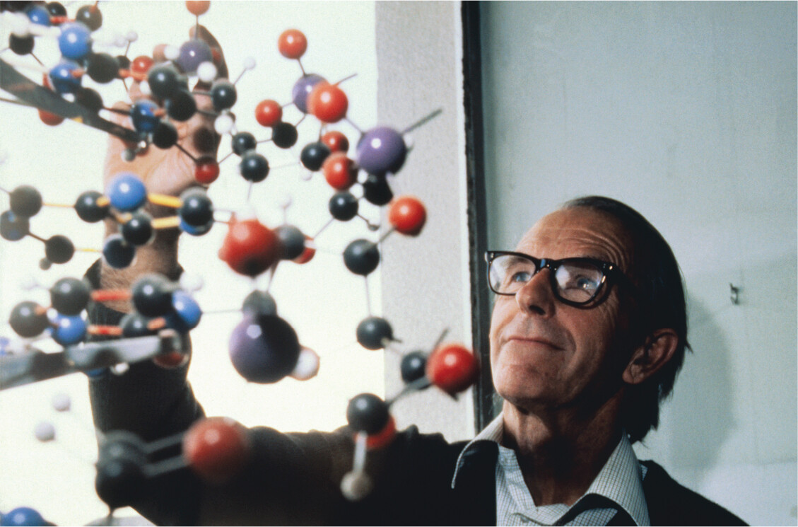
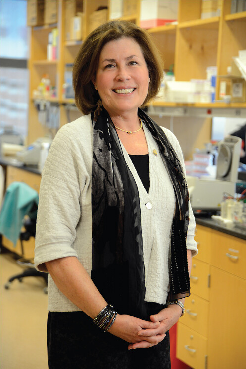
A method of DNA sequencing was devised by Frederick Sanger (1918–2013; Figure 1.27A) and was used to reveal the first genome of a virus in 1977. This method uses a bacterial enzyme, DNA polymerase. Sanger shared the 1980 Nobel Prize in Chemistry with Walter Gilbert and Paul Berg. The first genome sequence of a cellular microbe was obtained in 1995 for Haemophilus influenzae, a bacterium that causes ear infections and meningitis in children. The H. influenzae genome has nearly 2 million base pairs that specify about 1,700 genes. It was sequenced by a team of scientists led by Hamilton Smith and Craig Venter, who devised a special computational strategy for assembling large amounts of sequence data. This strategy was later applied to sequencing the human genome. Another group of scientists, led by Claire Fraser (Figure 1.27B), sequenced the genomes of many microbes, including Bacillus anthracis, the cause of anthrax, and Treponema pallidum, the cause of syphilis. Today, entire communities of microbial genomes are sequenced at once. The sequence of DNA from a microbial community is called a metagenome (discussed in Chapter 9).
Figure 1.27Microbial Genome Sequencers
More information
A photo of Frederick Sanger reaching up towards a hanging D N A model. The D N A model is 3 D and is made of balls and sticks that represent atoms and bonds. Sanger is dressed in a suit and wears glasses. His dark hair is combed neatly. He is smiling as he adjusts the model.
A. Frederick Sanger helped devise the method of DNA sequence analysis that is the basis of modern genome sequencing.More information
A photo of Claire Fraser standing in a laboratory. Fraser is smiling at the camera and holds her hands at her waist. She is dressed in formal attire. Behind her, the lab benches and shelves are covered in various pieces of lab equipment.
B. Claire Fraser led a team that completed the sequences of many microbial genomes.
Microbial Discoveries Transform Medicine and Industry
Twentieth-century microbiology transformed the practice of medicine and generated entire industries of biotechnology and bioremediation (the use of living organisms to decontaminate pollutants). After the discovery of penicillin, Americans poured millions of dollars of private and public funds into medical research. The March of Dimes campaign for private donations to prevent polio led to a vaccine that has nearly eliminated the disease. With the end of World War II, research on microbes and other aspects of biology drew increasing financial support from US government agencies—such as the National Institutes of Health and the National Science Foundation—and from governments of other countries, particularly the European nations and Japan. Further support came from private foundations such as the Pasteur Institute, the Wellcome Trust, and the Howard Hughes Medical Institute.
Today, microbial molecules continue to enable advances in medicine that until now could only have been imagined in science fiction. Human genome sequences are being used to determine the gene variants of given patients and thus optimize their treatment according to their genes. It will be important to ensure that genomic research addresses people of all kinds of genetic heritage, to sustain health equity and to avoid perpetuating health disparities (discussed in Chapter 26).
The tools from microbial molecular biology provide surprising ways to edit human DNA. Most important are these innovations:
The AIDS virus HIV was engineered to make DNA carrier molecules that can insert genes into cells of the human body. These DNA carriers, called lentivectors, are the basis of CAR-T (chimeric antigen receptor T-cell) therapy. In 2017, CAR-T therapy was approved to treat pediatric patients with acute lymphoblastic leukemia (see Chapter 12). Now CAR-T is being developed for other cancers.
CRISPR (clustered regularly interspaced short palindromic repeats) is a DNA editing tool that was engineered from the protein-RNA complex that defends bacteria against infection by viruses (bacteriophages). In 2019, the first therapeutic use of CRISPR was approved for cancer therapy. (See Chapter 9 for more on CRISPR.)
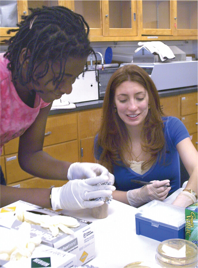
Careers in diverse fields attract student microbiologists (Figure 1.28; Table 1.2). Nurses, physician assistants, pharmacists, and other health care professionals use microbiological science to keep patients healthy and diagnose their illnesses. Public health workers apply statistical models to manage the health of communities and nations. Forensic technologists use DNA and microbial analysis to solve crimes. In environmental science, newly discovered microbes provide innovative ways to bioremediate wastes and control insect pests. On a global level, the challenges of pollution and climate change increasingly involve contributions of environmental microbiology to human health.
Figure 1.28 Microbiology Students at Kenyon College Prepare for Careers in Nursing and Medical Research
More information
A photo of two students working at a desk in a microbiology lab room. The students are smiling, but are focused on their work. They are both wearing disposable gloves. One student appears to be documenting information in a lab notebook and the other student appears to be analyzing the contents of a broth culture. A few agar plates and some lab equipment sit on the desk before them.
Table 1.2
Careers Involving Microbiology
Profession
Primary Activity
Nursing
Providing comprehensive care for patients with illness
Physician assistant
Practicing medicine under supervision of a licensed physician
Medical laboratory science
Diagnosing, treating, and developing therapies for human diseases
Veterinary laboratory science
Diagnosing, treating, and developing therapies for animal diseases
Public health
Monitoring, assessing, and promoting policies influencing the health of populations
Food and industrial microbiology
Developing microbial and industrial food products; addressing food contamination and bioremediation
Agricultural microbiology
Managing plant pathogens and plant-associated microbes
Environmental microbiology
Environmental remediation through microbial processes
Forensic microbiology
Analyzing microbial strains as evidence in criminal investigations
Experimental microbiology
Investigating fundamental questions about microbial form and function, genetics, and ecology
Microbiology: The Human Experience is your guide to microbes and the enormous impact they have on human life. Our focus is medical by design, but we also explore intriguing connections between microbes, Earth’s ecology, and human existence. Part I of the book lays the foundation for studying microorganisms and for understanding infectious disease. Chapter 1 provides the human time line of discovery. Next, Chapter 2 introduces the basic concepts of microbial disease, and then Chapter 3 describes the tools of microscopy that enable us to see cells and their inner workings. Chapter 4 explains the fundamental biochemistry needed to understand microbial life (and infectious disease), while Chapter 5 reveals the intricate structures of the microbes themselves. Chapter 6 introduces microbial growth and control within the human body and human-related environments. Throughout, we invite readers to share with us the excitement of discovery in microbiology and the potential applications of that knowledge to improving human health.
SECTION SUMMARY
The alpha helix structure of protein was discovered by Herman Branson and colleagues using X-ray diffraction crystallography.
The structure and function of DNA were revealed by a series of experiments in the twentieth century. The double-helical structure was shown by Rosalind Franklin through X-ray crystallography, and the base-pairing model was devised by James Watson and Francis Crick.
Genome sequence determination has shaped the study of biology in the twenty-first century.
Microbial discoveries transformed medicine and industry. Biotechnology enables the production of new kinds of pharmaceuticals and industrial products.
Microbiology today offers many different careers in the health field and in industry.